alabama ems patient care protocols eighth edition january 2016€¦ · adph oems patient care...
TRANSCRIPT
ADPH OEMS PATIENT CARE PROTOCOLS EIGHTH EDITION, JANUARY 2016
ALABAMA EMS PATIENT CARE PROTOCOLS
EIGHTH EDITION JANUARY 2016
OEMS
ADPH OEMS PATIENT CARE PROTOCOLS EIGHTH EDITION, JANUARY 2016
PATIENT CARE PROTOCOLS TABLE OF CONTENTS
Preface 1 Section 1: Policies 3 1.01 Scope of Practice 4 1.02 Communications 8 1.03 Death in the Field 9 1.04 Disputes Regarding Patient Care 11 1.05 Documentation of Care 12 1.06 Do Not Attempt to Resuscitate (DNAR) 14 1.07 Medical Direction Hospital 15 1.08 Medical Management of the Scene 17 1.08 Medical Professionals at the Scene 19 1.10 Medication and Procedure Categories 20 1.11 Optional Medications and Procedures 21 1.12 Patient Rights 22 1.13 Physician Medical Direction 24 1.14 Refusal of Care or Transport 25 1.15 Time at the Scene 26 1.16 Trauma System 27 Section 2: Operational Guidelines 30 2.01 Cancellation/Slow Down 31 2.02 Crime Scene Response 32 2.03 Hazardous Materials 34 2.04 Helicopter EMS 37 2.05 Staging for High Risk Response 39 Section 3: Treatment Protocols 40 3.01 General Patient Care 41 3.02 Abdominal Pain 42 3.03 Adrenal Insufficiency 44 3.04 Allergic Reaction 46 3.05 Altered Mental Status 48 3.06 Amputation 50 3.07 Bites and Envenomations 52 3.08 Burns 54 3.09 Cardiac Arrest, Adult 59 3.10 Cardiac Arrest, Pediatric 63 3.11 Cardiac Dysrhythmias, Adult 67 3.12 Cardiac Dysrhythmias, Pediatric 69 3.13 Chest Pain or Acute Coronary Syndrome (ACS) 71
ADPH OEMS PATIENT CARE PROTOCOLS EIGHTH EDITION, JANUARY 2016
3.14 Childbirth 73 3.15 Congestive Heart Failure 75 3.16 Electromuscular Incapacitation Devices (Taser®) 77 3.17 Fractures and Dislocations 79 3.18 Head Trauma 81 3.19 Hypertensive Emergencies 83 3.20 Hyperthermia 84 3.21 Hypoglycemia 85 3.22 Hypothermia 86 3.23 Influenza/Respiratory Illness 88 3.24 Nausea and Vomiting 90 3.25 Near Drowning 91 3.26 Newborn 92 3.27 Poisons and Overdoses 94 3.28 Preeclampsia/Eclampsia 97 3.29 Respiratory Distress 98 3.30 Seizure 100 3.31 Shock 102 3.32 Spinal Injury 103 3.33 Stroke 107 3.34 Syncope 111 3.35 Vaginal Bleeding 112 Section 4: Procedures 113 4.01 Blind Insertion Airway Devices (BIAD) 114 4.02 Capnography 115 4.03 Cardioversion (Synchronized) 116 4.04 Chest Decompression 117 4.05 Continuous Positive Airway Pressure (CPAP) 119 4.06 ECG (12-Lead) 121 4.07 Endotracheal Intubation (Oral) 122 4.08 Endotracheal Intubation (Nasal) 124 4.09 External Pacing 126 4.10 Hemostatic Agents 127 4.11 Intraosseous Therapy 128 4.12 Intravenous Therapy 130 4.13 Patient Restraint 131 4.14 Rectal Diazepam Administration 132 4.15 Transportation of Pediatric Patients 133 Section 5: Medications 135 5.01 Adenosine 136 5.02 Albuterol and Ipratropium 137
ADPH OEMS PATIENT CARE PROTOCOLS EIGHTH EDITION, JANUARY 2016
5.03 Amiodarone 138 5.04 Aspirin 139 5.05 Atropine Sulfate 140 5.06 Calcium Chloride 141 5.07 Dextrose 142 5.08 Diazepam 143 5.09 Diphenhydramine 144 5.10 Dopamine 145 5.11 Epinephrine 147 5.12 Fentanyl 148 5.13 Furosemide 149 5.14 Glucagon 150 5.15 Haloperidol 151 5.16 Hydroxocobalamin (Cyanokit) 152 5.17 Lidocaine 153 5.18 Lorazepam 154 5.19 Magnesium Sulfate 155 5.20 Midazolam 156 5.21 Morphine Sulfate 157 5.22 Naloxone 158 5.23 Nitroglycerin 159 5.24 Nitrous Oxide 160 5.25 Normal Saline 161 5.26 Ondansetron 162 5.27 Oxygen 163 5.28 Sodium Bicarbonate 164 5.29 Thiamine 165 Section 6: Acceptable EMS Equipment and Devices 166 6.01 Blind Insertion Airway Devices (BIAD) 167 6.02 Bougie (for Difficult Intubation) 167 6.03 Hemostatic Agents 167 6.04 Intraosseous Needle Insertion Devices 167 Section 7: Disaster 168 7.01 Respiratory Illness/Influenza Mass Casualty Emergency 169 7.02 Search and Rescue Marking System 172 7.03 Triage of Mass Casualties 173 Section 8: Forms 176 8.01 Chest Decompression Report 177 8.02 Do Not Attempt Resuscitation (DNAR) Form 178 8.03 Request to Be Transported to a Hospital on Divert 180
ADPH OEMS PATIENT CARE PROTOCOLS EIGHTH EDITION, JANUARY 2016
8.04 Thrombolytic Checklist (STEMI) 181 8.05 Thrombolytic Checklist (Stroke) 182 Section 9: Expanded Scope of Practice 183 9.01 Rapid Sequence Intubation 184 9.02 Needle Cricothyroidotomy 190 9.03 Medications 192
ADPH EMS PROTOCOLS EIGHTH EDITION, JANUARY 2016Preface
ADPH OEMS PATIENT CARE PROTOCOLS 1 EIGHTH EDITION, JANUARY 2016
KEY POINTS
These protocols are intended to guide Emergency Medical Services Personnel (EMSP) in the response and management of emergency situations and the care and treatment of patients. Anyone who wants to change the protocols can make a request in writing to the State Emergency Medical Control Committee, or an EMSP may make the request by email to: Dr. William Crawford, State EMS Medical Director Alabama State Emergency Medical Control Committee c/o Office of EMS Alabama Department of Public Health (ADPH) P.O. Box 303017 Montgomery, AL 36130-3017 Or [email protected] This manual contains ALL the medications and procedures allowed for EMSP in Alabama. EMSP are responsible for their actions within the respective scope of practice of the license that they hold. Online Medical Direction (OLMD) cannot order EMSP to perform procedures or administer medications that are not presented in these protocols. EMSP should respectfully decline any orders which would cause them to violate their scope of practice. The medication section of this manual is provided for information purposes only. EMSP may administer medications only as listed in the protocol unless OLMD orders a deviation. This manual also serves as a reference for physicians providing OLMD to EMSP. Treatment direction which is more appropriate to the patient’s condition than the protocol should be provided by the physician as long as the EMSP scope of practice is not exceeded. Treatment direction includes basic care, advanced procedures, and medication administration. OLMD can expect an EMSP to respectfully decline any orders which would cause them to violate their scope of practice. Pediatric information is differentiated by label and font characteristics. Anything pertaining to pediatric patients will be presented in Green Bold Tahoma Font. Unless otherwise noted in a protocol, a pediatric patient is defined as someone 15 years old or younger.

ADPH EMS PROTOCOLS EIGHTH EDITION, JANUARY 2016Preface
ADPH OEMS PATIENT CARE PROTOCOLS 2 EIGHTH EDITION, JANUARY 2016
PROTOCOL UPDATES
The ADPH EMS Protocols are revised through updates performed by request of the State Emergency Medical Control Committee (SEMCC) or the Office of EMS (OEMS) Director. Individual protocols and guidelines are updated through REVISIONS. Each protocol can be revised individually and the revision letter and revision date are noted on the protocol in the upper right hand corner. Periodically, the revisions are incorporated into the manual and a new Edition is released. The new EDITION number and date are printed on the cover and the lower right footnote.
ADPH EMS PROTOCOLS EIGHTH EDITION JANUARY 2016 Policies 1
ADPH OEMS PATIENT CARE PROTOCOLS 3 EIGHTH EDITION, JANUARY 2016
POLICIES EIGHTH EDITION JANUARY 2016Scope of Practice 1.01
ADPH OEMS PATIENT CARE PROTOCOLS 4 EIGHTH EDITION, JANUARY 2016
KEY POINTS
Licensed Emergency Medical Services Personnel (EMSPs) are authorized to perform procedures and administer medications as defined by these protocols. Each level of EMSP, as defined by the EMS Rules, has a specific list of authorized procedures and medications as defined by that level’s scope of practice. EMSPs are prohibited from performing any procedure or utilizing any medication not approved by the State Board of Health even though they may have been taught these medications and procedures in their EMSP curriculum. Lower level EMSPs can assist higher level EMSPs with patient care activities as long as the lower level EMSP does not exceed his/her Scope of Practice regarding administration of medications or performance of procedures. Ultimately, the higher level EMSP is responsible for patient care and documentation.
POLICIES EIGHTH EDITION JANUARY 2016Scope of Practice (continued) 1.01
ADPH OEMS PATIENT CARE PROTOCOLS 5 EIGHTH EDITION, JANUARY 2016
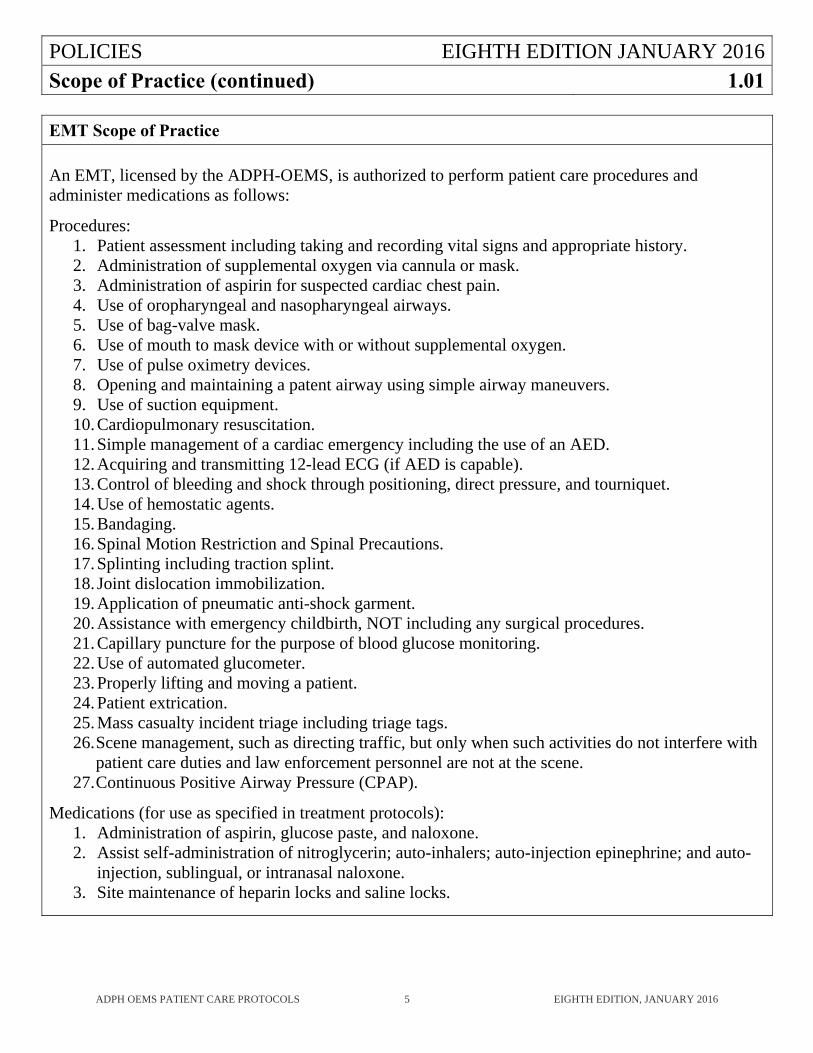
EMT Scope of Practice
An EMT, licensed by the ADPH-OEMS, is authorized to perform patient care procedures and administer medications as follows: Procedures:
1. Patient assessment including taking and recording vital signs and appropriate history. 2. Administration of supplemental oxygen via cannula or mask. 3. Administration of aspirin for suspected cardiac chest pain. 4. Use of oropharyngeal and nasopharyngeal airways. 5. Use of bag-valve mask. 6. Use of mouth to mask device with or without supplemental oxygen. 7. Use of pulse oximetry devices. 8. Opening and maintaining a patent airway using simple airway maneuvers. 9. Use of suction equipment. 10. Cardiopulmonary resuscitation. 11. Simple management of a cardiac emergency including the use of an AED. 12. Acquiring and transmitting 12-lead ECG (if AED is capable). 13. Control of bleeding and shock through positioning, direct pressure, and tourniquet. 14. Use of hemostatic agents. 15. Bandaging. 16. Spinal Motion Restriction and Spinal Precautions. 17. Splinting including traction splint. 18. Joint dislocation immobilization. 19. Application of pneumatic anti-shock garment. 20. Assistance with emergency childbirth, NOT including any surgical procedures. 21. Capillary puncture for the purpose of blood glucose monitoring. 22. Use of automated glucometer. 23. Properly lifting and moving a patient. 24. Patient extrication. 25. Mass casualty incident triage including triage tags. 26. Scene management, such as directing traffic, but only when such activities do not interfere with
patient care duties and law enforcement personnel are not at the scene. 27. Continuous Positive Airway Pressure (CPAP).
Medications (for use as specified in treatment protocols):
1. Administration of aspirin, glucose paste, and naloxone. 2. Assist self-administration of nitroglycerin; auto-inhalers; auto-injection epinephrine; and auto-
injection, sublingual, or intranasal naloxone. 3. Site maintenance of heparin locks and saline locks.
POLICIES EIGHTH EDITION JANUARY 2016Scope of Practice (continued) 1.01
ADPH OEMS PATIENT CARE PROTOCOLS 6 EIGHTH EDITION, JANUARY 2016
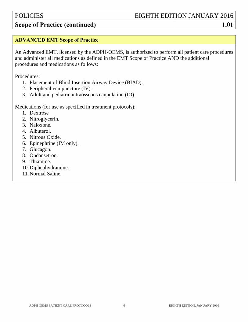
ADVANCED EMT Scope of Practice
An Advanced EMT, licensed by the ADPH-OEMS, is authorized to perform all patient care procedures and administer all medications as defined in the EMT Scope of Practice AND the additional procedures and medications as follows: Procedures:
1. Placement of Blind Insertion Airway Device (BIAD). 2. Peripheral venipuncture (IV). 3. Adult and pediatric intraosseous cannulation (IO).
Medications (for use as specified in treatment protocols):
1. Dextrose 2. Nitroglycerin. 3. Naloxone. 4. Albuterol. 5. Nitrous Oxide. 6. Epinephrine (IM only). 7. Glucagon. 8. Ondansetron. 9. Thiamine. 10. Diphenhydramine. 11. Normal Saline.
POLICIES EIGHTH EDITION JANUARY 2016Scope of Practice (continued) 1.01
ADPH OEMS PATIENT CARE PROTOCOLS 7 EIGHTH EDITION, JANUARY 2016
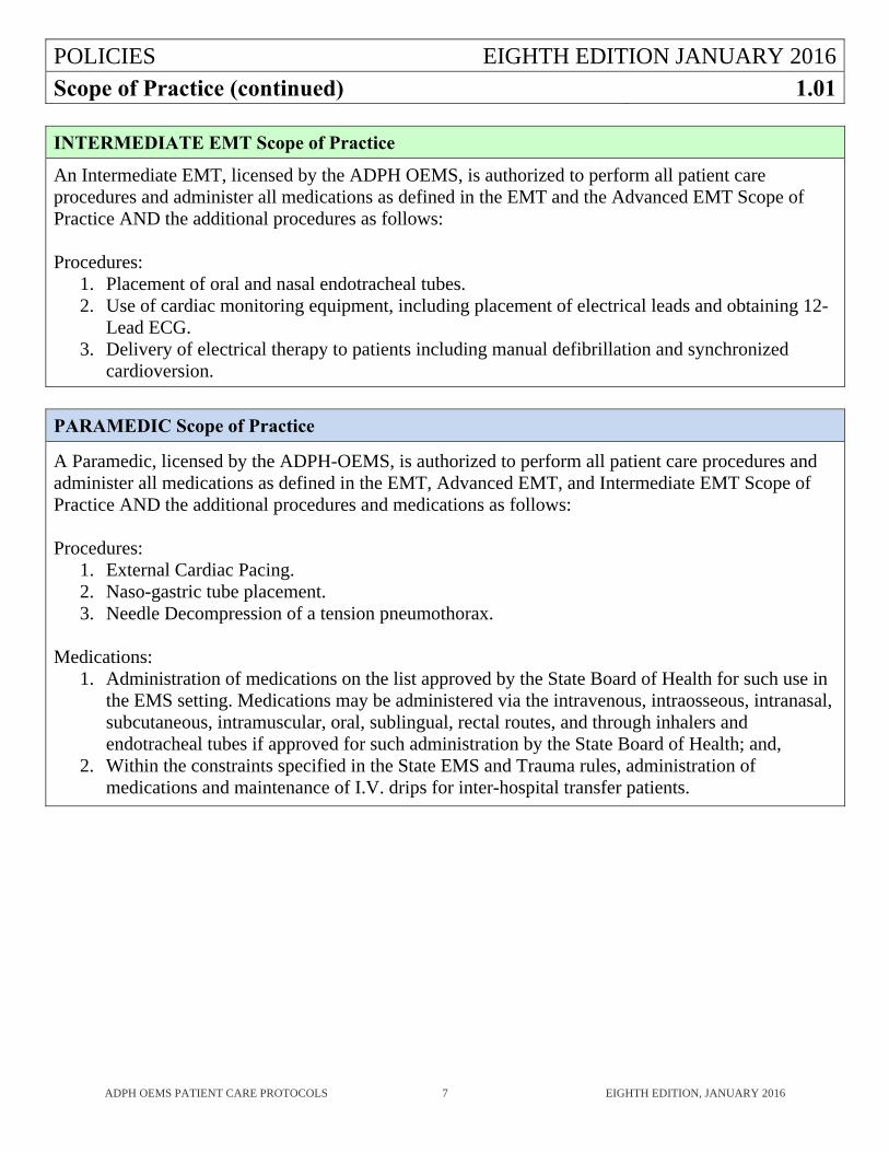
INTERMEDIATE EMT Scope of Practice
An Intermediate EMT, licensed by the ADPH OEMS, is authorized to perform all patient care procedures and administer all medications as defined in the EMT and the Advanced EMT Scope of Practice AND the additional procedures as follows: Procedures:
1. Placement of oral and nasal endotracheal tubes. 2. Use of cardiac monitoring equipment, including placement of electrical leads and obtaining 12-
Lead ECG. 3. Delivery of electrical therapy to patients including manual defibrillation and synchronized
cardioversion.
PARAMEDIC Scope of Practice
A Paramedic, licensed by the ADPH-OEMS, is authorized to perform all patient care procedures and administer all medications as defined in the EMT, Advanced EMT, and Intermediate EMT Scope of Practice AND the additional procedures and medications as follows: Procedures:
1. External Cardiac Pacing. 2. Naso-gastric tube placement. 3. Needle Decompression of a tension pneumothorax.
Medications:
1. Administration of medications on the list approved by the State Board of Health for such use in the EMS setting. Medications may be administered via the intravenous, intraosseous, intranasal, subcutaneous, intramuscular, oral, sublingual, rectal routes, and through inhalers and endotracheal tubes if approved for such administration by the State Board of Health; and,
2. Within the constraints specified in the State EMS and Trauma rules, administration of medications and maintenance of I.V. drips for inter-hospital transfer patients.
POLICIES EIGHTH EDITION JANUARY 2016Communications 1.02
ADPH OEMS PATIENT CARE PROTOCOLS 8 EIGHTH EDITION, JANUARY 2016
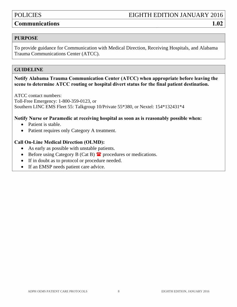
PURPOSE
To provide guidance for Communication with Medical Direction, Receiving Hospitals, and Alabama Trauma Communications Center (ATCC).
GUIDELINE
Notify Alabama Trauma Communication Center (ATCC) when appropriate before leaving the scene to determine ATCC routing or hospital divert status for the final patient destination. ATCC contact numbers: Toll-Free Emergency: 1-800-359-0123, or Southern LINC EMS Fleet 55: Talkgroup 10/Private 55*380, or Nextel: 154*132431*4 Notify Nurse or Paramedic at receiving hospital as soon as is reasonably possible when:
Patient is stable. Patient requires only Category A treatment.
Call On-Line Medical Direction (OLMD):
As early as possible with unstable patients. Before using Category B (Cat B) procedures or medications. If in doubt as to protocol or procedure needed. If an EMSP needs patient care advice.
POLICIES EIGHTH EDITION JANUARY 2016
Death in the Field 1.03
ADPH OEMS PATIENT CARE PROTOCOLS 9 EIGHTH EDITION, JANUARY 2016
PURPOSE
To establish guidelines for determining when resuscitative efforts should not be initiated or should be terminated.
GUIDELINE
WITHHOLDING RESUSCITATIVE EFFORTS 1. Determining death in the field (DIF) without initiating resuscitative efforts should be considered
under any of the following conditions: a. Decapitation. b. Massive crush injury or evisceration of the heart, lung, or brain. c. Incineration. d. Rigor Mortis in a warm environment. e. Venous pooling in dependent body parts (dependent lividity). f. Decomposition. g. Patient qualifies as a “DNAR” patient (see DNAR Protocol 1.06). h. A pulseless, apneic patient in a mass casualty incident, multiple-patient scene, where the
resources of the system are required for the stabilization of living patients. i. A victim of blunt trauma with no vital signs in the field.
2. OLMD must be contacted and must confirm the withholding of resuscitative efforts. 3. If the patient is declared dead on scene, the body must not be moved until the proper authority
(such as law enforcement agencies, the coroner, the medical examiner, or their designee), has been notified (if not already on scene), and they agree to the movement of the body.
Traumatic Cardiac Arrest Special Considerations:
1. In deaths from blunt trauma, a monitor is not necessary to use in initial assessment of the patient unless the paramedic doubts death has occurred. If the monitor is used, only a recognizable QRS of at least eighty (80) per minute should be considered compatible with life in these trauma patients.
2. In cases of penetrating torso injury with no vital signs in the field, OLMD should be immediately contacted without delay. OLMD can determine whether to continue resuscitative efforts.
3. If OLMD stops resuscitation during transport, the patient must be taken to that OLMD physician to be pronounced dead. In some circumstances, it is possible that OLMD may not be working in the receiving facility. If the OLMD is not at the receiving facility and resuscitation is terminated during transport, you much notify the receiving facility as soon as possible.
POLICIES EIGHTH EDITION JANUARY 2016
Death in the Field (continued) 1.03
ADPH OEMS PATIENT CARE PROTOCOLS 10 EIGHTH EDITION, JANUARY 2016
GUIDELINE (continued)
DETERMINING DEATH IN CARDIAC MEDICAL ARREST 1. Cardiopulmonary resuscitation and advanced life support may be terminated by prehospital
personnel if all of the following criteria are met: a. Patient is in cardiac arrest at the time of arrival of advanced life support. b. Appropriate full advanced life support procedures, including Advanced Airway
placement, are performed for twenty minutes with no spontaneous pulse, and no evidence of neurologic function, unless earlier termination is appropriate as determined by OLMD.
c. OLMD approves termination of efforts. d. If OLMD stops resuscitation during transport, the patient must be taken to that OLMD
physician to be pronounced dead. In some circumstances, it is possible that OLMD may not be working in the receiving facility. If the OLMD is not at the receiving facility and resuscitation is terminated during transport, you must notify the receiving facility as soon as possible.
e. If the patient is declared dead on scene, the body must not be moved until the proper authority (such as law enforcement agencies, the coroner, the medical examiner, or their designee), has been notified (if not already on scene), and they agree to the movement of the body.
2. All patients in Ventricular Fibrillation should, in general, have full resuscitation continued and be transported, except when DNAR or other withholding resuscitative efforts apply. If in doubt, contact OLMD.
3. Termination will not be considered in any of the following circumstances: a. Patients with persistent ventricular fibrillation or pulseless ventricular tachycardia. b. Patients who have return of spontaneous pulse at any time during the resuscitative effort. c. Patients who exhibit neurologic function. d. Patients who arrest after the arrival of advanced life support.
DOCUMENTATION 1. All patient care provided should be documented with procedure and time. 2. In non-traumatic deaths, all non-resuscitation or stopped resuscitation cases should have an
ECG rhythm strip that shows the patient’s rhythm. 3. All conversations with physicians should be fully documented with physician’s name, times,
and instructions. 4. If resuscitation is withheld on scene, and the coroner or medical examiner is not coming to the
scene, if possible, obtain name and address of the deceased, name, address, and phone number of a family member, and name and phone number of patient’s private physician.
PRECAUTIONS 1. Most victims of electrocution, lightning, and drowning should have resuscitative efforts begun
and be transported to the hospital. 2. Hypothermic patients should be treated using the Hypothermia protocol (3.22). 3. Consider the needs of survivors when discontinuing resuscitation.
POLICIES EIGHTH EDITION JANUARY 2016
Disputes Regarding Patient Care 1.04
ADPH OEMS PATIENT CARE PROTOCOLS 11 EIGHTH EDITION, JANUARY 2016
PURPOSE
To describe how EMS personnel should resolve disputes with each other or other medical professionals at emergency scenes, upon hospital arrival, or anytime the patient is in the care of the EMS provider.
GUIDELINE
Disagreements about care should be handled in a professional manner so as not to detract from patient care.
The ADPH EMS Patient Care Protocols should be followed whenever possible and should be the basis for resolving disputes.
If there is a dispute between EMS personnel or medical professionals concerning the care of a patient, OLMD should be contacted in order to resolve the dispute.
Written reports should be prepared concerning any dispute arising at the scene, with a copy sent to the Off-Line Medical Director of each service and pertinent regional EMS agency or ADPH OEMS.
POLICIES EIGHTH EDITION JANUARY 2016
Documentation of Care 1.05
ADPH OEMS PATIENT CARE PROTOCOLS 12 EIGHTH EDITION, JANUARY 2016
PURPOSE
1. Each EMS provider shall ensure that an accurate and complete patient care report is prepared
for each instance in which:
a. A patient was assessed.
b. Medical care was rendered.
c. A patient was transported.
d. A patient was pronounced dead at the scene.
e. A patient was transferred to another licensed service.
f. A patient was transferred from one medical facility to another.
g. The person or persons for whom EMS was dispatched refused treatment, transport, or
both.
2. Documentation should include at least:
a. Patient problem presented.
b. History.
c. Primary Survey.
d. Vital signs including pulse oximetry, with time.
e. Secondary Survey.
f. Treatment provided and time.
g. ECG strip, if monitored.
h. Capnography strip, if monitored.
i. Any change in condition of patient.
j. OLMD contact.
k. Any deviation from protocol.
POLICIES EIGHTH EDITION JANUARY 2016
Documentation of Care (continued) 1.05
ADPH OEMS PATIENT CARE PROTOCOLS 13 EIGHTH EDITION, JANUARY 2016
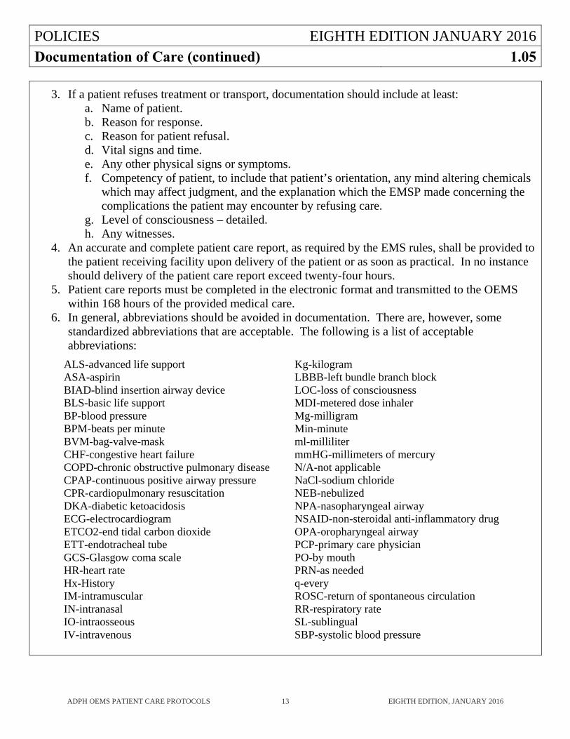
3. If a patient refuses treatment or transport, documentation should include at least: a. Name of patient. b. Reason for response. c. Reason for patient refusal. d. Vital signs and time. e. Any other physical signs or symptoms. f. Competency of patient, to include that patient’s orientation, any mind altering chemicals
which may affect judgment, and the explanation which the EMSP made concerning the complications the patient may encounter by refusing care.
g. Level of consciousness – detailed. h. Any witnesses.
4. An accurate and complete patient care report, as required by the EMS rules, shall be provided to the patient receiving facility upon delivery of the patient or as soon as practical. In no instance should delivery of the patient care report exceed twenty-four hours.
5. Patient care reports must be completed in the electronic format and transmitted to the OEMS within 168 hours of the provided medical care.
6. In general, abbreviations should be avoided in documentation. There are, however, some standardized abbreviations that are acceptable. The following is a list of acceptable abbreviations:
ALS-advanced life support ASA-aspirin BIAD-blind insertion airway device BLS-basic life support BP-blood pressure BPM-beats per minute BVM-bag-valve-mask CHF-congestive heart failure COPD-chronic obstructive pulmonary disease CPAP-continuous positive airway pressure CPR-cardiopulmonary resuscitation DKA-diabetic ketoacidosis ECG-electrocardiogram ETCO2-end tidal carbon dioxide ETT-endotracheal tube GCS-Glasgow coma scale HR-heart rate Hx-History IM-intramuscular IN-intranasal IO-intraosseous IV-intravenous
Kg-kilogram LBBB-left bundle branch block LOC-loss of consciousness MDI-metered dose inhaler Mg-milligram Min-minute ml-milliliter mmHG-millimeters of mercury N/A-not applicable NaCl-sodium chloride NEB-nebulized NPA-nasopharyngeal airway NSAID-non-steroidal anti-inflammatory drug OPA-oropharyngeal airway PCP-primary care physician PO-by mouth PRN-as needed q-every ROSC-return of spontaneous circulation RR-respiratory rate SL-sublingual SBP-systolic blood pressure
POLICIES EIGHTH EDITION JANUARY 2016
Do Not Attempt to Resuscitate (DNAR) 1.06
ADPH OEMS PATIENT CARE PROTOCOLS 14 EIGHTH EDITION, JANUARY 2016
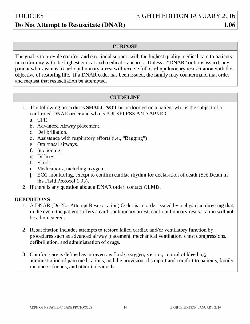
PURPOSE
The goal is to provide comfort and emotional support with the highest quality medical care to patients in conformity with the highest ethical and medical standards. Unless a “DNAR” order is issued, any patient who sustains a cardiopulmonary arrest will receive full cardiopulmonary resuscitation with the objective of restoring life. If a DNAR order has been issued, the family may countermand that order and request that resuscitation be attempted.
GUIDELINE
1. The following procedures SHALL NOT be performed on a patient who is the subject of a confirmed DNAR order and who is PULSELESS AND APNEIC. a. CPR. b. Advanced Airway placement. c. Defibrillation. d. Assistance with respiratory efforts (i.e., “Bagging”) e. Oral/nasal airways. f. Suctioning. g. IV lines. h. Fluids. i. Medications, including oxygen. j. ECG monitoring, except to confirm cardiac rhythm for declaration of death (See Death in
the Field Protocol 1.03). 2. If there is any question about a DNAR order, contact OLMD.
DEFINITIONS
1. A DNAR (Do Not Attempt Resuscitation) Order is an order issued by a physician directing that, in the event the patient suffers a cardiopulmonary arrest, cardiopulmonary resuscitation will not be administered.
2. Resuscitation includes attempts to restore failed cardiac and/or ventilatory function by procedures such as advanced airway placement, mechanical ventilation, chest compressions, defibrillation, and administration of drugs.
3. Comfort care is defined as intravenous fluids, oxygen, suction, control of bleeding, administration of pain medications, and the provision of support and comfort to patients, family members, friends, and other individuals.
POLICIES EIGHTH EDITION JANUARY 2016Medical Direction Hospitals 1.07
ADPH OEMS PATIENT CARE PROTOCOLS 15 EIGHTH EDITION, JANUARY 2016
KEY POINTS
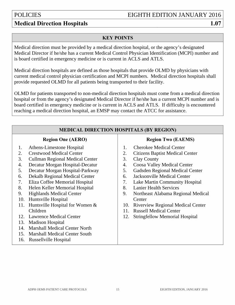
Medical direction must be provided by a medical direction hospital, or the agency’s designated Medical Director if he/she has a current Medical Control Physician Identification (MCPI) number and is board certified in emergency medicine or is current in ACLS and ATLS. Medical direction hospitals are defined as those hospitals that provide OLMD by physicians with current medical control physician certification and MCPI numbers. Medical direction hospitals shall provide requested OLMD for all patients being transported to their facility. OLMD for patients transported to non-medical direction hospitals must come from a medical direction hospital or from the agency’s designated Medical Director if he/she has a current MCPI number and is board certified in emergency medicine or is current in ACLS and ATLS. If difficulty is encountered reaching a medical direction hospital, an EMSP may contact the ATCC for assistance.
MEDICAL DIRECTION HOSPITALS (BY REGION)
Region One (AERO)
1. Athens-Limestone Hospital 2. Crestwood Medical Center 3. Cullman Regional Medical Center 4. Decatur Morgan Hospital-Decatur 5. Decatur Morgan Hospital-Parkway 6. Dekalb Regional Medical Center 7. Eliza Coffee Memorial Hospital 8. Helen Keller Memorial Hospital 9. Highlands Medical Center 10. Huntsville Hospital 11. Huntsville Hospital for Women &
Children 12. Lawrence Medical Center 13. Madison Hospital 14. Marshall Medical Center North 15. Marshall Medical Center South 16. Russellville Hospital
Region Two (EAEMS)
1. Cherokee Medical Center 2. Citizens Baptist Medical Center 3. Clay County 4. Coosa Valley Medical Center 5. Gadsden Regional Medical Center 6. Jacksonville Medical Center 7. Lake Martin Community Hospital 8. Lanier Health Services 9. Northeast Alabama Regional Medical
Center 10. Riverview Regional Medical Center 11. Russell Medical Center 12. Stringfellow Memorial Hospital
POLICIES EIGHTH EDITION JANUARY 2016
Medical Direction Hospitals (continued) 1.07
ADPH OEMS PATIENT CARE PROTOCOLS 16 EIGHTH EDITION, JANUARY 2016
MEDICAL DIRECTION HOSPITALS (BY REGION) (continued)
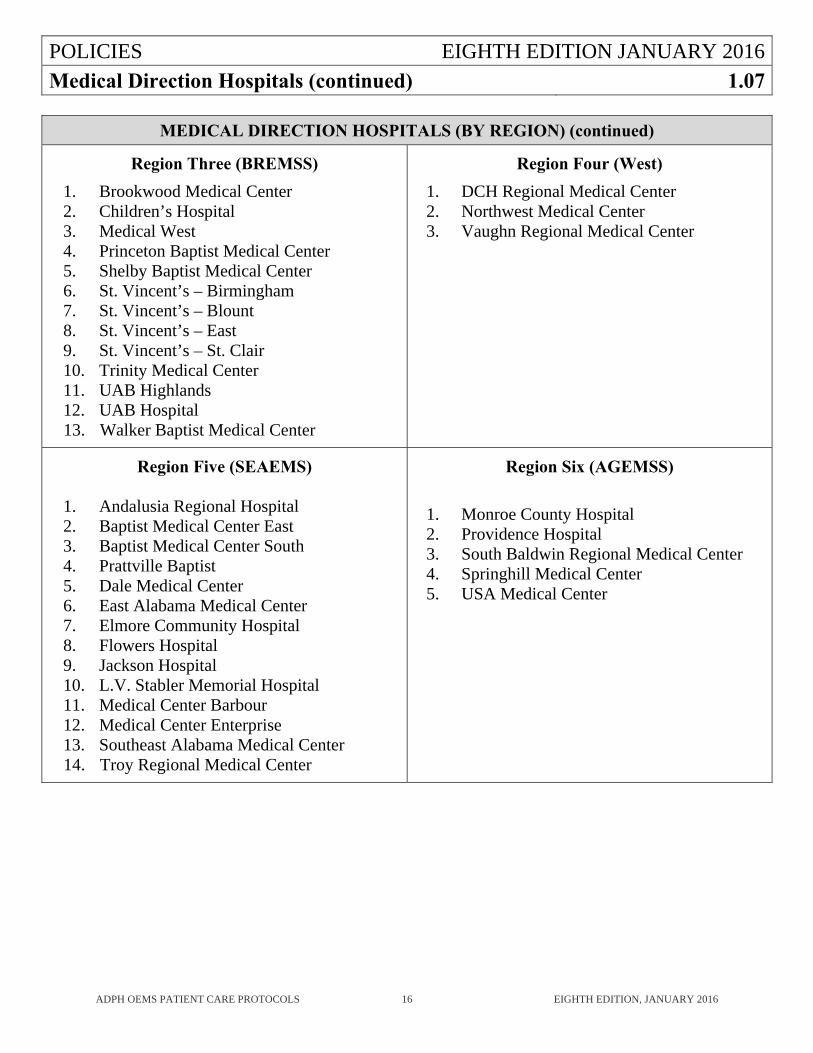
Region Three (BREMSS)
1. Brookwood Medical Center 2. Children’s Hospital 3. Medical West 4. Princeton Baptist Medical Center 5. Shelby Baptist Medical Center 6. St. Vincent’s – Birmingham 7. St. Vincent’s – Blount 8. St. Vincent’s – East 9. St. Vincent’s – St. Clair 10. Trinity Medical Center 11. UAB Highlands 12. UAB Hospital 13. Walker Baptist Medical Center
Region Four (West)
1. DCH Regional Medical Center 2. Northwest Medical Center 3. Vaughn Regional Medical Center
Region Five (SEAEMS)
1. Andalusia Regional Hospital 2. Baptist Medical Center East 3. Baptist Medical Center South 4. Prattville Baptist 5. Dale Medical Center 6. East Alabama Medical Center 7. Elmore Community Hospital 8. Flowers Hospital 9. Jackson Hospital 10. L.V. Stabler Memorial Hospital 11. Medical Center Barbour 12. Medical Center Enterprise 13. Southeast Alabama Medical Center 14. Troy Regional Medical Center
Region Six (AGEMSS)
1. Monroe County Hospital 2. Providence Hospital 3. South Baldwin Regional Medical Center 4. Springhill Medical Center 5. USA Medical Center
POLICIES EIGHTH EDITION JANUARY 2016
Medical Management of the Scene 1.08
ADPH OEMS PATIENT CARE PROTOCOLS 17 EIGHTH EDITION, JANUARY 2016
PURPOSE
To assist in determining who is in charge of patient care at the scene of an emergency.
GUIDELINE
1. The highest level EMSP on the first arriving ALS unit will assume responsibility for directing overall patient care and will continue this function unless relieved by the responding jurisdiction’s personnel. The responding jurisdiction’s personnel must be authorized such responsibilities by local, city, county, district ordinances or legislative acts, or must have been dispatched by the recognized dispatch agency. These personnel must also be of equal or higher EMSP license level.
2. It is the responsibility of the highest level EMSP on the scene to determine the appropriate level of care for transport of the patient. When the highest level EMSP on the scene determines that a lower level of care is appropriate for the patient, that EMSP may turn over patient care to an EMSP licensed at a lower level of care who is willing to accept patient care responsibilities.
3. An EMSP shall yield patient care responsibilities to an EMSP licensed at a higher level when directed to do so by the higher-level EMSP. An Advanced EMT, Intermediate EMT or Paramedic who is providing ALS care to a patient may be relieved by any other licensed Advanced EMT, Intermediate EMT or Paramedic authorized to provide the necessary level of care if the relieving EMSP is willing to assume patient care duties.
4. The responsibilities of the EMSP directing overall patient care include: a. Avoiding direct patient care activities if enough personnel are available. This EMSP must
watch over the entire patient care scene activities and be sure that the patient care activities are being accomplished in a rapid, efficient, appropriate, and timely manner. If there are only two EMSPs at the scene, the senior EMSP must do those patient care activities which will allow him/her to watch over the whole scene easily.
b. Assigning other EMSPs to provide patient care. c. Determining when transportation of the patient is to occur. d. Performing medical coordination with all agencies and personnel.
5. The EMSP directing overall patient care will be held responsible for general patient care activities performed at the scene, and he/she will be so identified on all patient care reports.
POLICIES EIGHTH EDITION JANUARY 2016
Medical Management of the Scene (continued) 1.08
ADPH OEMS PATIENT CARE PROTOCOLS 18 EIGHTH EDITION, JANUARY 2016
6. If a patient requires transport, and the Person-In-Charge (PIC) is from a non-transport agency, direction of patient care will be turned over to the transporting EMSPs when: (1) the patient is placed on the transporting unit’s gurney, unless PIC agency personnel accompany transport, or (2) at a time agreed upon by both EMSPs. Continued patient care will then become the responsibility of the transporting unit. The approximate time of transfer will be noted on all patient care forms. It is expected that an orderly transfer of information and cooperative management of patient needs will occur. When there are two agencies responding to a call, and a transfer of care occurs, there will be two PICs noted on all patient care forms: the first arriving PIC and the transporting PIC.
7. If a patient is transported to a hospital, the highest-level EMSP shall continue to provide care until relieved by appropriate hospital medical personnel.
8. Any disputes about patient care should be referred immediately to and resolved by the OLMD physician.
9. Patient care may be transferred to a flight nurse or physician for air transportation.
10. Patient care may also be transferred to a physician at the scene (see protocol for Medical Professionals at the Scene 1.09).
POLICIES EIGHTH EDITION JANUARY 2016Medical Professionals at the Scene 1.09
ADPH OEMS PATIENT CARE PROTOCOLS 19 EIGHTH EDITION, JANUARY 2016
PURPOSE
To define the role of medical professionals during a prehospital emergency.
GUIDELINE
Medical professionals at the scene of an emergency may provide assistance and shall be treated with professional courtesy.
Medical professionals who offer their assistance at the scene should be asked to identify themselves and their level of training. If the medical professional wishes to assist with care given to the patient after arrival of the EMS unit, the senior EMSP should inform him/her that it is ADPH/EMS policy that the medical professional provide proof of his/her identity.
The authority for medical direction of EMSP procedures rests with the written treatment protocols adopted by the Alabama Department of Public Health and OLMD.
A physician on-the-scene who is caring for a patient prior to the arrival of an EMS unit may retain medical responsibility for the patient if he/she so desires. The EMSP should tell the physician who wishes to supervise or direct patient care, that the physician must accompany the patient to the hospital to maintain continuity of patient care. The physician-on-the-scene shall have made available to him/her the services and equipment of the EMS unit, if requested. There should be full documentation of these events, including the physician’s name.
If a conflict arises about patient care or treatment protocols, the EMSP should contact OLMD for assistance.
POLICIES EIGHTH EDITION JANUARY 2016
Medications and Procedure Categories 1.10
ADPH OEMS PATIENT CARE PROTOCOLS 20 EIGHTH EDITION, JANUARY 2016
KEY POINTS Category A medications can be given and Category A procedures performed without prior physician contact. Category B medications and procedures, however, require contact with a physician prior to administration. Medication orders may be signed by an OLMD physician or by the service’s medical director.
POLICIES EIGHTH EDITION JANUARY 2016
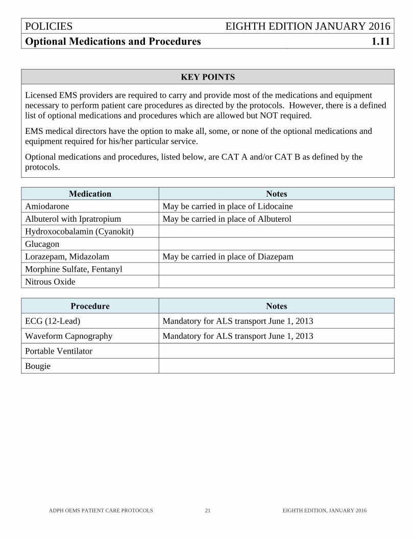
Optional Medications and Procedures 1.11
ADPH OEMS PATIENT CARE PROTOCOLS 21 EIGHTH EDITION, JANUARY 2016
KEY POINTS
Licensed EMS providers are required to carry and provide most of the medications and equipment necessary to perform patient care procedures as directed by the protocols. However, there is a defined list of optional medications and procedures which are allowed but NOT required. EMS medical directors have the option to make all, some, or none of the optional medications and equipment required for his/her particular service. Optional medications and procedures, listed below, are CAT A and/or CAT B as defined by the protocols.
Medication Notes
Amiodarone May be carried in place of Lidocaine
Albuterol with Ipratropium May be carried in place of Albuterol
Hydroxocobalamin (Cyanokit)
Glucagon
Lorazepam, Midazolam May be carried in place of Diazepam
Morphine Sulfate, Fentanyl
Nitrous Oxide
Procedure Notes
ECG (12-Lead) Mandatory for ALS transport June 1, 2013
Waveform Capnography Mandatory for ALS transport June 1, 2013
Portable Ventilator
Bougie
POLICIES EIGHTH EDITION JANUARY 2016
Patient Rights 1.12
ADPH OEMS PATIENT CARE PROTOCOLS 22 EIGHTH EDITION, JANUARY 2016
GUIDELINE
1. The EMS protocols are intended for use with a conscious, consenting patient, or an unconscious (implied consent) patient. An adult is considered to be of sound mind unless he/she is obviously under the influence of drugs or alcohol or has been determined by a judge to be incompetent. If the person is obviously under the influence of alcohol or drugs and yet refuses treatment, see three (3) below.
2. If a conscious, rational patient refuses treatment, comply with the patient’s request and document the refusal. If in the EMSP’s judgment a patient who has refused treatment (whether competent or incompetent) needs emergency care, contact OLMD.
3. If a patient may harm him/herself and refuses treatment, contact OLMD (and law enforcement if necessary). If the patient threatens harm to an EMSP, move from the close proximity of the patient, and from harm’s way. If the law enforcement officers are unable or unwilling to restrain the patient, the EMSP’s responsibility is completed with his/her notification of the law enforcement agency and OLMD.
4. If a patient’s family, physician, or nursing home refuses treatment for a patient, protocols are contained herein to deal with those situations.
5. An adult patient who is conscious and alert has the right to select a hospital to which he/she is to be transported, and neither the EMS service nor OLMD has the right to override that decision. If the hospital is on diversion status and the patient still insists on being transported to that hospital, the EMS provider must honor this request and OLMD cannot override this decision. If, in an EMSP’s judgment, transport to the patient’s chosen hospital will cause loss of life or limb, and the EMSP cannot convince the patient to allow transport to a more appropriate hospital, contact OLMD and ask him/her to speak to the patient. If the patient still insists on being transported to the inappropriate hospital, an EMSP must honor this request.
6. If the patient is unconscious or has altered mental status, the EMSP should normally take the patient to the hospital requested by the immediate family. If that hospital is on diversion or is not appropriate for the patient’s problem, contact OLMD and transport the patient to the hospital he/she orders. Patients who are unconscious and have unstable vital signs should be transported to the closest most appropriate emergency department which may be different from the family’s chosen hospital. If there is any doubt about which ED is the most appropriate destination, contact OLMD for guidance. Patients in cardiac arrest should always be transported to the closest emergency department, however, traumatic cardiac arrests may benefit from transport to a trauma center.
7. If the patient requests to be transported to a hospital out of the EMSP’s normal service area or that transport would leave the community without ambulance service, an EMSP may request a backup ambulance (or an ambulance from the hospital to which the patient requests to be transported) to transport the patient. This may require taking the patient (if unstable) to the nearest appropriate hospital while transportation is arranged. This is not a license to circumvent the Alabama Trauma System by always taking trauma patients to the local hospital instead of directly to the closest trauma center.
POLICIES EIGHTH EDITION JANUARY 2016
Patient Rights (continued) 1.12
ADPH OEMS PATIENT CARE PROTOCOLS 23 EIGHTH EDITION, JANUARY 2016
GUIDELINE (continued)
If an EMSP are unable to comply with the regional trauma plan, the EMSP must contact the Office of EMS to develop a plan to correct this.
8. When a minor may give consent generally: Public Health Laws of Alabama, 2006 edition, 22-8-4 states, “Any minor who is 14 years of age or older, or has graduated from high school, or is married, or having been married is divorced or is pregnant may give effective consent to any legally authorized medical, dental, or health or mental health services for himself or herself, and the consent of no other person shall be necessary.” (Acts 1971, No 2281, p. 3681, 1). An EMSP may treat and/or transport, under the doctrine of implied consent, a minor who requires immediate care to save his/her life or prevent serious injury. The age of adulthood in Alabama is 19 years old. If an unemancipated minor is old enough to consent but refuses (or their parent or legal guardian refuses) care that the EMSP thinks is needed, contact OLMD.
9. In other situations involving minors where no parental contact can be obtained, OLMD contact is mandatory. To err on the side of treatment is the safe approach. Careful documentation is important.
10. In situations when a patient is in the custody of law enforcement personnel, the patient is the responsibility of the law enforcement personnel. In these circumstances, the EMSP is expected to confer with law enforcement personnel and make a recommendation regarding the most appropriate care for the patient. However all decisions regarding these patients rest with law enforcement personnel, including destination hospital and consent or refusal of medical care. The law enforcement personnel are responsible for signing authorization for any refusal of care. If the situation arises in which the EMSP and law enforcement personnel disagree over the most appropriate care for the patient, OLMD should be contacted for consultation to ensure that law enforcement personnel have the most appropriate medical information available to allow them to make an informed decision regarding the patient’s care.
POLICIES EIGHTH EDITION JANUARY 2016
Physician Medical Direction 1.13
ADPH OEMS PATIENT CARE PROTOCOLS 24 EIGHTH EDITION, JANUARY 2016
KEY POINTS
Medical direction for medications and patient care procedures is provided under physician oversight. To provide on-line medical direction, a physician must have taken the medical direction course and hold a current medical direction physician identification number. The on-line medical direction physician must be skilled in and available for both adult and pediatric medical direction. Category A medications can be given and Category A procedures performed without prior physician contact. In such cases, only a report to a nurse or paramedic at the receiving hospital is necessary. Category B medications and procedures, however, require contact with a physician prior to administration. A report should be made to the physician in any case in which the patient is unstable. Medication orders may be signed by an OLMD physician or by the service’s medical director.
POLICIES EIGHTH EDITION JANUARY 2016
Refusal of Care or Transport 1.14
ADPH OEMS PATIENT CARE PROTOCOLS 25 EIGHTH EDITION, JANUARY 2016
PURPOSE
To specify when a patient may refuse care and provide guidance for addressing refusals of care.
GUIDELINE
1. For the alert, conscious patient who requests no transport or treatment, but in the EMSP’s judgment the patient needs to be transported to the hospital or treated, then the EMSP shall: a. Contact OLMD and try to establish communication between the patient and OLMD. If
communication cannot be established, the EMSP shall explain the risks and benefits of transport and treatment, but the EMSP shall accept the right of the competent adult patient to refuse treatment and transport.
b. In all events, the EMSP shall follow the patient’s directions regarding transport and treatment.
c. In all events, the EMSP shall document the patient status. This process must include patient competence.
2. For the ill patient who is unable to control his or her own decision, (unconscious, incapacitated,
etc.) and where care is refused: a. If physically possible, BLS care at the EMT level will be followed during attempts to
establish communication. b. The EMSP will contact OLMD and establish contact between the patient’s family and the
OLMD. After this contact has been made, the EMSP will follow the orders of the OLMD physician.
c. In all events, the EMSP shall document this process (to include patient competence).
POLICIES EIGHTH EDITION JANUARY 2016
Time at the Scene 1.15
ADPH OEMS PATIENT CARE PROTOCOLS 26 EIGHTH EDITION, JANUARY 2016
PURPOSE
To delineate on-scene time limitations.
GUIDELINE
1. If at any time an EMSP cannot provide or protect a patient airway within five minutes after patient encounter and initiating emergency medical care, he/she is required to transport the patient immediately.
2. If at any time an EMSP predicts that he/she will be on the scene, or has been on the scene for 30
minutes after patient encounter and initiating emergency medical care, he/she is required to contact OLMD. a. Communicate pertinent patient history. b. Communicate treatment given. c. Ask whether the patient should be transported immediately or other care should be given. d. Anticipate answering the question: “What further needs to be done?”
3. For cases involving significant trauma, time spent on the scene should be ten (10) minutes or
less where extrication has been accomplished, and the patient can be moved away from the site.
POLICIES EIGHTH EDITION JANUARY 2016
Trauma System 1.16
ADPH OEMS PATIENT CARE PROTOCOLS 27 EIGHTH EDITION, JANUARY 2016
PURPOSE
To provide patient entry criteria and system guidance for the Alabama Trauma System.
GUIDELINE
ALABAMA TRAUMA SYSTEM ENTRY CRITERIA Physiological Criteria:
1. A systolic BP <90 mm/Hg in an adult or child 6 years or older <80 mm/Hg in a child five or younger. This includes any trauma related cardiac arrest that will be treated or transported to the hospital.
2. Respiratory distress - rate < 10 or >29 in adults, or <20 or >60 in a newborn. <20 or >40 in a child three years or younger. <12 or >29 in a child four years or older.
3. Head trauma with Glasgow Coma Scale score of 13 or less or head trauma with any neurologic changes in a child five years or younger.
Anatomical Criteria:
1. The patient has a flail chest. 2. The patient has two or more obvious proximal long bone fractures (humerus, femur). 3. The patient has penetrating trauma to the head, neck, torso, or extremities proximal to the elbow
or knee. 4. The patient has in the same body area a combination of trauma and burns (partial and full
thickness) of fifteen percent or greater. 5. See Burns Protocol (3.08) for criteria to enter a burned patient into the trauma system. 6. The patient has an amputation proximal to the wrist or ankle. 7. The patient has one or more limbs which are paralyzed. 8. The patient has a pelvic fracture, as evidenced by a positive “pelvic movement” exam. 9. The patient has a crushed, degloved, mangled, or pulseless extremity. 10. The patient has an open or depressed skull fracture.
Mechanism of the patient injury:
1. A patient with the same method of restraint and in the same seating area as a dead victim. 2. Ejection of the patient from an enclosed vehicle. 3. Motorcycle/bicycle/ATV crash with the patient being thrown at least ten feet from the
motorcycle/bicycle. 4. Auto versus pedestrian with significant impact with the patient thrown, or run over by a vehicle. 5. An unbroken fall of twenty feet or more onto a hard surface. Unbroken fall of 10 feet or
3 times the height of the child onto a hard surface.
POLICIES EIGHTH EDITION JANUARY 2016Trauma System (continued) 1.16
ADPH OEMS PATIENT CARE PROTOCOLS 28 EIGHTH EDITION, JANUARY 2016
GUIDELINE
ALABAMA TRAUMA SYSTEM ENTRY CRITERIA
EMSP Discretion: 1. If the EMSP is convinced that the patient could have a severe injury which is not yet obvious, the
patient should be entered into the Alabama Trauma System. 2. The EMT’s suspicion of severity of trauma/injury may be raised by the following factors:
a. Age >55 b. Age <five c. Environment (hot/cold) d. Patient’s previous medical history e. Insulin dependent diabetes or other metabolic disorder f. Bleeding disorder or currently taking anticoagulant medication (coumadin, heparin) g. COPD/Emphysema h. Renal failure on dialysis i. Pregnancy j. Child with congenital disorder k. Extrication time >20 minutes with heavy tools utilized l. Motorcycle crash m. Head trauma with history of more than momentary loss of consciousness.
ENTERING A PATIENT INTO THE ALABAMA TRAUMA SYSTEM EMS Providers should call the Alabama Trauma Communications Center (ATCC) to determine patient destination.
ATCC contact numbers: Toll-Free Emergency: 1-800-359-0123, or Southern LINC EMS Fleet 55: Talkgroup 10/Private 55*380, or Nextel: 154*132431*4
The initial unit on-scene should enter the patient into the Alabama Trauma System but if they have not done so, it becomes the responsibility of the transporting service (ground or air) before the receiving facility is selected.
POLICIES EIGHTH EDITION JANUARY 2016
Trauma System (continued) 1.16
ADPH OEMS PATIENT CARE PROTOCOLS 29 EIGHTH EDITION, JANUARY 2016
GUIDELINE (continued)
ENTERING A PATIENT INTO THE ALABAMA TRAUMA SYSTEM (continued) For helicopter EMS (HEMS) it is preferable to request a preliminary receiving facility from ATCC prior to arrival on the scene and then later enter the patient into the ATCC as soon as is logistically possible. After assessing a trauma situation and making the determination that the patient should be entered into the Alabama Trauma System, the EMSP licensed at the highest level should contact the ATCC at the earliest practical time before the receiving facility is selected and provide the following information. The highest level EMSP on the scene may delegate the call to ATCC to a lower level EMSP if patient care duties require the higher level EMSP’s attention:
1) EMSP service 2) Location of Trauma Scene 3) Age and Sex of the patient(s) 4) Reason for Entry and Mechanism of Injury 5) Patient assessment
a) Airway Status b) Vital signs and GCS c) Areas of Injury d) Environmental issues or co-morbid factors
6) Transportation type 7) Transportation timing
ATCC will provide a unique identification number that must be entered into the e-PCR.
Notify the ATCC of any change in the patient’s condition. The receiving trauma center or ATCC should be updated by the transporting unit 5-10 minutes out. This update should only consist of any patient changes and patient’s current condition. A repeat of information used to enter the patient into the Alabama Trauma System is not necessary since this information will be relayed by the ATCC to the receiving trauma center.
After the patient is delivered to the trauma center, the transporting provider should call the ATCC with the Patient Care Report times.
ADPH EMS PROTOCOLS EIGHTH EDITION
Operational Guidelines 2
ADPH OEMS PATIENT CARE PROTOCOLS 30 EIGHTH EDITION, JANUARY 2016
These operations guidelines are intended to direct the actions of EMS personnel when there are no duly authorized local operations guidelines utilized by an EMS service or agency.
When there is conflict between the local operational standards and those listed in this document, then the local standards take precedence.
It is expected that if a scene conflict or jurisdictional disagreement occurs, OLMD will be consulted and his/her directions followed.
OPERATIONAL GUIDELINES EIGHTH EDITION JANUARY 2016
Cancellation/Slow Down 2.01
ADPH OEMS PATIENT CARE PROTOCOLS 31 EIGHTH EDITION, JANUARY 2016
PURPOSE
The first unit on the scene or dispatch may recommend that other responding units slow down or discontinue their response. It is recognized that it is in the best interest of patient care and the public to slow or cancel units responding with lights and siren to calls, when it is determined by competent personnel that the situation does not require such a rapid response.
GUIDELINE
BLS units and rapid responders may recommend ALS units to slow to non-emergency traffic when a patient does not appear in their opinion to require advanced life support. They may cancel ALS units when there is no patient, or a patient refuses care or transport.
ALS units may recommend slowing or canceling other responders once the patient has been evaluated at the scene and a determination is made that no other units are required, or no other units are required emergency.
Advanced Life Support for the purpose of this policy is IV administration, medication therapy, advanced airway management, cardiac monitoring, or cardiac defibrillation.
Decisions on slowing down and cancellations shall be solely based on medical or trauma criteria.
OPERATIONAL GUIDELINES EIGHTH EDITION JANUARY 2016
Crime Scene Response 2.02
ADPH OEMS PATIENT CARE PROTOCOLS 32 EIGHTH EDITION, JANUARY 2016
PURPOSE
The safety of EMSP and emergency care for the victim remain the primary goals in all crime scene operations, however, preservation of the scene remains the most important secondary goal. Never compromise patient care to preserve a crime scene. If the EMSP is part of an organized Tactical EMS arrangement with law enforcement units, such as SWAT teams, the EMSP will follow those operational guidelines, as approved by his/her Medical Director.
GUIDELINE
1. EMSP should not approach any scene suspected of involving violence, unless law enforcement states that the scene is reasonably secure. EMSP should not approach any crime scene in which law enforcement personnel are not present, in which law enforcement personnel are in defensive positions, or when weapons are being presented by law enforcement personnel.
2. EMSP should approach every call with caution while being observant. This is particularly true
of scenes that may involve a crime against person or property. Noise and light discipline should be used with emergency warning equipment shut down some distance from the incident. a. A portable radio to call for assistance is recommended. b. Never stand directly in front of doors when knocking for entry. c. If a weapon is involved, try to secure the weapon unless the weapon is still in the assailant’s
possession. The weapon should be secured in such a way that it does not jeopardize the patient’s life or the EMSP’s life. Weapons are potential evidence and should not be compromised if at all possible.
d. If the EMSP’s life is in danger, it may be necessary to leave the patient. Always have a plan for escape.
3. All information regarding a call should be gathered. Calls involving crimes in progress, the use
of weapons, or any suspicious call in high crime areas, should be treated with caution. If possible, EMSP should wear soft body armor on calls of this nature and while operating in high crime areas.
4. When approaching a crime scene with law enforcement present, ask for the best route of approach and avoid destroying what may be valuable evidence. Use only one route in and out of scene and disturb only what is absolutely necessary. a. Avoid disturbing tire tracks or foot prints and avoid blood on surfaces. b. Do not disturb items on the scene unless absolutely necessary. c. Do not cut or treat through holes made by projectiles or other objects in clothing. d. Remove any medical items brought into the scene. e. When possible, place any victim to be transported on a clean sheet. When the victim is
removed at the hospital, retain the sheet for law enforcement personnel. This is particularly important in crimes in which trace evidence may be transferred from the suspect to the
OPERATIONAL GUIDELINES EIGHTH EDITION JANUARY 2016
Crime Scene Response (continued) 2.02
ADPH OEMS PATIENT CARE PROTOCOLS 33 EIGHTH EDITION, JANUARY 2016
GUIDELINE (continued)
victim. Retain, preferably wrapped in a clean sheet or placed in an unused paper bag, any clothing or other items removed by EMS personnel while in the ambulance. Do not place blood-contaminated items in a plastic bag as this may ruin their value as evidence.
5. Do not touch or handle items, particularly weapons, found at a crime scene unless absolutely necessary. Do not handle expended bullets or casings with metal forceps if they should be found in clothing or on a sheet. Retain them in the sheet or clothing in which they are found and notify law enforcement personnel. It is required that EMS personnel enter a crime scene to confirm obvious death. However, this procedure can be accomplished with minimal scene disturbance. Coordinate with law enforcement personnel in preserving the crime scene to the greatest extent possible.
6. Be aware of any statements made by victims, suspects or others present at a crime scene. Make certain to scan the scene, noting how it appears upon arrival, particularly the victim, and remember any changes made to the crime scene during patient assessment and/or treatment.
7. Following the incident, record detailed notes regarding actions and observations made during the incident. Any statements made outside the presence of law enforcement personnel by the victim or suspect should be carefully recorded, and a copy given to law enforcement investigators.
8. If a scene appears suspicious, then await the arrival of law enforcement personnel before approaching.
9. A detailed report that covers all aspects of the EMSP’s involvement at the crime scene is important in case he/she is later called to testify in court. These narratives should cover the EMSP’s observations and conversations with persons present at the scene, location of response vehicles and equipment, who was present, furniture weapons or clothing that has been moved, items that were handled by EMSPs, and his/her route to the victim. This narrative should be a separate report from the Patient Care Report.
OPERATIONAL GUIDELINES EIGHTH EDITION JANUARY 2016
Hazardous Materials 2.03
ADPH OEMS PATIENT CARE PROTOCOLS 34 EIGHTH EDITION, JANUARY 2016
PURPOSE
1. EMSP may be first on the scene of a hazardous materials situation. This protocol is intended to guide EMSP who do not normally function in hazardous materials scenes and are trained only to the awareness level. This protocol is intended to compliment any existing hazardous materials guidelines of fire agencies. If the two protocols are in conflict, fire department protocol takes precedence.
2. Based on information from dispatch, if the scene to which an EMSP is responding is a known or suspected hazardous materials situation, stage and wait for the hazardous materials personnel.
3. When scene size-up suggests that hazardous materials are involved, stage and wait for the hazardous materials personnel.
4. All scenes should be considered as being a potential hazardous materials situation.
GUIDELINE
Approach 1. Utilize a cautionary approach at all times. 2. The reported location may be inaccurate and response into a contaminated area might occur. 3. Approach upwind and upgrade if possible. If unable to approach from upwind/upgrade,
approach at 90 degrees to wind/grade, if possible, with safety in mind. 4. Position vehicle well away from problem and headed away from incident. 5. Communicate the EMSP’s actions or intended actions to EMS Dispatch. 6. If an EMSP is the first responder on-scene, confirm that fire and police have been notified. 7. The agency responsible for hazardous materials response may respond with different levels of
personnel and equipment based upon the information received. Do not always expect a hazardous materials team to respond.
8. If an EMSP is the first responder on-scene, his/her first priority is scene isolation. KEEP OTHERS AWAY! KEEP UNNECESSARY EQUIPMENT FROM BECOMING CONTAMINATED.
9. If the EMSP believes that he/she, or the vehicle, are contaminated, stage in an isolated area. Person in Charge
1. If the EMSP is the first medical person on the scene, he/she should assume the role of PIC of medical care (not necessarily scene control) until a hazardous materials trained EMSP arrives.
2. The EMSP will direct all patient care. 3. The EMSP, in concert with the incident commander, will determine the method of transport of
the exposed patient (air vs. ground). 4. The EMSP will determine who will provide care during transport.
OPERATIONAL GUIDELINES EIGHTH EDITION JANUARY 2016
Hazardous Materials (continued) 2.03
ADPH OEMS PATIENT CARE PROTOCOLS 35 EIGHTH EDITION, JANUARY 2016
GUIDELINE (continued)
Patient Care for the Contaminated Patient 1. Types of incidents which may require decontamination of the patient:
a. Radiation. b. Biological hazards. c. Chemical. d. Toxic Substances.
2. Contamination can occur through: a. Smoke. b. Direct contact. c. Vapor. d. Run-off.
3. Transporting contaminated patients should be a serious concern to those involved. Patients who have been in contact with, or who are even suspected of having been in contact with, a hazardous substance, should be transported for evaluation.
4. The hazardous materials team must be contacted about removal of contaminated clothing and packaging of the patient with regard to the EMSP and the patient’s protection.
5. Determine the hazardous substance involved, and provide treatment as directed by the EMSP in charge.
6. Be aware that many hazardous materials incident scenes are also crime scenes. Follow Guideline 2.02 Crime Scene Response when appropriate.
Ambulance Preparation 1. The EMSP shall determine the process needed for ambulance preparation. 2. Remove any supplies and equipment that would not be needed for immediate patient care. 3. Seal cabinets, and drape interior, including floor and squad bench, with plastic or visqueen (if
available from hazardous materials team). 4. Prepare stretcher by removing foam pad and placing down long backboard. Cover with plastic
and tape in place, if needed (if available from hazardous materials team).
Transport and Arrival at the Hospital. 1. If an ambulance has transported a patient from an incident that is subsequently determined to
involve hazardous materials exposure, scene personnel must immediately relay all relevant information to the transporting unit(s) and/or receiving facility(s) involved.
2. OLMD and the receiving hospital should be contacted as soon as possible. The EMSP should communicate the material involved, degree of exposure, decontamination procedures used, and patient condition.
3. The ambulance should park in an area away from the emergency department, or go directly to a decontamination center or area.
4. Patient(s) should not be brought into the emergency department before the EMSP receive permission from the hospital staff.
OPERATIONAL GUIDELINES EIGHTH EDITION JANUARY 2016
Hazardous Materials (continued) 2.03
ADPH OEMS PATIENT CARE PROTOCOLS 36 EIGHTH EDITION, JANUARY 2016
GUIDELINE (continued)
5. Once the patient(s) has been released to the hospital, follow the EMSP direction and, if necessary, double bag the plastic sheeting used to cover the gurney and the floor into plastic bags. Double bag any equipment that is contaminated.
6. After unloading patient from ambulance, check with the fire department incident commander to see where the ambulance can be safely decontaminated, and whether or not there is equipment available for this purpose. Do not begin decontamination until after consultation with the Hazardous Materials Team Leader.
7. Following decontamination recommendation from the hazardous materials team, decontaminate the ambulance and personnel before returning to the incident scene. If returning to the incident scene, bring bags containing contaminated materials, equipment, clothing, etc., and turn them over to the hazardous materials team.
EMSP Exposure
1. If an EMSP is exposed, or is concerned with the possibility of exposure, medical help should be sought immediately.
2. Report all exposures to the hazardous materials team, Poison Center, and risk manager or supervisor.
3. Do not return to service until cleared to do so by the fire department.
OPERATIONAL GUIDELINES EIGHTH EDITION JANUARY 2016
Helicopter EMS 2.04
ADPH OEMS PATIENT CARE PROTOCOLS 37 EIGHTH EDITION, JANUARY 2016
PURPOSE
To provide guidance regarding the use of Helicopter EMS services (HEMS).
GUIDELINE
Helicopter EMS should be utilized when transportation by air will significantly reduce total transport time for patients with time-dependent illness or injury or when the patient requires potentially lifesaving prehospital interventions that cannot be provided by the responding EMS service.
When HEMS is requested, the HEMS service that can respond to the scene in the shortest time should be called. If a HEMS service cannot respond to a call and a second service is requested, the requesting agency must notify the second service that the call has already been refused and why. At no time should HEMS be dispatched to a pre-hospital scene without dispatch of ground EMS unless there are no other available EMS units. HEMS units may augment ground EMS services when the number of critically ill or injured patients requiring transport exceeds the transport capabilities of available ground EMS services.
Early activation of Helicopter EMS is defined as dispatch of an EMS helicopter to a patient care scene based on the information received through 911 operators or first responders on the scene.
Situations in which HEMS may be needed include, but are not limited to: 1. Patients who meet entry criteria for the Alabama Trauma System (1.16) 2. Multiple victim incidents with severe illness or injuries 3. Severe burns and explosions 4. New onset focal weakness, paralysis, or aphasia (suspected stroke) 5. ST Elevation MI or suspected acute coronary syndrome 6. Near drowning 7. Medical Emergencies such as severe dyspnea, airway obstruction, or shock when in the EMSPs
best medical judgment HEMS would be the most appropriate form of transportation 8. Report of serious injury in a patient whose location would be difficult to access by ground
ambulance but is more accessible by helicopter When there is a question about whether or not HEMS is the most appropriate means of transportation for the patient, contact OLMD for further guidance.
When a HEMS service has been early-activated or placed on stand-by status, it is the responsibility of the ground EMS service to determine if air transport is the most appropriate means of transportation for the patient and relay that information to the HEMS service through their dispatch mechanism. If HEMS has been activated and the most highly certified EMSP on the scene determines in his/her best professional judgment that air transport will not provide significant benefit to the patient, then HEMS should be cancelled as soon as possible. A HEMS request made by an ALS agency may be cancelled ONLY by the agency making the initial request.

OPERATIONAL GUIDELINES EIGHTH EDITION JANUARY 2016
Helicopter EMS 2.04
ADPH OEMS PATIENT CARE PROTOCOLS 38 EIGHTH EDITION, JANUARY 2016
Upon arrival to the scene, if the HEMS crew determines that the patient does not meet criteria for air transport or that patient, weather, or aircraft issues preclude use of the helicopter for transport then the flight crew may request ground transportation for that patient and transfer care of the patient to the ground EMS crew in accordance with the Medical Management of the Scene Policy (1.08). This shall NOT constitute abandonment as defined by EMS rules. An EMS service should not unduly delay transport of a patient while waiting for HEMS to arrive. If HEMS is delayed beyond their stated estimated time of arrival (e.g. >10 minutes), it is the responsibility of the helicopter service to notify the ground EMS crew of the delay. If the patient is packaged and ready for transport, it is acceptable for the ground EMS service to reassign the landing zone to a mutually agreeable site that is closer to the destination facility and initiate patient transport. The helicopter may intercept the ground ambulance at this agreed upon alternate landing site or when appropriate the ground EMS crew may complete the transport. When a new landing zone at a mutually agreeable site is chosen, the ground EMS crew must communicate this change by voice with the HEMS agency that is responding to the scene. Hospitals may be used as a landing site and patients will not be considered to have arrived at the hospital when the patient does not enter the hospital for evaluation and/or care.
OPERATIONAL GUIDELINES EIGHTH EDITION JANUARY 2016
Staging For High Risk Response 2.05
ADPH OEMS PATIENT CARE PROTOCOLS 39 EIGHTH EDITION, JANUARY 2016
PURPOSE
To establish guidelines for the response of private and public EMS responders to incidents which involve violence, or are anticipated to be potentially violent in nature.
GUIDELINE
1. When to stage: a. Any time dispatch directs them to do so. b. Any time a violent incident might expose EMS personnel to danger. c. Any call at the EMS unit’s discretion.
2. How to stage: a. Stage approximately two blocks from the incident address in urban areas and ½ mile from
the incident address in rural areas and out of the line of sight. b. Announce arrival in staging and location. c. Additional responding EMS units will respond to the same staging location if possible
(avoid traveling past incident address). d. Unless traffic hazard, turn off headlights and all warning devices. e. Turn on four-way flashers. f. Once staged, EMS units will not enter the scene until the scene is declared secure by law
enforcement or dispatch.
NOTE It shall not be assumed that the mere presence of law enforcement on scene means that medical responders may now proceed safely into the call location. If law enforcement is on scene, call dispatch to request verification that EMS units may proceed onto the scene or stage. This may be modified depending on local situations.
ADPH EMS PROTOCOLS EIGHTH EDITION
TREATMENT PROTOCOLS 3
ADPH OEMS PATIENT CARE PROTOCOLS 40 EIGHTH EDITION, JANUARY 2016
Each Treatment Protocol begins with sections titled History and Physical Exam, and Key Points. These sections include information that is useful to all EMSPs. The third section of each protocol is titled Treatment. This section is divided into two columns. The left column includes general treatment information that does not specify Scope of Practice for each intervention. The right column is divided into four levels that correspond to the levels of EMSP licensure in Alabama. This section specifies treatments that are suitable for each level of EMSP and are color-coded.
EMT approved treatments are listed on the top in the white field. Advanced-EMT approved treatments are listed next in the yellow field. Intermediate-EMT approved treatments are listed third in the green field. Paramedic approved treatments are listed last in the blue field.
Each EMSP can perform and is responsible for the treatments listed in the right column of the treatment protocol appropriate to their Scope of Practice IN ADDITION TO all the treatments listed in the Scope of Practice for all levels of lesser training. For example, an EMT may perform those treatments listed under EMT. An Advanced-EMT may perform those treatments listed under EMT and Advanced-EMT. Intermediate-EMTs may perform all treatments listed under EMT, Advanced-EMT, and Intermediate-EMT. Paramedics may perform all treatments listed. All providers are required to understand and operate within their Scope of Practice as noted in the Scope of Practice Policy (1.01). All levels of providers are responsible to utilize online medical direction (OLMD) when indicated. It may be appropriate to treat a patient using more than one Treatment Protocol.
TREATMENT PROTOCOLS EIGHTH EDITION JANUARY 2016
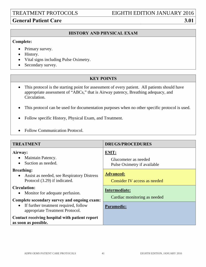
General Patient Care 3.01
ADPH OEMS PATIENT CARE PROTOCOLS 41 EIGHTH EDITION, JANUARY 2016
HISTORY AND PHYSICAL EXAM
Complete:
Primary survey. History. Vital signs including Pulse Oximetry. Secondary survey.
KEY POINTS
This protocol is the starting point for assessment of every patient. All patients should have appropriate assessment of “ABCs,” that is Airway patency, Breathing adequacy, and Circulation.
This protocol can be used for documentation purposes when no other specific protocol is used. Follow specific History, Physical Exam, and Treatment.
Follow Communication Protocol.
TREATMENT DRUGS/PROCEDURES
Airway: Maintain Patency. Suction as needed.
Breathing: Assist as needed, see Respiratory Distress
Protocol (3.29) if indicated.
Circulation: Monitor for adequate perfusion.
Complete secondary survey and ongoing exam: If further treatment required, follow
appropriate Treatment Protocol.
Contact receiving hospital with patient report as soon as possible.
EMT:
Glucometer as needed Pulse Oximetry if available
Advanced:
Consider IV access as needed
Intermediate:
Cardiac monitoring as needed
Paramedic:
TREATMENT PROTOCOLS EIGHTH EDITION JANUARY 2016
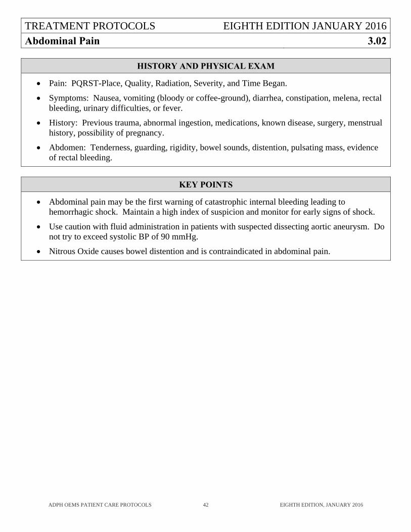
Abdominal Pain 3.02
ADPH OEMS PATIENT CARE PROTOCOLS 42 EIGHTH EDITION, JANUARY 2016
HISTORY AND PHYSICAL EXAM
Pain: PQRST-Place, Quality, Radiation, Severity, and Time Began.
Symptoms: Nausea, vomiting (bloody or coffee-ground), diarrhea, constipation, melena, rectal bleeding, urinary difficulties, or fever.
History: Previous trauma, abnormal ingestion, medications, known disease, surgery, menstrual history, possibility of pregnancy.
Abdomen: Tenderness, guarding, rigidity, bowel sounds, distention, pulsating mass, evidence of rectal bleeding.
KEY POINTS
Abdominal pain may be the first warning of catastrophic internal bleeding leading to hemorrhagic shock. Maintain a high index of suspicion and monitor for early signs of shock.
Use caution with fluid administration in patients with suspected dissecting aortic aneurysm. Do not try to exceed systolic BP of 90 mmHg.
Nitrous Oxide causes bowel distention and is contraindicated in abdominal pain.
TREATMENT PROTOCOLS EIGHTH EDITION JANUARY 2016
Abdominal Pain (continued) 3.02
ADPH OEMS PATIENT CARE PROTOCOLS 43 EIGHTH EDITION, JANUARY 2016
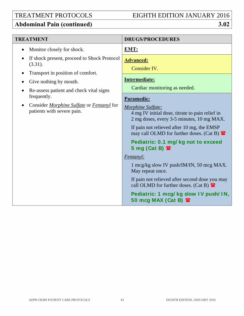
TREATMENT DRUGS/PROCEDURES
Monitor closely for shock.
If shock present, proceed to Shock Protocol (3.31).
Transport in position of comfort.
Give nothing by mouth.
Re-assess patient and check vital signs frequently.
Consider Morphine Sulfate or Fentanyl for patients with severe pain.
EMT:
Advanced:
Consider IV.
Intermediate:
Cardiac monitoring as needed.
Paramedic:
Morphine Sulfate: 4 mg IV initial dose, titrate to pain relief in 2 mg doses, every 3-5 minutes, 10 mg MAX.
If pain not relieved after 10 mg, the EMSP may call OLMD for further doses. (Cat B)
Pediatric: 0.1 mg/kg not to exceed 5 mg (Cat B)
Fentanyl:
1 mcg/kg slow IV push/IM/IN, 50 mcg MAX. May repeat once.
If pain not relieved after second dose you may call OLMD for further doses. (Cat B)
Pediatric: 1 mcg/kg slow IV push/IN, 50 mcg MAX (Cat B)
TREATMENT PROTOCOLS EIGHTH EDITION JANUARY 2016
Adrenal Insufficiency 3.03
ADPH OEMS PATIENT CARE PROTOCOLS 44 EIGHTH EDITION, JANUARY 2016
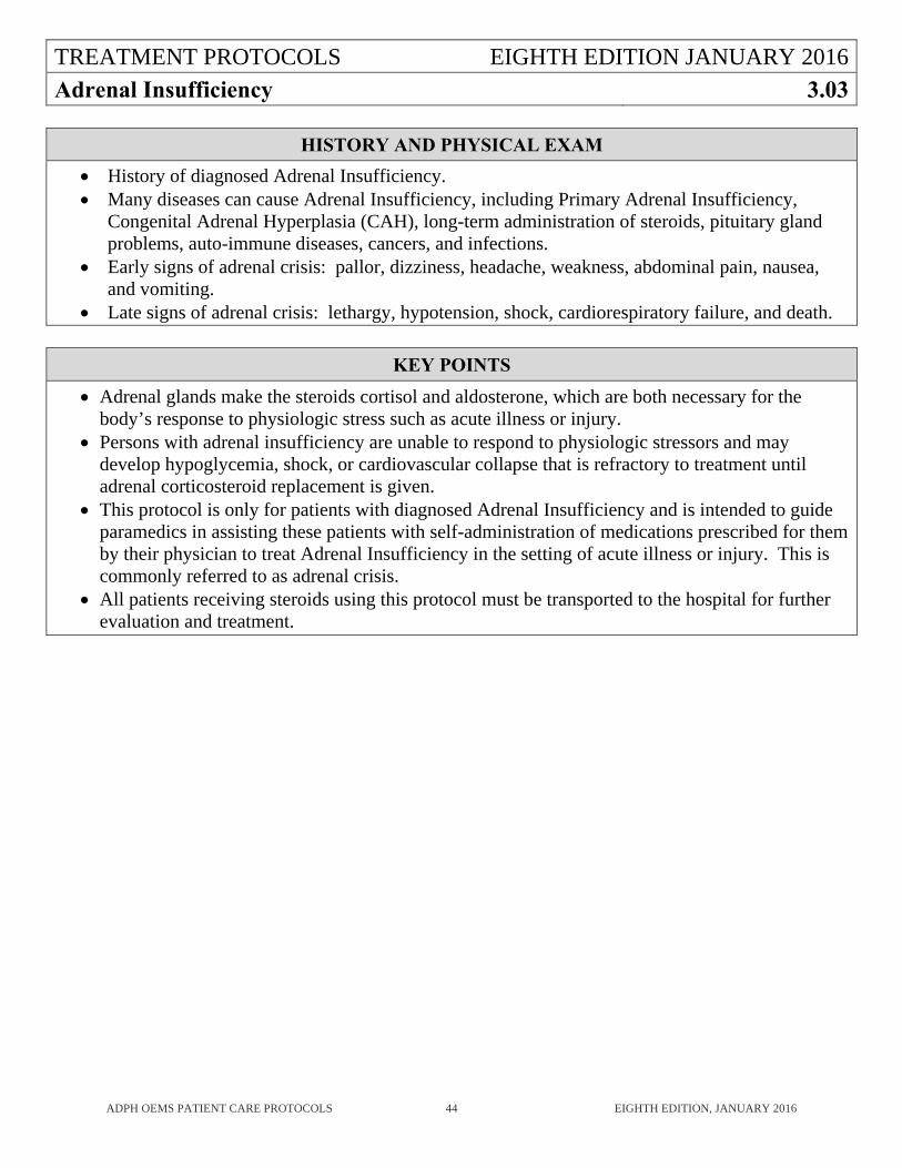
HISTORY AND PHYSICAL EXAM
History of diagnosed Adrenal Insufficiency. Many diseases can cause Adrenal Insufficiency, including Primary Adrenal Insufficiency,
Congenital Adrenal Hyperplasia (CAH), long-term administration of steroids, pituitary gland problems, auto-immune diseases, cancers, and infections.
Early signs of adrenal crisis: pallor, dizziness, headache, weakness, abdominal pain, nausea, and vomiting.
Late signs of adrenal crisis: lethargy, hypotension, shock, cardiorespiratory failure, and death.
KEY POINTS
Adrenal glands make the steroids cortisol and aldosterone, which are both necessary for the body’s response to physiologic stress such as acute illness or injury.
Persons with adrenal insufficiency are unable to respond to physiologic stressors and may develop hypoglycemia, shock, or cardiovascular collapse that is refractory to treatment until adrenal corticosteroid replacement is given.
This protocol is only for patients with diagnosed Adrenal Insufficiency and is intended to guide paramedics in assisting these patients with self-administration of medications prescribed for them by their physician to treat Adrenal Insufficiency in the setting of acute illness or injury. This is commonly referred to as adrenal crisis.
All patients receiving steroids using this protocol must be transported to the hospital for further evaluation and treatment.
TREATMENT PROTOCOLS EIGHTH EDITION JANUARY 2016
Adrenal Insufficiency (continued) 3.03
ADPH OEMS PATIENT CARE PROTOCOLS 45 EIGHTH EDITION, JANUARY 2016
TREATMENT DRUGS/PROCEDURES
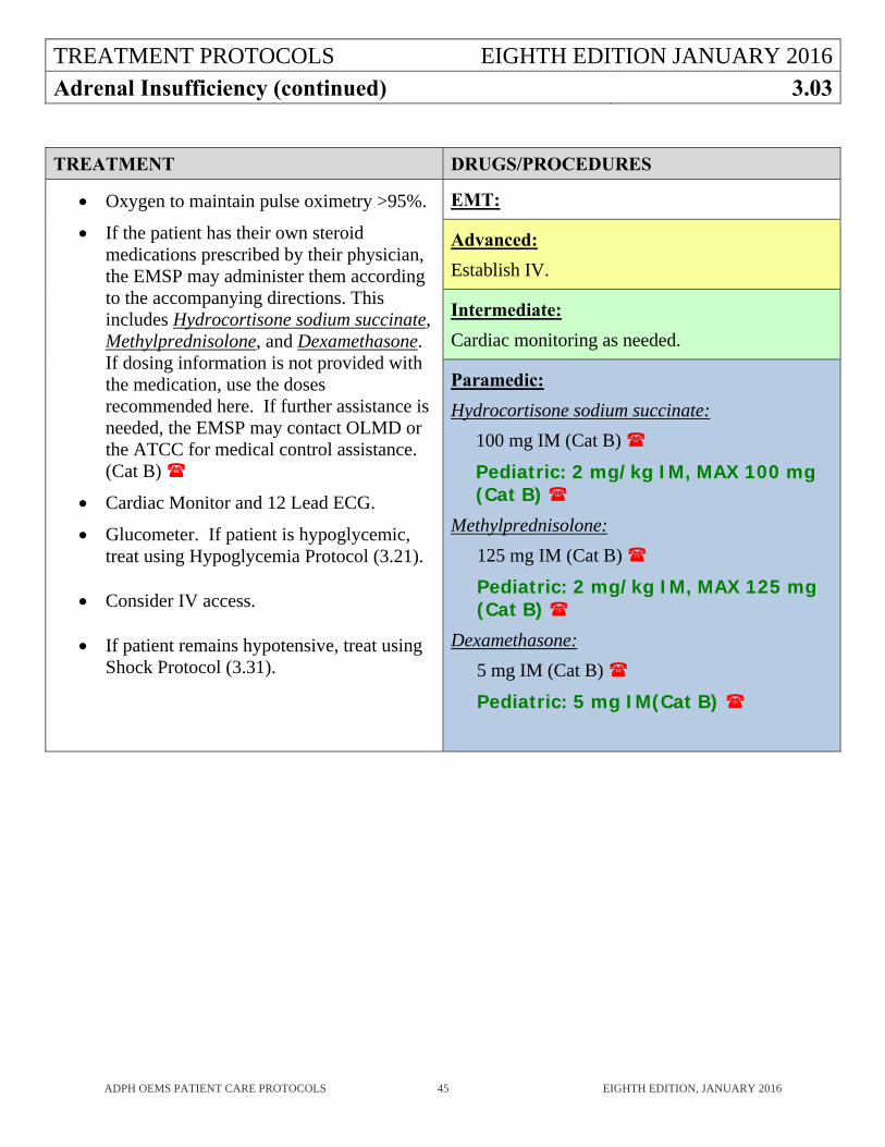
Oxygen to maintain pulse oximetry >95%.
If the patient has their own steroid medications prescribed by their physician, the EMSP may administer them according to the accompanying directions. This includes Hydrocortisone sodium succinate, Methylprednisolone, and Dexamethasone. If dosing information is not provided with the medication, use the doses recommended here. If further assistance is needed, the EMSP may contact OLMD or the ATCC for medical control assistance. (Cat B)
Cardiac Monitor and 12 Lead ECG.
Glucometer. If patient is hypoglycemic, treat using Hypoglycemia Protocol (3.21).
Consider IV access.
If patient remains hypotensive, treat using Shock Protocol (3.31).
EMT:
Advanced:
Establish IV.
Intermediate:
Cardiac monitoring as needed.
Paramedic:
Hydrocortisone sodium succinate:
100 mg IM (Cat B)
Pediatric: 2 mg/kg IM, MAX 100 mg (Cat B)
Methylprednisolone:
125 mg IM (Cat B)
Pediatric: 2 mg/kg IM, MAX 125 mg (Cat B)
Dexamethasone:
5 mg IM (Cat B)
Pediatric: 5 mg IM(Cat B)
TREATMENT PROTOCOLS EIGHTH EDITION JANUARY 2016
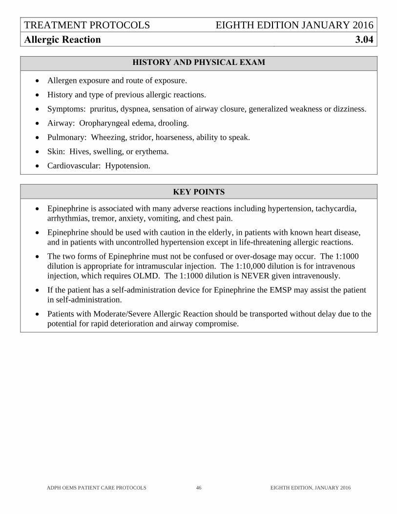
Allergic Reaction 3.04
ADPH OEMS PATIENT CARE PROTOCOLS 46 EIGHTH EDITION, JANUARY 2016
HISTORY AND PHYSICAL EXAM
Allergen exposure and route of exposure.
History and type of previous allergic reactions.
Symptoms: pruritus, dyspnea, sensation of airway closure, generalized weakness or dizziness.
Airway: Oropharyngeal edema, drooling.
Pulmonary: Wheezing, stridor, hoarseness, ability to speak.
Skin: Hives, swelling, or erythema.
Cardiovascular: Hypotension.
KEY POINTS
Epinephrine is associated with many adverse reactions including hypertension, tachycardia, arrhythmias, tremor, anxiety, vomiting, and chest pain.
Epinephrine should be used with caution in the elderly, in patients with known heart disease, and in patients with uncontrolled hypertension except in life-threatening allergic reactions.
The two forms of Epinephrine must not be confused or over-dosage may occur. The 1:1000 dilution is appropriate for intramuscular injection. The 1:10,000 dilution is for intravenous injection, which requires OLMD. The 1:1000 dilution is NEVER given intravenously.
If the patient has a self-administration device for Epinephrine the EMSP may assist the patient in self-administration.
Patients with Moderate/Severe Allergic Reaction should be transported without delay due to the potential for rapid deterioration and airway compromise.
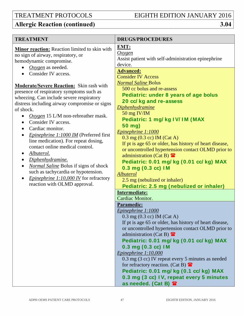
TREATMENT PROTOCOLS EIGHTH EDITION JANUARY 2016
Allergic Reaction (continued) 3.04
ADPH OEMS PATIENT CARE PROTOCOLS 47 EIGHTH EDITION, JANUARY 2016
TREATMENT DRUGS/PROCEDURES
Minor reaction: Reaction limited to skin with no sign of airway, respiratory, or hemodynamic compromise.
Oxygen as needed. Consider IV access.
Moderate/Severe Reaction: Skin rash with presence of respiratory symptoms such as wheezing. Can include severe respiratory distress including airway compromise or signs of shock.
Oxygen 15 L/M non-rebreather mask. Consider IV access. Cardiac monitor. Epinephrine 1:1000 IM (Preferred first
line medication). For repeat dosing, contact online medical control.
Albuterol. Diphenhydramine. Normal Saline Bolus if signs of shock
such as tachycardia or hypotension. Epinephrine 1:10,000 IV for refractory
reaction with OLMD approval.
EMT: Oxygen Assist patient with self-administration epinephrine device. Advanced: Consider IV Access Normal Saline Bolus
500 cc bolus and re-assess Pediatric: under 8 years of age bolus 20 cc/kg and re-assess
Diphenhydramine 50 mg IV/IM Pediatric: 1 mg/kg IV/IM (MAX 50 mg)
Epinephrine 1:1000 0.3 mg (0.3 cc) IM (Cat A) If pt is age 65 or older, has history of heart disease, or uncontrolled hypertension contact OLMD prior to administration (Cat B) Pediatric: 0.01 mg/kg (0.01 cc/kg) MAX 0.3 mg (0.3 cc) IM
Albuterol 2.5 mg (nebulized or inhaler) Pediatric: 2.5 mg (nebulized or inhaler)
Intermediate: Cardiac Monitor. Paramedic: Epinephrine 1:1000
0.3 mg (0.3 cc) IM (Cat A) If pt is age 65 or older, has history of heart disease, or uncontrolled hypertension contact OLMD prior to administration (Cat B) Pediatric: 0.01 mg/kg (0.01 cc/kg) MAX 0.3 mg (0.3 cc) IM
Epinephrine 1:10,000 0.3 mg (3 cc) IV repeat every 5 minutes as needed for refractory reaction. (Cat B) Pediatric: 0.01 mg/kg (0.1 cc/kg) MAX 0.3 mg (3 cc) IV, repeat every 5 minutes as needed. (Cat B)
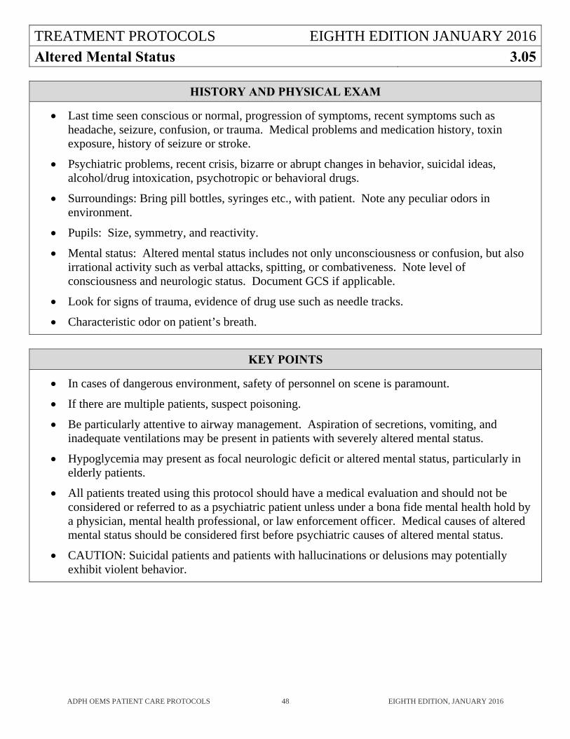
TREATMENT PROTOCOLS EIGHTH EDITION JANUARY 2016
Altered Mental Status 3.05
ADPH OEMS PATIENT CARE PROTOCOLS 48 EIGHTH EDITION, JANUARY 2016
HISTORY AND PHYSICAL EXAM
Last time seen conscious or normal, progression of symptoms, recent symptoms such as headache, seizure, confusion, or trauma. Medical problems and medication history, toxin exposure, history of seizure or stroke.
Psychiatric problems, recent crisis, bizarre or abrupt changes in behavior, suicidal ideas, alcohol/drug intoxication, psychotropic or behavioral drugs.
Surroundings: Bring pill bottles, syringes etc., with patient. Note any peculiar odors in environment.
Pupils: Size, symmetry, and reactivity.
Mental status: Altered mental status includes not only unconsciousness or confusion, but also irrational activity such as verbal attacks, spitting, or combativeness. Note level of consciousness and neurologic status. Document GCS if applicable.
Look for signs of trauma, evidence of drug use such as needle tracks.
Characteristic odor on patient’s breath.
KEY POINTS
In cases of dangerous environment, safety of personnel on scene is paramount.
If there are multiple patients, suspect poisoning.
Be particularly attentive to airway management. Aspiration of secretions, vomiting, and inadequate ventilations may be present in patients with severely altered mental status.
Hypoglycemia may present as focal neurologic deficit or altered mental status, particularly in elderly patients.
All patients treated using this protocol should have a medical evaluation and should not be considered or referred to as a psychiatric patient unless under a bona fide mental health hold by a physician, mental health professional, or law enforcement officer. Medical causes of altered mental status should be considered first before psychiatric causes of altered mental status.
CAUTION: Suicidal patients and patients with hallucinations or delusions may potentially exhibit violent behavior.
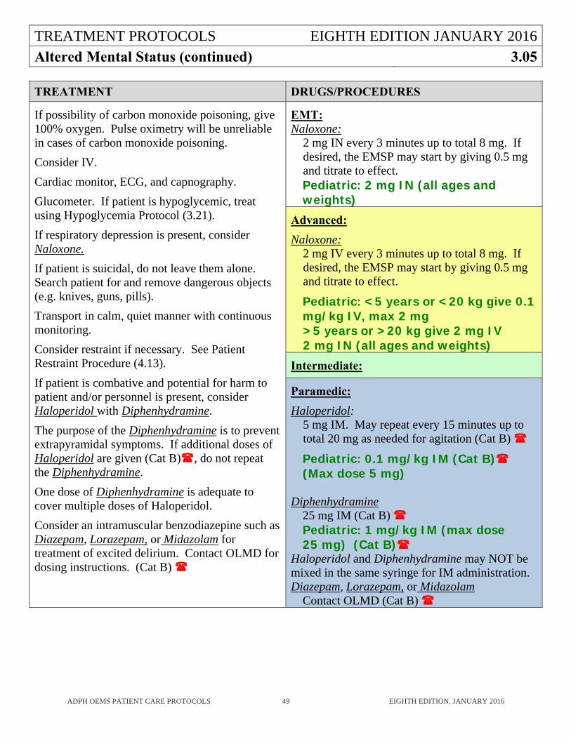
TREATMENT PROTOCOLS EIGHTH EDITION JANUARY 2016
Altered Mental Status (continued) 3.05
ADPH OEMS PATIENT CARE PROTOCOLS 49 EIGHTH EDITION, JANUARY 2016
TREATMENT DRUGS/PROCEDURES
If possibility of carbon monoxide poisoning, give 100% oxygen. Pulse oximetry will be unreliable in cases of carbon monoxide poisoning.
Consider IV.
Cardiac monitor, ECG, and capnography.
Glucometer. If patient is hypoglycemic, treat using Hypoglycemia Protocol (3.21).
If respiratory depression is present, consider Naloxone.
If patient is suicidal, do not leave them alone. Search patient for and remove dangerous objects (e.g. knives, guns, pills).
Transport in calm, quiet manner with continuous monitoring.
Consider restraint if necessary. See Patient Restraint Procedure (4.13).
If patient is combative and potential for harm to patient and/or personnel is present, consider Haloperidol with Diphenhydramine.
The purpose of the Diphenhydramine is to prevent extrapyramidal symptoms. If additional doses of Haloperidol are given (Cat B), do not repeat the Diphenhydramine.
One dose of Diphenhydramine is adequate to cover multiple doses of Haloperidol.
Consider an intramuscular benzodiazepine such as Diazepam, Lorazepam, or Midazolam for treatment of excited delirium. Contact OLMD for dosing instructions. (Cat B)
EMT: Naloxone:
2 mg IN every 3 minutes up to total 8 mg. If desired, the EMSP may start by giving 0.5 mg and titrate to effect. Pediatric: 2 mg IN (all ages and weights)
Advanced:
Naloxone: 2 mg IV every 3 minutes up to total 8 mg. If desired, the EMSP may start by giving 0.5 mg and titrate to effect.
Pediatric: <5 years or <20 kg give 0.1 mg/kg IV, max 2 mg >5 years or >20 kg give 2 mg IV 2 mg IN (all ages and weights)
Intermediate:
Paramedic:
Haloperidol: 5 mg IM. May repeat every 15 minutes up to total 20 mg as needed for agitation (Cat B)
Pediatric: 0.1 mg/kg IM (Cat B) (Max dose 5 mg)
Diphenhydramine
25 mg IM (Cat B) Pediatric: 1 mg/kg IM (max dose 25 mg) (Cat B)
Haloperidol and Diphenhydramine may NOT be mixed in the same syringe for IM administration. Diazepam, Lorazepam, or Midazolam
Contact OLMD (Cat B)
TREATMENT PROTOCOLS EIGHTH EDITION JANUARY 2016
Amputation 3.06
ADPH OEMS PATIENT CARE PROTOCOLS 50 EIGHTH EDITION, JANUARY 2016
HISTORY AND PHYSICAL EXAM
Timing and mechanism of amputation. History of bleeding disorder, including blood thinning medications. Amount of blood loss. Note structural attachments in partial amputations.
KEY POINTS
Do not immerse the amputated part in liquid or dry ice. Time is of the greatest importance to assure viability. If the extrication time will be prolonged,
consider sending the amputated part ahead to be surgically prepared for reimplantation. If bleeding cannot be controlled by direct pressure and elevation, a tourniquet should be applied
as close as practical to the injury site. The tourniquet should not be covered. Note on the patient the time of tourniquet application and document in the record.
If the amputated part is recovered and appears to be reimplantable, consider transport to a hospital with reimplantation capability. If required, contact ATCC for assistance in locating a hospital with reimplantation capabilities. OLMD should be consulted if there is any question concerning the viability of the amputated part of the transport distance.
Amputations proximal to the wrists or ankles must be entered into the Alabama Trauma System where applicable.
TREATMENT PROTOCOLS EIGHTH EDITION JANUARY 2016
Amputation (continued) 3.06
ADPH OEMS PATIENT CARE PROTOCOLS 51 EIGHTH EDITION, JANUARY 2016
TREATMENT DRUGS/PROCEDURES
Control bleeding. If bleeding cannot be controlled using
direct pressure and elevation, consider using a tourniquet. If the tourniquet does not control the bleeding, consider using a Hemostatic Agent.
Consider IV. If shock present, proceed to Shock Protocol
(3.31). Consider Morphine Sulfate, Fentanyl, or
Nitrous Oxide for treatment of pain.
Amputation category: Stump: Control bleeding and cover with
sterile dressing. Amputated Part: Wrap in sterile dressing
moistened with sterile saline and place in a plastic bag. Place the bag in ice water. Transport the part with the patient if possible.
Partial Amputation: Control bleeding. Saturate wound with sterile saline and cover with dry sterile dressing. Splint in anatomical position.
EMT: Hemostatic Agent
Advanced: Consider IV.
Nitrous Oxide: (Cat B) Pediatric: (Cat B)
Intermediate:
Paramedic: Morphine Sulfate:
4 mg IV initial dose, titrate to pain relief in 2 mg doses, every 3-5 minutes, 10 mg MAX.
If pain not relieved after 10 mg, call OLMD for further doses.(Cat B)
Pediatric: 0.1 mg/kg not to exceed 5 mg (Cat B)
Fentanyl:
1 mcg/kg slow IV push/IM/IN, 50 mcg MAX. May repeat once. If pain not relieved after second dose you may call OLMD for further doses. (Cat B) Pediatric: 1 mcg/kg slow IV push/IN, 50 mcg MAX (Cat B)
TREATMENT PROTOCOLS EIGHTH EDITION JANUARY 2016
Bites and Envenomations 3.07
ADPH OEMS PATIENT CARE PROTOCOLS 52 EIGHTH EDITION, JANUARY 2016
HISTORY AND PHYSICAL EXAM
Type of bite/sting and description of creature, rabies status of creature. Timing, location, size of bite/sting. Previous reaction to bite/sting. Rash, wound, soft tissue swelling, redness, amount of pain. Evidence of allergic reaction such as itching, hives, difficulty breathing, wheezing, hypotension
or shock.
KEY POINTS
Human bites have higher infection rates than animal bites due to normal mouth bacteria. Cat bites may progress to infection rapidly due to specific bacteria in their mouths. Carnivore bites (such as dogs) have potential for progression to infection and risk of Rabies
exposure. Venomous snakes in this area are generally of the pit viper family: rattlesnake, copperhead,
cottonmouth water moccasin. Coral snake bites are rare. The amount of envenomation is variable. It is no longer recommended to use tourniquets or venom extractors to treat snakebites.
Black widow spider bites tend to be minimally painful at first, but over a few hours patients develop severe muscular pain and abdominal rigidity.
Brown recluse spider bites are minimally painful, but progress to tissue necrosis over the course of a few days.
Jellyfish stings can be very painful. Treat by flushing the skin with salty ocean water and carefully removing any visible tentacles with tweezers. Do not use freshwater to flush the skin as this will cause undischarged nematocysts to rupture and release their toxins.
Stingray spine punctures can be extremely painful. Impaled barbs should be left in place for transport. The wound can be immersed in non-scalding hot water to tolerance for 30 minutes which attenuates the heat labile venom of the stingray.
While identification of the creature is important, remember that safety of the EMSP is more important than killing and/or identifying the creature. Consider taking a photo of the creature to show to the receiving physician if a camera is available to the EMSP.
TREATMENT PROTOCOLS EIGHTH EDITION JANUARY 2016
Bites and Envenomations (continued) 3.07
ADPH OEMS PATIENT CARE PROTOCOLS 53 EIGHTH EDITION, JANUARY 2016
TREATMENT DRUGS/PROCEDURES
Control bleeding. Consider IV. If shock present, proceed to Shock Protocol
(3.30). If allergic reaction identified, proceed to
Allergic Reaction Protocol (3.04). Consider Morphine Sulfate, Fentanyl, or
Nitrous Oxide for treatment of pain. Flush jellyfish stings with salty ocean
water; carefully remove visible tentacles with tweezers.
Stingray stings can be soaked in non-scalding hot water to tolerance, if hot water is available.
EMT:
Advanced:
Consider IV.
Nitrous Oxide: (Cat B) Pediatric: (Cat B)
Intermediate:
Paramedic: Morphine Sulfate:
4 mg IV initial dose, titrate to pain relief in 2 mg doses, every 3-5 minutes, 10mg MAX.
If pain not relieved after 10 mg, call OLMD for further doses.(Cat B)
Pediatric: 0.1 mg/kg not to exceed 5 mg (Cat B)
Fentanyl: 1 mcg/kg slow IV push/IM/IN, 50 mcg MAX. May repeat once. If pain not relieved after second dose you may call OLMD for further doses. (Cat B) Pediatric: 1 mcg/kg slow IV push/IN, 50 mcg MAX (Cat B)
TREATMENT PROTOCOLS EIGHTH EDITION JANUARY 2016
Burns 3.08
ADPH OEMS PATIENT CARE PROTOCOLS 54 EIGHTH EDITION, JANUARY 2016
HISTORY AND PHYSICAL EXAM Environmental Hazards - Smoke, toxic chemicals or fumes, potential for explosion, electrical
sources, etc. Type of exposure - Any information concerning products involved should be collected at the
scene if possible. Note if patient was in a closed space and if inhalation of smoke or fumes occurred.
Duration of exposure. Associated trauma or blast injury. History of loss of consciousness. Past medical history - especially cardiac or pulmonary disorders. Identify severity of burns (superficial- reddened only; partial thickness- blistered areas; full
thickness - scarred or leathery areas) and extent of burns (refer to the rule of nines). Associated trauma - Burns associated with explosion have great potential for other injuries.
TREATMENT PROTOCOLS EIGHTH EDITION JANUARY 2016
Burns (continued) 3.08
ADPH OEMS PATIENT CARE PROTOCOLS 55 EIGHTH EDITION, JANUARY 2016
KEY POINTS
Inhalation exposure can cause airway compromise. Note presence of stridor, facial swelling, carbonaceous sputum, singed nasal hair or drooling. Be prepared to support patient or secure the airway if necessary via endotracheal intubation.
Smoke or chemical exposure can cause bronchospasm. Note presence of wheezing. Carbon monoxide poisoning routinely will cause dyspnea. Pulse oximeter gives false high reading in presence of carbon monoxide poisoning or cyanide poisoning.
Large burns will cause severe fluid loss. Note tachycardia, signs of volume depletion and hypotension.
Carbon monoxide will cause cerebral anoxia. Check for headache, confusion, or decreased level of consciousness.
Scene hazards - electrical wires, chemical fumes, carbon monoxide or fire. Do not attempt rescue in hazardous environment unless trained in this area.
Unconsciousness - always consider the possibility of occult head or cervical spine injury. Suspect the possibility of carbon monoxide exposure. Pulse oximeter is unreliable if carbon monoxide is present. If unconscious from smoke inhalation consider use of Cyanokit.
Do not induce hypothermia by applying cold or moist dressing to burned areas as the body loses excessive heat through burned skin.
Consider the possibility of abuse when certain burns are encountered. These include cigarette burns, iron burns, grill burns, and any burns in the elderly or children where the described mechanism of injury appears to be unlikely.
Cardiac involvement - consider the potential for myocardial injury, ischemia, and arrhythmia in any patient with electrical or inhalation injury.
Avoid initiating IVs in burned areas except in extreme circumstances. Transport - Do not delay the transport of the seriously burned patient to administer volume
boluses of fluid. Fluid loss occurs over the course of hours. Initiate fluids en route if burns are extensive, or the potential for airway compromise exists.
Patients with the following types of burns need to be treated at a burn specialty center. These patients should be entered into the Alabama Trauma System where applicable:
a. Partial-thickness burns of >10% Total Body Surface Area (TBSA). b. Burns to the face, hands, feet, genitalia, perineum, or major joints. c. Third-degree burns. d. Electrical burns, including lightning injury. e. Chemical burns. f. Inhalation injury. g. Burns in patients with co-morbid medical conditions. h. Burns >15% TBSA with concomitant trauma.
TREATMENT PROTOCOLS EIGHTH EDITION JANUARY 2016
Burns (continued) 3.08
ADPH OEMS PATIENT CARE PROTOCOLS 56 EIGHTH EDITION, JANUARY 2016
TREATMENT DRUGS/PROCEDURES
Stop the burning process by removing burning clothing and cooling with adequate available sterile water.
Brush off dry chemicals if present on skin before flushing with large amounts of water.
Liquid chemical should be flushed with copious amounts of normal saline.
Eyes may be irrigated with normal saline.
Cover affected areas with a dry burn sheet.
Cardiac monitor for patients with electrical or significant inhalational injury.
If patient is wheezing, consider Albuterol.
Start large bore IV for all electrical burns, significant chemical exposures, inhalational exposures, any loss of consciousness, potential for other associated trauma, or severe burns.
Normal Saline for burns >20% with at least partial thickness involvement and hospital arrival time will be >20 min.
If shock present, proceed to Shock Protocol (3.31).
Consider Morphine Sulfate, Fentanyl, or Nitrous Oxide for treatment of pain.
If known cyanide exposure or if patient is a smoke inhalation victim who shows clinical evidence of closed-space smoke exposure and is either comatose, in shock, or in cardiac arrest, consider Hydroxocobolamin.
EMT: Oxygen 12-15 L/M non-rebreather mask for all significant burns or inhalation injuries.
Advanced: Consider Advanced Airway: Blind Insertion Airway Device. Pediatric: Usually not indicated in pediatric patients (Cat B)
Albuterol 2.5 mg (nebulized or inhaler). Pediatric: 2.5 mg (nebulized or inhaler).
Consider IV. Normal Saline
250-500 cc/hr. Pediatric: 20 cc/kg bolus and reassess
Nitrous Oxide: (Cat B) Pediatric: (Cat B)
Intermediate: Consider Advanced Airway: Endotracheal tube.
Pediatric: Usually not indicated in pediatric patients (Cat B)
Cardiac Monitor
Paramedic: Morphine Sulfate:
4 mg IV initial dose, titrate to pain relief in 2 mg doses, every 3-5 minutes, 10 mg MAX. If pain not relieved after 10 mg, call OLMD for further doses. (Cat B) Pediatric: 0.1 mg/kg not to exceed 4 mg (Cat B)
Fentanyl: 1 mcg/kg slow IV push/IM/IN, 50 mcg/kg MAX. May repeat once. If pain not relieved after second dose, you may call OLMD for further doses. (Cat B) Pediatric: 1 mcg/kg slow IV push/IN, 50 mcg MAX (Cat B)
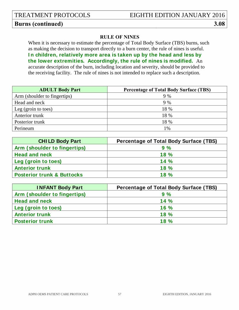
TREATMENT PROTOCOLS EIGHTH EDITION JANUARY 2016
Burns (continued) 3.08
ADPH OEMS PATIENT CARE PROTOCOLS 57 EIGHTH EDITION, JANUARY 2016
RULE OF NINES When it is necessary to estimate the percentage of Total Body Surface (TBS) burns, such as making the decision to transport directly to a burn center, the rule of nines is useful. In children, relatively more area is taken up by the head and less by the lower extremities. Accordingly, the rule of nines is modified. An accurate description of the burn, including location and severity, should be provided to the receiving facility. The rule of nines is not intended to replace such a description.
ADULT Body Part Percentage of Total Body Surface (TBS) Arm (shoulder to fingertips) 9 % Head and neck 9 % Leg (groin to toes) 18 % Anterior trunk 18 % Posterior trunk 18 % Perineum 1%
CHILD Body Part Percentage of Total Body Surface (TBS)
Arm (shoulder to fingertips) 9 % Head and neck 18 % Leg (groin to toes) 14 % Anterior trunk 18 % Posterior trunk & Buttocks 18 %
INFANT Body Part Percentage of Total Body Surface (TBS)
Arm (shoulder to fingertips) 9 % Head and neck 14 % Leg (groin to toes) 16 % Anterior trunk 18 % Posterior trunk 18 %
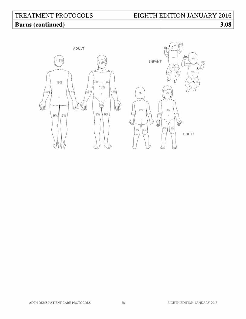
TREATMENT PROTOCOLS EIGHTH EDITION JANUARY 2016
Burns (continued) 3.08
ADPH OEMS PATIENT CARE PROTOCOLS 58 EIGHTH EDITION, JANUARY 2016
TREATMENT PROTOCOLS EIGHTH EDITION JANUARY 2016
Cardiac Arrest (Adult) 3.09
ADPH OEMS PATIENT CARE PROTOCOLS 59 EIGHTH EDITION, JANUARY 2016
HISTORY AND PHYSICAL EXAM
Downtime and circumstances, was arrest witnessed? Was bystander CPR performed? Preceding symptoms?
Patient’s past medical history, medications, allergies.
Determine level of responsiveness, quality of respiratory effort, presence of pulses.
Cardiac rhythm analysis.
Reversible causes of cardiac arrest: Hypovolemia, Hypoxia, Acidosis, Hypokalemia, Hyperkalemia, Hypothermia, Tension Pneumothorax, Cardiac Tamponade, Toxins, Pulmonary Thromboembolism, Acute MI.
KEY POINTS
Performance of high quality chest compressions at a rate of 100-120 compressions/minute and 2 inches depth allowing for full chest recoil combined with early defibrillation are the most critical elements of the resuscitation. Consider use of a metronome to ensure proper chest compression rate.
Once resuscitative efforts are begun, they should be continued until arrival at the receiving hospital or until a joint decision has been made with OLMD that resuscitation should cease. See policy Death in the Field (1.03).
Remember to treat the patient and not the monitor. Treatment decisions must be made considering the patient’s condition, not just the rhythm on the monitor.
Patients with penetrating torso injury and cardiac arrest can sometimes survive. The priority for these patients, as opposed to patients with other etiologies of cardiac arrest, is rapid transport and NOT chest compressions. Chest compressions may still be performed, but should not delay transport. These patients should receive IV fluids according to the Shock protocol (3.31).
If the patient in cardiac arrest has a venous port or other central venous access device, the EMSP may use it.
If quantitative waveform capnography <10 mm Hg, attempt to improve CPR quality.
Consider treatment for opiate overdose per Poisons and Overdoses Protocol (3.27) if opiate overdose is suspected in the cardiac arrest patient.
TREATMENT PROTOCOLS EIGHTH EDITION JANUARY 2016
Cardiac Arrest (Adult) (continued) 3.09
ADPH OEMS PATIENT CARE PROTOCOLS 60 EIGHTH EDITION, JANUARY 2016
TREATMENT DRUGS/PROCEDURES
Ventricular Fibrillation/Pulseless Ventricular Tachycardia
CPR with minimal interruption to chest compressions.
Bag-valve-mask ventilation with 100% oxygen. Avoid excessive ventilation.
Cardiac Monitor or AED.
Defibrillation. Consider escalating energy settings if first shock is unsuccessful.
Establish IV/IO Access.
Provide continuous chest compressions, alternating 2 min cycles of chest compressions with defibrillation and drug therapies.
Epinephrine every 3-5 min.
Amiodarone or Lidocaine.
Magnesium Sulfate for torsades de pointes.
Consider Advanced Airway.
Treat reversible causes.
*Follow American Heart Association (AHA) Guidelines
EMT: CPR Bag-Valve-Mask Ventilation 30:2 compressions to ventilations rate. 100% Oxygen AED
Advanced: Advanced Airway: Blind Insertion Airway Device. 8-10 breaths/minute with continuous chest compressions.
Intermediate: Defibrillation
Biphasic: use manufacturers recommended setting Monophasic: 360 J
Advanced Airway: Endotracheal tube. 8-10 breaths/minute with continuous chest compressions.
Paramedic: Epinephrine :
1 mg IV/IO every 3-5 min
Amiodarone: 1st dose: 300 mg IV/IO 2nd dose: 150 mg IV/IO
Lidocaine: 1st dose: 1.5 mg/kg IV/IO 2nd dose: 0.75 mg/kg IV/IO MAX 3 mg/kg
Magnesium Sulfate: 2 gm in 250 cc NS IV/IO
TREATMENT PROTOCOLS EIGHTH EDITION JANUARY 2016
Cardiac Arrest (Adult) (continued) 3.09
ADPH OEMS PATIENT CARE PROTOCOLS 61 EIGHTH EDITION, JANUARY 2016
TREATMENT DRUGS/PROCEDURES
Asystole/Pulseless Electrical Activity
CPR with minimal interruptions to chest compressions.
Bag-valve-mask ventilation with 100% oxygen. Avoid excessive ventilation.
Cardiac Monitor or AED. Establish IV/IO Access. Provide continuous chest compressions,
alternating 2 min cycles of chest compressions with drug therapies.
Epinephrine every 3-5 min. Consider Advanced Airway. Treat reversible causes. Consider Sodium Bicarbonate particularly
in prolonged cardiac arrest, known cocaine, aspirin, or tricyclic antidepressant toxicity, or renal failure patients who may have hyperkalemia (high potassium).
Consider Calcium Chloride particularly in renal failure patients who may have hyperkalemia (high potassium).
*Follow AHA Guidelines
EMT: CPR Bag-Valve-Mask Ventilation 30:2 compressions to ventilations rate. 100% Oxygen AED
Advanced: Advanced Airway: Blind Insertion Airway Device. 8-10 breaths/minute with continuous chest compressions.
Intermediate: Advanced Airway: Endotracheal tube. 8-10 breaths/minute with continuous chest compressions.
Paramedic: Epinephrine :
1 mg IV/IO every 3-5 min Sodium Bicarbonate:
1 mEq/kg IV/IO Calcium Chloride:
1gm (10 cc of 10% solution) IV/IO
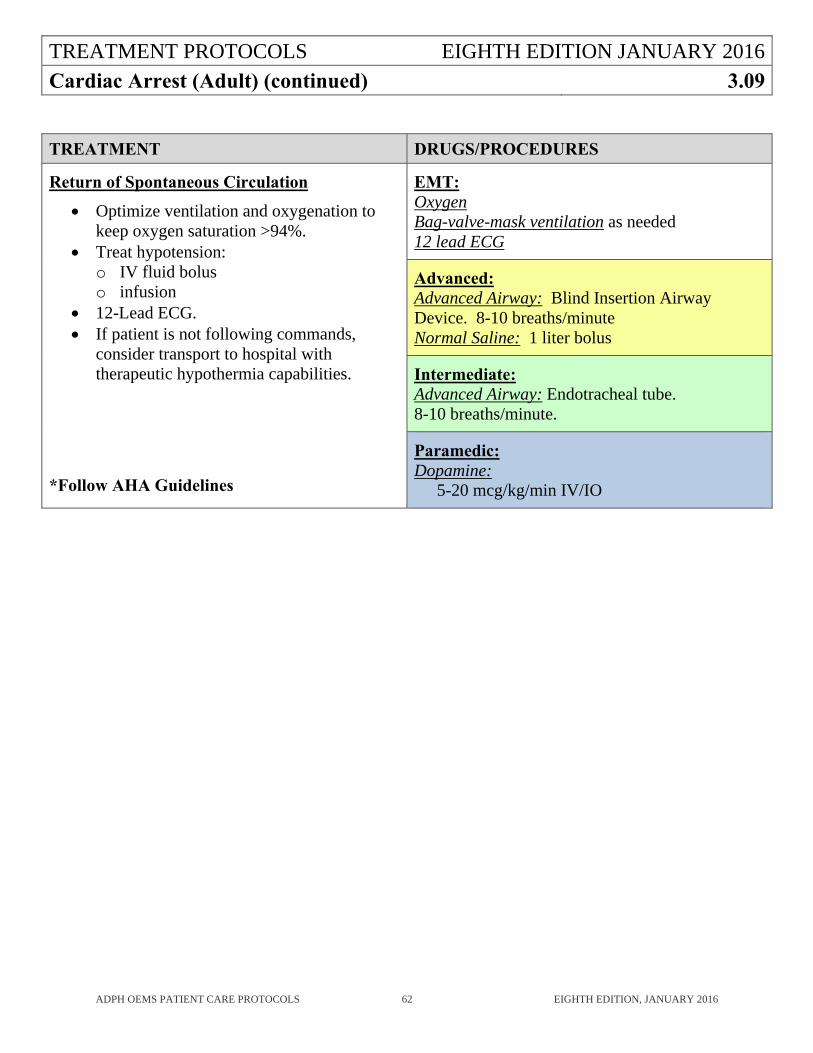
TREATMENT PROTOCOLS EIGHTH EDITION JANUARY 2016
Cardiac Arrest (Adult) (continued) 3.09
ADPH OEMS PATIENT CARE PROTOCOLS 62 EIGHTH EDITION, JANUARY 2016
TREATMENT DRUGS/PROCEDURES
Return of Spontaneous Circulation
Optimize ventilation and oxygenation to keep oxygen saturation >94%.
Treat hypotension: o IV fluid bolus o infusion
12-Lead ECG. If patient is not following commands,
consider transport to hospital with therapeutic hypothermia capabilities.
*Follow AHA Guidelines
EMT: Oxygen Bag-valve-mask ventilation as needed 12 lead ECG
Advanced: Advanced Airway: Blind Insertion Airway Device. 8-10 breaths/minute Normal Saline: 1 liter bolus
Intermediate: Advanced Airway: Endotracheal tube. 8-10 breaths/minute.
Paramedic: Dopamine:
5-20 mcg/kg/min IV/IO
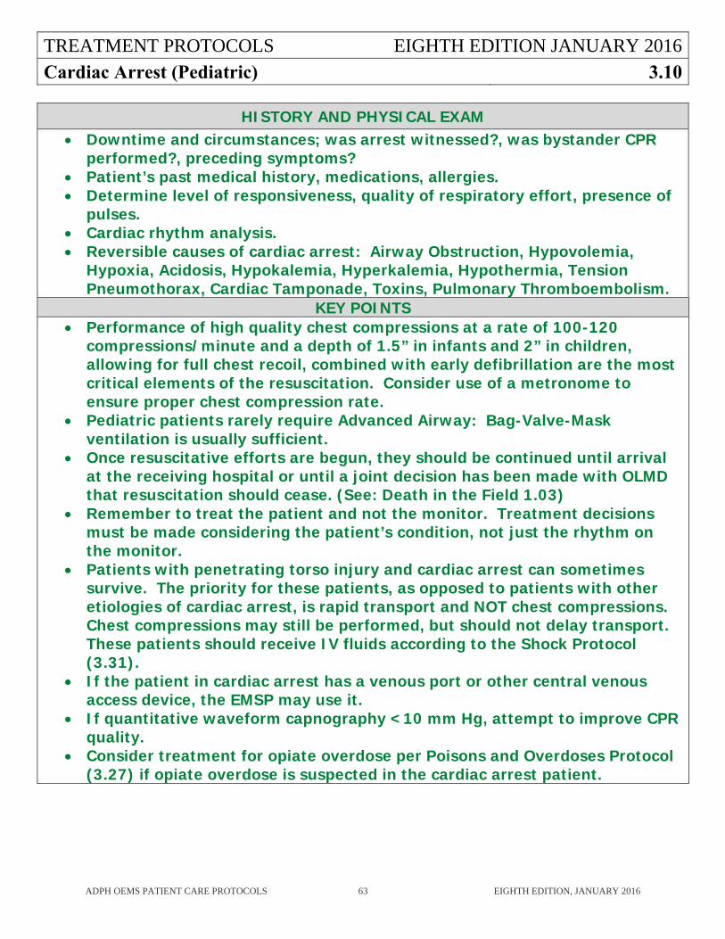
TREATMENT PROTOCOLS EIGHTH EDITION JANUARY 2016
Cardiac Arrest (Pediatric) 3.10
ADPH OEMS PATIENT CARE PROTOCOLS 63 EIGHTH EDITION, JANUARY 2016
HISTORY AND PHYSICAL EXAM Downtime and circumstances; was arrest witnessed?, was bystander CPR
performed?, preceding symptoms? Patient’s past medical history, medications, allergies. Determine level of responsiveness, quality of respiratory effort, presence of
pulses. Cardiac rhythm analysis. Reversible causes of cardiac arrest: Airway Obstruction, Hypovolemia,
Hypoxia, Acidosis, Hypokalemia, Hyperkalemia, Hypothermia, Tension Pneumothorax, Cardiac Tamponade, Toxins, Pulmonary Thromboembolism.
KEY POINTS Performance of high quality chest compressions at a rate of 100-120
compressions/minute and a depth of 1.5” in infants and 2” in children, allowing for full chest recoil, combined with early defibrillation are the most critical elements of the resuscitation. Consider use of a metronome to ensure proper chest compression rate.
Pediatric patients rarely require Advanced Airway: Bag-Valve-Mask ventilation is usually sufficient.
Once resuscitative efforts are begun, they should be continued until arrival at the receiving hospital or until a joint decision has been made with OLMD that resuscitation should cease. (See: Death in the Field 1.03)
Remember to treat the patient and not the monitor. Treatment decisions must be made considering the patient’s condition, not just the rhythm on the monitor.
Patients with penetrating torso injury and cardiac arrest can sometimes survive. The priority for these patients, as opposed to patients with other etiologies of cardiac arrest, is rapid transport and NOT chest compressions. Chest compressions may still be performed, but should not delay transport. These patients should receive IV fluids according to the Shock Protocol (3.31).
If the patient in cardiac arrest has a venous port or other central venous access device, the EMSP may use it.
If quantitative waveform capnography <10 mm Hg, attempt to improve CPR quality.
Consider treatment for opiate overdose per Poisons and Overdoses Protocol (3.27) if opiate overdose is suspected in the cardiac arrest patient.
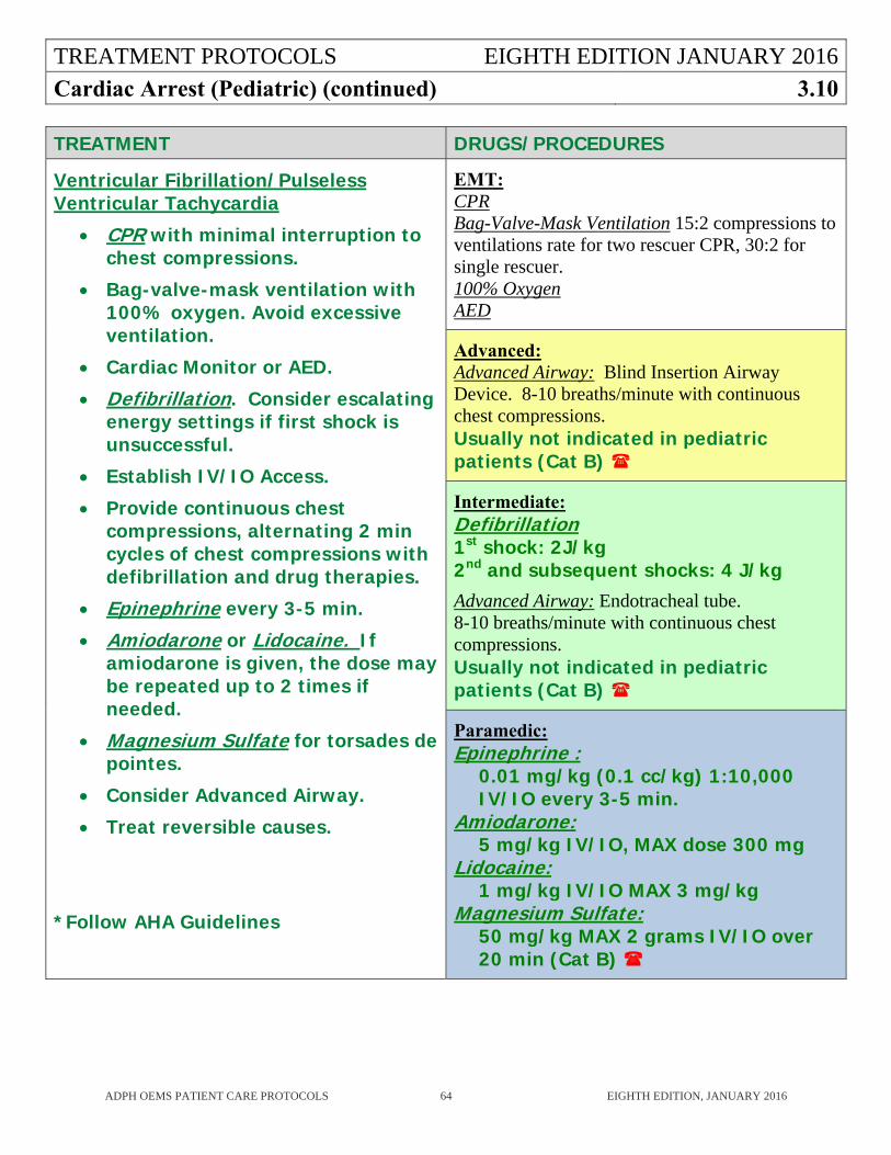
TREATMENT PROTOCOLS EIGHTH EDITION JANUARY 2016
Cardiac Arrest (Pediatric) (continued) 3.10
ADPH OEMS PATIENT CARE PROTOCOLS 64 EIGHTH EDITION, JANUARY 2016
TREATMENT DRUGS/PROCEDURES
Ventricular Fibrillation/Pulseless Ventricular Tachycardia
CPR with minimal interruption to chest compressions.
Bag-valve-mask ventilation with 100% oxygen. Avoid excessive ventilation.
Cardiac Monitor or AED.
Defibrillation. Consider escalating energy settings if first shock is unsuccessful.
Establish IV/IO Access.
Provide continuous chest compressions, alternating 2 min cycles of chest compressions with defibrillation and drug therapies.
Epinephrine every 3-5 min.
Amiodarone or Lidocaine. If amiodarone is given, the dose may be repeated up to 2 times if needed.
Magnesium Sulfate for torsades de pointes.
Consider Advanced Airway.
Treat reversible causes.
*Follow AHA Guidelines
EMT: CPR Bag-Valve-Mask Ventilation 15:2 compressions to ventilations rate for two rescuer CPR, 30:2 for single rescuer. 100% Oxygen AED
Advanced: Advanced Airway: Blind Insertion Airway Device. 8-10 breaths/minute with continuous chest compressions. Usually not indicated in pediatric patients (Cat B)
Intermediate: Defibrillation 1st shock: 2J/kg 2nd and subsequent shocks: 4 J/kg Advanced Airway: Endotracheal tube. 8-10 breaths/minute with continuous chest compressions. Usually not indicated in pediatric patients (Cat B)
Paramedic: Epinephrine :
0.01 mg/kg (0.1 cc/kg) 1:10,000 IV/IO every 3-5 min.
Amiodarone: 5 mg/kg IV/IO, MAX dose 300 mg
Lidocaine: 1 mg/kg IV/IO MAX 3 mg/kg
Magnesium Sulfate: 50 mg/kg MAX 2 grams IV/IO over 20 min (Cat B)
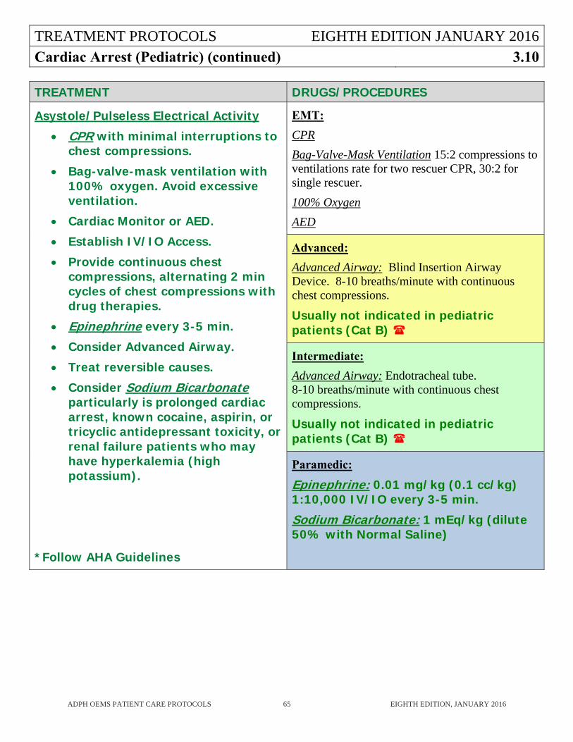
TREATMENT PROTOCOLS EIGHTH EDITION JANUARY 2016
Cardiac Arrest (Pediatric) (continued) 3.10
ADPH OEMS PATIENT CARE PROTOCOLS 65 EIGHTH EDITION, JANUARY 2016
TREATMENT DRUGS/PROCEDURES
Asystole/Pulseless Electrical Activity
CPR with minimal interruptions to chest compressions.
Bag-valve-mask ventilation with 100% oxygen. Avoid excessive ventilation.
Cardiac Monitor or AED.
Establish IV/IO Access.
Provide continuous chest compressions, alternating 2 min cycles of chest compressions with drug therapies.
Epinephrine every 3-5 min.
Consider Advanced Airway.
Treat reversible causes.
Consider Sodium Bicarbonate particularly is prolonged cardiac arrest, known cocaine, aspirin, or tricyclic antidepressant toxicity, or renal failure patients who may have hyperkalemia (high potassium).
*Follow AHA Guidelines
EMT:
CPR
Bag-Valve-Mask Ventilation 15:2 compressions to ventilations rate for two rescuer CPR, 30:2 for single rescuer.
100% Oxygen
AED
Advanced:
Advanced Airway: Blind Insertion Airway Device. 8-10 breaths/minute with continuous chest compressions.
Usually not indicated in pediatric patients (Cat B)
Intermediate:
Advanced Airway: Endotracheal tube. 8-10 breaths/minute with continuous chest compressions.
Usually not indicated in pediatric patients (Cat B)
Paramedic:
Epinephrine: 0.01 mg/kg (0.1 cc/kg) 1:10,000 IV/IO every 3-5 min. Sodium Bicarbonate: 1 mEq/kg (dilute 50% with Normal Saline)
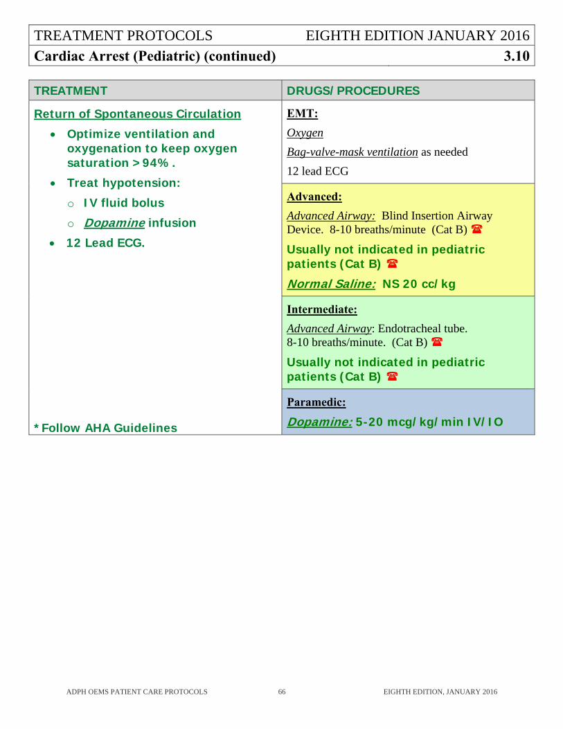
TREATMENT PROTOCOLS EIGHTH EDITION JANUARY 2016
Cardiac Arrest (Pediatric) (continued) 3.10
ADPH OEMS PATIENT CARE PROTOCOLS 66 EIGHTH EDITION, JANUARY 2016
TREATMENT DRUGS/PROCEDURES
Return of Spontaneous Circulation
Optimize ventilation and oxygenation to keep oxygen saturation >94%.
Treat hypotension:
o IV fluid bolus
o Dopamine infusion
12 Lead ECG.
*Follow AHA Guidelines
EMT:
Oxygen
Bag-valve-mask ventilation as needed
12 lead ECG
Advanced:
Advanced Airway: Blind Insertion Airway Device. 8-10 breaths/minute (Cat B)
Usually not indicated in pediatric patients (Cat B)
Normal Saline: NS 20 cc/kg
Intermediate:
Advanced Airway: Endotracheal tube. 8-10 breaths/minute. (Cat B)
Usually not indicated in pediatric patients (Cat B)
Paramedic:
Dopamine: 5-20 mcg/kg/min IV/IO
TREATMENT PROTOCOLS EIGHTH EDITION JANUARY 2016
Cardiac Dysrhythmia (Adult) 3.11
ADPH OEMS PATIENT CARE PROTOCOLS 67 EIGHTH EDITION, JANUARY 2016
HISTORY AND PHYSICAL EXAM
Chief Complaint, onset sudden or gradual.
Related symptoms such as palpitations, dizziness, chest pain, syncope, dyspnea.
Past medical history and medications.
Look for evidence of low cardiac output such as altered level of consciousness, presence of shock syndrome, signs of congestive heart failure.
KEY POINTS
Cardiac dysrhythmias may not require treatment in the field if the patient has no signs of impaired perfusion.
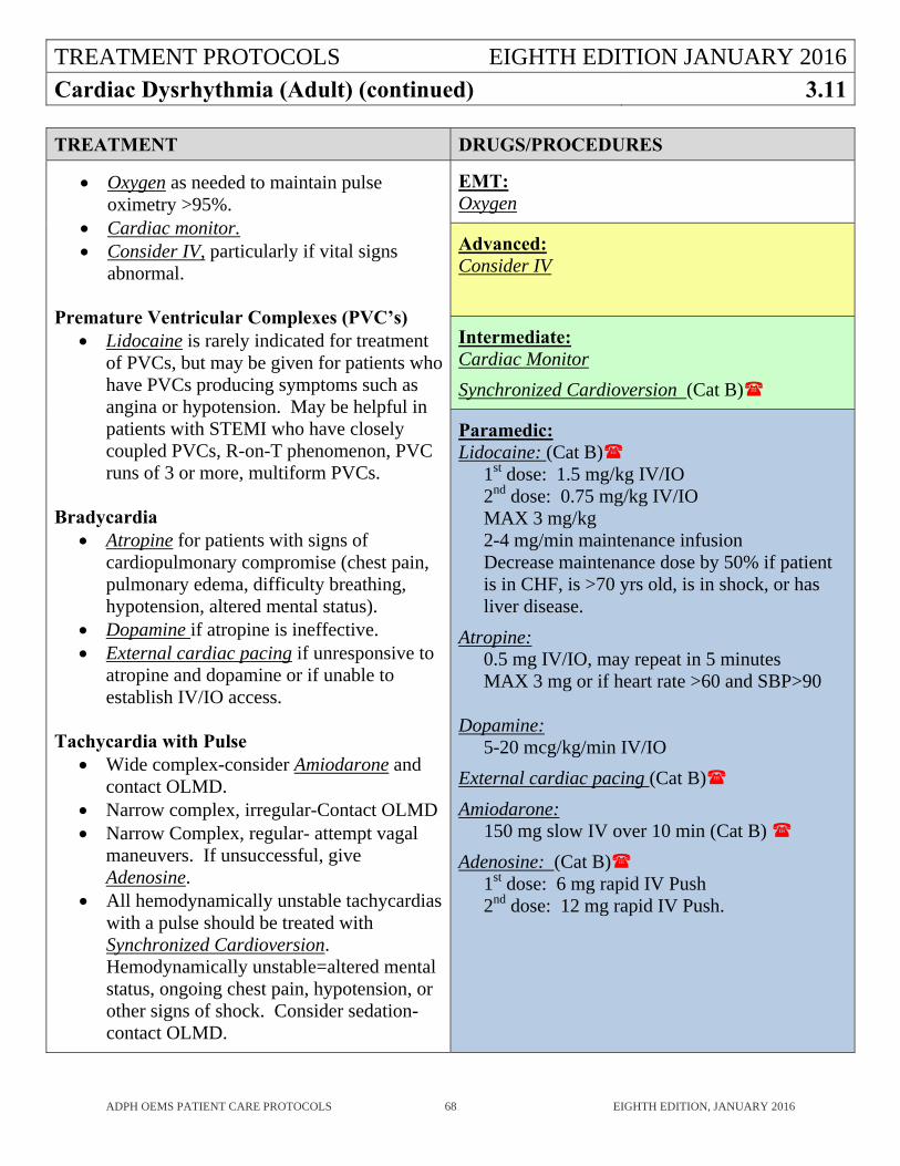
TREATMENT PROTOCOLS EIGHTH EDITION JANUARY 2016
Cardiac Dysrhythmia (Adult) (continued) 3.11
ADPH OEMS PATIENT CARE PROTOCOLS 68 EIGHTH EDITION, JANUARY 2016
TREATMENT DRUGS/PROCEDURES
Oxygen as needed to maintain pulse oximetry >95%.
Cardiac monitor. Consider IV, particularly if vital signs
abnormal.
Premature Ventricular Complexes (PVC’s) Lidocaine is rarely indicated for treatment
of PVCs, but may be given for patients who have PVCs producing symptoms such as angina or hypotension. May be helpful in patients with STEMI who have closely coupled PVCs, R-on-T phenomenon, PVC runs of 3 or more, multiform PVCs.
Bradycardia
Atropine for patients with signs of cardiopulmonary compromise (chest pain, pulmonary edema, difficulty breathing, hypotension, altered mental status).
Dopamine if atropine is ineffective. External cardiac pacing if unresponsive to
atropine and dopamine or if unable to establish IV/IO access.
Tachycardia with Pulse
Wide complex-consider Amiodarone and contact OLMD.
Narrow complex, irregular-Contact OLMD Narrow Complex, regular- attempt vagal
maneuvers. If unsuccessful, give Adenosine.
All hemodynamically unstable tachycardias with a pulse should be treated with Synchronized Cardioversion. Hemodynamically unstable=altered mental status, ongoing chest pain, hypotension, or other signs of shock. Consider sedation-contact OLMD.
EMT: Oxygen
Advanced: Consider IV
Intermediate: Cardiac Monitor
Synchronized Cardioversion (Cat B)
Paramedic: Lidocaine: (Cat B)
1st dose: 1.5 mg/kg IV/IO 2nd dose: 0.75 mg/kg IV/IO MAX 3 mg/kg 2-4 mg/min maintenance infusion Decrease maintenance dose by 50% if patient is in CHF, is >70 yrs old, is in shock, or has liver disease.
Atropine: 0.5 mg IV/IO, may repeat in 5 minutes MAX 3 mg or if heart rate >60 and SBP>90
Dopamine: 5-20 mcg/kg/min IV/IO
External cardiac pacing (Cat B)
Amiodarone: 150 mg slow IV over 10 min (Cat B)
Adenosine: (Cat B) 1st dose: 6 mg rapid IV Push 2nd dose: 12 mg rapid IV Push.
TREATMENT PROTOCOLS EIGHTH EDITION JANUARY 2016
Cardiac Dysrhythmia (Pediatric) 3.12
ADPH OEMS PATIENT CARE PROTOCOLS 69 EIGHTH EDITION, JANUARY 2016
HISTORY AND PHYSICAL EXAM
Chief Complaint, onset sudden or gradual.
Related symptoms such as palpitations, dizziness, chest pain, syncope, dyspnea.
Past medical history and medications.
Look for evidence of low cardiac output such as altered level of consciousness, presence of shock syndrome, signs of congestive heart failure.
KEY POINTS
Cardiac dysrhythmias may not require treatment in the field if the patient has no signs of impaired perfusion.
Bradycardia in children is usually due to respiratory causes.
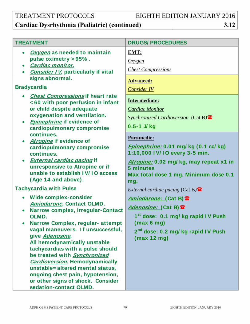
TREATMENT PROTOCOLS EIGHTH EDITION JANUARY 2016
Cardiac Dysrhythmia (Pediatric) (continued) 3.12
ADPH OEMS PATIENT CARE PROTOCOLS 70 EIGHTH EDITION, JANUARY 2016
TREATMENT DRUGS/PROCEDURES
Oxygen as needed to maintain pulse oximetry >95%.
Cardiac monitor. Consider IV, particularly if vital
signs abnormal.
Bradycardia
Chest Compressions if heart rate <60 with poor perfusion in infant or child despite adequate oxygenation and ventilation.
Epinephrine if evidence of cardiopulmonary compromise continues.
Atropine if evidence of cardiopulmonary compromise continues.
External cardiac pacing if unresponsive to Atropine or if unable to establish IV/IO access (Age 14 and above).
Tachycardia with Pulse
Wide complex-consider Amiodarone, Contact OLMD.
Narrow complex, irregular-Contact OLMD.
Narrow Complex, regular- attempt vagal maneuvers. If unsuccessful, give Adenosine. All hemodynamically unstable tachycardias with a pulse should be treated with Synchronized Cardioversion. Hemodynamically unstable=altered mental status, ongoing chest pain, hypotension, or other signs of shock. Consider sedation-contact OLMD.
EMT:
Oxygen
Chest Compressions
Advanced:
Consider IV
Intermediate:
Cardiac Monitor
Synchronized Cardioversion (Cat B)
0.5-1 J/kg
Paramedic:
Epinephrine: 0.01 mg/kg (0.1 cc/kg) 1:10,000 IV/IO every 3-5 min.
Atropine: 0.02 mg/kg, may repeat x1 in 5 minutes Max total dose 1 mg, Minimum dose 0.1 mg. External cardiac pacing (Cat B)
Amiodarone: (Cat B)
Adenosine: (Cat B)
1st dose: 0.1 mg/kg rapid IV Push (max 6 mg)
2nd dose: 0.2 mg/kg rapid IV Push (max 12 mg)
TREATMENT PROTOCOLS EIGHTH EDITION JANUARY 2016Chest Pain or Suspected Acute Coronary Syndrome (ACS)
3.13
ADPH OEMS PATIENT CARE PROTOCOLS 71 EIGHTH EDITION, JANUARY 2016
HISTORY AND PHYSICAL EXAM
Assess Pain (Onset, Place, Quality, Radiation, Severity, Time began). Associated symptoms: nausea, vomiting, diaphoresis, shortness of breath. History: cardiac or pulmonary events, medications, syncope. Risk Factors: Family history, smoking, obesity, diabetes, hypertension, high cholesterol. Vital signs no less than every 10 minutes and after each medication. Symmetry of pulses. Signs of Congestive Heart Failure such as neck vein distention, peripheral edema, or pulmonary
edema. Examine abdomen.
KEY POINTS
This protocol is for adults. Contact OLMD for chest pain in pediatric patients. Chest wall tenderness does not rule out cardiac ischemia. ST segment elevation MI (STEMI) can only be accurately diagnosed by acquiring a 12 lead ECG. Have a high index of suspicion for cardiac disease in women, diabetics, and all patients >50 years
old who have any symptoms that might be attributed to acute coronary syndrome (e.g. nausea, neck, jaw, or arm pain, chest pain, diaphoresis, syncope).
12-Lead ECG should be performed on all patients with chest pain, epigastric discomfort, or suspected acute coronary syndrome before leaving the scene.
In patients with STEMI, time to reperfusion is critical. The performance of a 12-lead ECG at the scene and its transmittal to the receiving hospital can significantly shorten the time to reperfusing treatment. Minimize scene times after performing and transmitting the 12-lead ECG when possible. Consider transporting patients with STEMI to hospital with available catheterization lab for percutaneous coronary intervention (PCI). If unsure of appropriate destination hospital, contact OLMD.
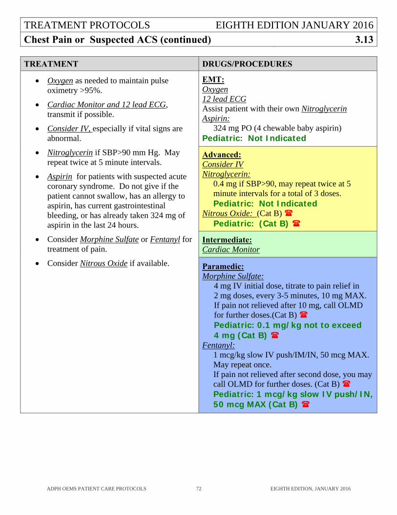
TREATMENT PROTOCOLS EIGHTH EDITION JANUARY 2016
Chest Pain or Suspected ACS (continued) 3.13
ADPH OEMS PATIENT CARE PROTOCOLS 72 EIGHTH EDITION, JANUARY 2016
TREATMENT DRUGS/PROCEDURES
Oxygen as needed to maintain pulse oximetry >95%.
Cardiac Monitor and 12 lead ECG, transmit if possible.
Consider IV, especially if vital signs are abnormal.
Nitroglycerin if SBP>90 mm Hg. May repeat twice at 5 minute intervals.
Aspirin for patients with suspected acute coronary syndrome. Do not give if the patient cannot swallow, has an allergy to aspirin, has current gastrointestinal bleeding, or has already taken 324 mg of aspirin in the last 24 hours.
Consider Morphine Sulfate or Fentanyl for treatment of pain.
Consider Nitrous Oxide if available.
EMT: Oxygen 12 lead ECG Assist patient with their own Nitroglycerin Aspirin:
324 mg PO (4 chewable baby aspirin) Pediatric: Not Indicated
Advanced: Consider IV Nitroglycerin:
0.4 mg if SBP>90, may repeat twice at 5 minute intervals for a total of 3 doses. Pediatric: Not Indicated
Nitrous Oxide: (Cat B) Pediatric: (Cat B)
Intermediate: Cardiac Monitor
Paramedic: Morphine Sulfate:
4 mg IV initial dose, titrate to pain relief in 2 mg doses, every 3-5 minutes, 10 mg MAX. If pain not relieved after 10 mg, call OLMD for further doses.(Cat B) Pediatric: 0.1 mg/kg not to exceed 4 mg (Cat B)
Fentanyl: 1 mcg/kg slow IV push/IM/IN, 50 mcg MAX. May repeat once. If pain not relieved after second dose, you may call OLMD for further doses. (Cat B) Pediatric: 1 mcg/kg slow IV push/IN, 50 mcg MAX (Cat B)
TREATMENT PROTOCOLS EIGHTH EDITION JANUARY 2016
Childbirth 3.14
ADPH OEMS PATIENT CARE PROTOCOLS 73 EIGHTH EDITION, JANUARY 2016
HISTORY AND PHYSICAL EXAM
History of pregnancy: Due date, last menstrual period, is this a known multiple gestation?
Does the patient feel that she is in labor or about to deliver (e.g. rectal or vaginal pressure)?
Recent symptoms such as pain or contractions? Timing and regularity? Vaginal bleeding, ruptured membranes, urge to push?
Medical history: medications, medical problems, age, number of prior pregnancies.
Vital signs and fetal heart rate if possible.
Contractions and relaxation of uterus.
Where privacy is possible, inspect perineum for vaginal bleeding or fluid (note color and presence of meconium), crowning (check during contraction), abnormal presentation (foot, arm, cord, or breech).
KEY POINTS
Do not delay transport particularly for patients with previous cesarean section, known imminent multiple births, abnormal presenting parts, excessive bleeding, and premature labor.
In case of prolapsed umbilical cord, place the mother in Trendelenburg or knee chest position. Elevated presenting body part to relieve pressure on the cord and keep the cord moist with saline gauze if it is exposed. Do not delay transport.
If thick meconium is present, aggressively suction and consider intubation for neonate. (See Newborn Protocol 3.26).
If a non-viable premature fetus is delivered and the fetus is available, place the fetus in a clean container and transport to the hospital with the mother. Remember to treat the fetus with the same respect as the EMSP would treat any deceased patient.
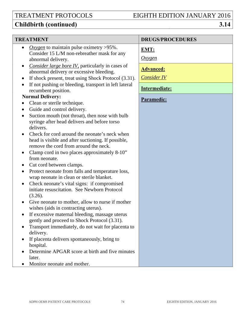
TREATMENT PROTOCOLS EIGHTH EDITION JANUARY 2016
Childbirth (continued) 3.14
ADPH OEMS PATIENT CARE PROTOCOLS 74 EIGHTH EDITION, JANUARY 2016
TREATMENT DRUGS/PROCEDURES
Oxygen to maintain pulse oximetry >95%. Consider 15 L/M non-rebreather mask for any abnormal delivery.
Consider large bore IV, particularly in cases of abnormal delivery or excessive bleeding.
If shock present, treat using Shock Protocol (3.31). If not pushing or bleeding, transport in left lateral
recumbent position. Normal Delivery: Clean or sterile technique. Guide and control delivery. Suction mouth (not throat), then nose with bulb
syringe after head delivers and before torso delivers.
Check for cord around the neonate’s neck when head is visible and after suctioning. If possible, remove the cord from around the neck.
Clamp cord in two places approximately 8-10” from neonate.
Cut cord between clamps. Protect neonate from falls and temperature loss,
wrap neonate in clean or sterile blanket. Check neonate’s vital signs: if compromised
initiate resuscitation. See Newborn Protocol (3.26).
Give neonate to mother, allow to nurse if mother wishes (aids in contracting uterus).
If excessive maternal bleeding, massage uterus gently and proceed to Shock Protocol (3.31).
Transport immediately, do not wait for placenta to delivery.
If placenta delivers spontaneously, bring to hospital.
Determine APGAR score at birth and five minutes later.
Monitor neonate and mother.
EMT:
Oxygen
Advanced:
Consider IV
Intermediate:
Paramedic:
TREATMENT PROTOCOLS EIGHTH EDITION JANUARY 2016
Congestive Heart Failure 3.15
ADPH OEMS PATIENT CARE PROTOCOLS 75 EIGHTH EDITION, JANUARY 2016
HISTORY AND PHYSICAL EXAM
History: Acuity of onset of symptoms? Obtain careful history of fever, chills, and purulent sputum products.
Past history: Chronic lung or heart problems? Medications or home oxygen? Associated symptoms: Chest pain, paresthesias of mouth or hands. Vital signs including pulse oximetry. If patient is usually on supplemental oxygen, note their
pulse oximetry on their usual amount of oxygen. Level of consciousness. Cyanosis. Signs of congestive heart failure: distended neck veins, pulmonary edema, possible wheezing,
possible blood-tinged or frothy sputum, and peripheral edema.
KEY POINTS
Accurate assessment of breath sounds is crucial. Use caution when treating congestive heart failure patients with albuterol since a side-effect is
tachycardia, which may worsen the congestive heart failure.
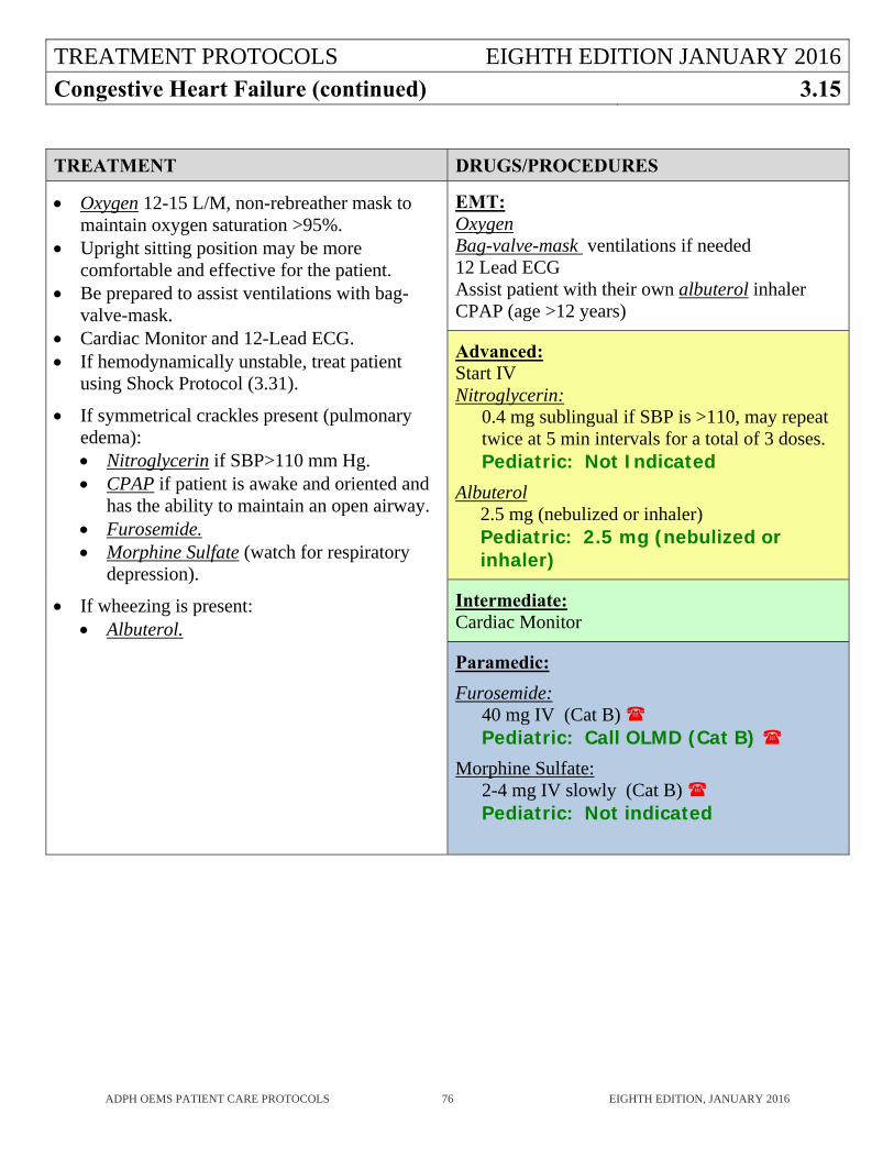
TREATMENT PROTOCOLS EIGHTH EDITION JANUARY 2016
Congestive Heart Failure (continued) 3.15
ADPH OEMS PATIENT CARE PROTOCOLS 76 EIGHTH EDITION, JANUARY 2016
TREATMENT DRUGS/PROCEDURES
Oxygen 12-15 L/M, non-rebreather mask to maintain oxygen saturation >95%.
Upright sitting position may be more comfortable and effective for the patient.
Be prepared to assist ventilations with bag-valve-mask.
Cardiac Monitor and 12-Lead ECG. If hemodynamically unstable, treat patient
using Shock Protocol (3.31).
If symmetrical crackles present (pulmonary edema): Nitroglycerin if SBP>110 mm Hg. CPAP if patient is awake and oriented and
has the ability to maintain an open airway. Furosemide. Morphine Sulfate (watch for respiratory
depression).
If wheezing is present: Albuterol.
EMT: Oxygen Bag-valve-mask ventilations if needed 12 Lead ECG Assist patient with their own albuterol inhaler CPAP (age >12 years)
Advanced: Start IV Nitroglycerin:
0.4 mg sublingual if SBP is >110, may repeat twice at 5 min intervals for a total of 3 doses. Pediatric: Not Indicated
Albuterol 2.5 mg (nebulized or inhaler) Pediatric: 2.5 mg (nebulized or inhaler)
Intermediate: Cardiac Monitor
Paramedic:
Furosemide: 40 mg IV (Cat B) Pediatric: Call OLMD (Cat B)
Morphine Sulfate: 2-4 mg IV slowly (Cat B) Pediatric: Not indicated
TREATMENT PROTOCOLS EIGHTH EDITION JANUARY 2016
Electromuscular Incapacitation Device (Taser®) 3.16
ADPH OEMS PATIENT CARE PROTOCOLS 77 EIGHTH EDITION, JANUARY 2016
HISTORY AND PHYSICAL EXAM
What was the patient doing that required use of the TASER®? Past History: Illicit drug use types and frequency. Medical problems and medications?
Psychotropic or behavioral drugs. Previous psychiatric disorders? If the device uses a barb (TASER®), are the barbs (2) still penetrating the skin? Are the barbs in a sensitive area such as the eye, eyelid, ear, nose, neck, female breast, or
genitalia? Are the wires still attached to the barbs? Do not touch the barbs or wires. Do not step on the
wires. The EMSP may safely touch the patient while the barbs and wires are attached. Take vital signs if safe and possible (patient cooperative). Note pupil size, symmetry, and
reactivity. If safe and possible, apply cardiac monitor and document rhythm strip. Mental status. Document status each time vital signs are taken. Characteristic odor on breath? Medical alert tag?
KEY POINTS
Law enforcement may request EMSPs to evaluate a patient who was the target of an electromuscular incapacitation device. The important issue is not removal of barbs but rather what caused the patient to be so combative that he/she had to be restrained using an electromuscular incapacitation device. Deaths have been recorded after use of these devices, however, it has always been due to the underlying cause of the combative behavior (psychosis, drugs, hypoglycemia, brain tumor, etc.).
Patients with normal vital signs who have returned to a normal mental status do not require transportation to the hospital unless physician assistance is required for barb removal or some other reason is present mandating hospital transport. If there is any doubt about whether or not transport is required, contact OLMD.
TREATMENT PROTOCOLS EIGHTH EDITION JANUARY 2016Electromuscular Incapacitation Device (Taser®) (continued)
3.16
ADPH OEMS PATIENT CARE PROTOCOLS 78 EIGHTH EDITION, JANUARY 2016
TREATMENT DRUGS/PROCEDURES
If vital signs are abnormal, apply cardiac monitor and obtain 12 lead ECG.
Consider IV if vital signs are abnormal. If patient exhibits Altered Mental Status,
treat using Altered Mental Status protocol (3.05).
If continued patient restraint is necessary, see Patient Restraint procedure (4.13). If the patient is under arrest, law enforcement should accompany the patient to the hospital.
Removal of barbs:
Treat barbs as contaminated needles. Confirm that the TASER has been shut off
and that the wires have been removed from the barbs.
Remove one barb at a time. Grab barb firmly and pull straight out in a
quick motion, using two fingers of the EMSP’s free hand on either side of the barb as a brace.
Clean the area with betadine or alcohol and apply a dressing.
Dispose of the barb in a sharps container or, if requested, give to law enforcement personnel.
Barbs in the eye, eyelid, ear, nose, neck, female breast, or genitalia should be transported to the hospital for physician removal.
EMT: 12 Lead ECG
Advanced: Consider IV
Intermediate: Cardiac Monitor
Paramedic:
TREATMENT PROTOCOLS EIGHTH EDITION JANUARY 2016
Fractures and Dislocations 3.17
ADPH OEMS PATIENT CARE PROTOCOLS 79 EIGHTH EDITION, JANUARY 2016
HISTORY AND PHYSICAL EXAM
History of trauma and mechanism of injury. Localized Tenderness, Instability, and Crepitation. Pulses, Motor Function, and Sensation. Obvious deformity, angulation, deep lacerations, and exposed bone fragments.
KEY POINTS
Fractures do not necessarily lead to deformity or loss of function, e.g., impacted fractures may cause pain but little or no deformity or loss of function.
Extremity injuries benefit from appropriate care, but are of low priority in a patient with multiple injuries.
Patients with fractures of two or more proximal long bones or pelvic fractures should be entered into the Alabama Trauma System where applicable.
TREATMENT PROTOCOLS EIGHTH EDITION JANUARY 2016
Fractures and Dislocations (continued) 3.17
ADPH OEMS PATIENT CARE PROTOCOLS 80 EIGHTH EDITION, JANUARY 2016
TREATMENT DRUGS/PROCEDURES
Oxygen as needed to maintain oxygen saturation >95%.
If vitals are stable, consider large bore IV. If vitals are unstable- proceed to Shock
Protocol (3.31). Consider spinal motion restriction. Examine for additional injuries. Treat
higher priority injuries first. If a high index of suspicion of pelvic or
femur fractures, start large bore IV with normal saline. Monitor closely for signs of shock.
Apply sterile dressings to open fractures. Splint and apply axial traction as needed. Elevate simple fractures. Apply ice or cold
packs if time and extent of other injuries allow.
Transport as necessary. Monitor circulation (pulse and skin temperature), neurological, and motor function in affected extremity.
If the patient has severe incapacitating pain, consider analgesia: o Morphine Sulfate. o Fentanyl. o Nitrous Oxide.
EMT: Oxygen
Advanced: Consider IV
Nitrous Oxide: (Cat B) Pediatric: (Cat B)
Intermediate:
Paramedic:
Morphine Sulfate: 4 mg IV initial dose, titrate to pain relief in 2 mg doses, every 3-5 minutes, 10 mg MAX.
If pain not relieved after 10 mg, call OLMD for further doses.(Cat B)
Pediatric: 0.1 mg/kg not to exceed 4 mg (Cat B)
Fentanyl:
1 mcg/kg slow IV push/IM/IN, 50 mcg MAX. May repeat once. If pain not relieved after second dose you may call OLMD for further doses. (Cat B) Pediatric: 1 mcg/kg slow IV push/IN, 50 mcg MAX (Cat B)
TREATMENT PROTOCOLS EIGHTH EDITION JANUARY 2016
Head Trauma 3.18
ADPH OEMS PATIENT CARE PROTOCOLS 81 EIGHTH EDITION, JANUARY 2016
HISTORY AND PHYSICAL EXAM
History: Mechanism of injury, Level of Consciousness changes. Past medical history. Protective devices: helmet or seat belts. Evaluate airway patency, breathing capability, and gross injuries to extremities and trunk. Document Glasgow Coma Scale (Document all 3 component scores, as well as the total: Eyes,
Verbal, and Motor). Pupil position and response to light stimulation. External evidence of head trauma, (e.g. bleeding from ears, CSF draining from the head or
mouth, scalp lacerations).
KEY POINTS
Notify OLMD of changes in the patient’s GCS score in relation to time intervals. Always consider cervical spine injury in patients with head trauma. Head injury does not cause shock in adults. If shock is present in an adult patient with head
trauma, consider that there is probably another cause of shock. Head injury can cause shock in infants. Other causes of alteration of level of consciousness should be ruled out. Hypoventilation can cause cerebral edema. Maintain rate of 8 breaths per minute or, if using
capnography, maintain CO2 35-45. Call OLMD if signs of cerebral herniation (extensor posturing, dilated or nonreactive pupils, or
decrease in GCS of >2 if the initial was <9) Hyperventilation (rate 20 bpm) is (Cat B). Head injury in itself is not a contraindication to air medical transport. Patients with Head Injury and GCS <13 or prolonged loss of consciousness should be entered
into the Alabama Trauma System where applicable.
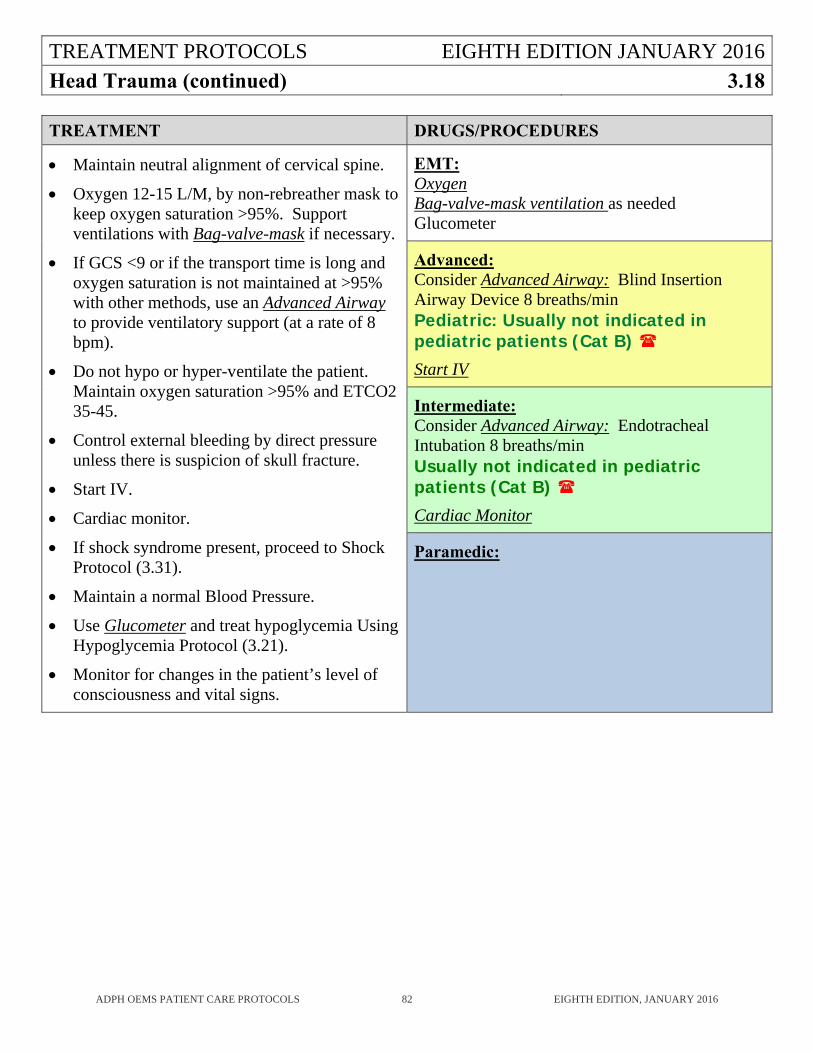
TREATMENT PROTOCOLS EIGHTH EDITION JANUARY 2016
Head Trauma (continued) 3.18
ADPH OEMS PATIENT CARE PROTOCOLS 82 EIGHTH EDITION, JANUARY 2016
TREATMENT DRUGS/PROCEDURES
Maintain neutral alignment of cervical spine.
Oxygen 12-15 L/M, by non-rebreather mask to keep oxygen saturation >95%. Support ventilations with Bag-valve-mask if necessary.
If GCS <9 or if the transport time is long and oxygen saturation is not maintained at >95% with other methods, use an Advanced Airway to provide ventilatory support (at a rate of 8 bpm).
Do not hypo or hyper-ventilate the patient. Maintain oxygen saturation >95% and ETCO2 35-45.
Control external bleeding by direct pressure unless there is suspicion of skull fracture.
Start IV.
Cardiac monitor.
If shock syndrome present, proceed to Shock Protocol (3.31).
Maintain a normal Blood Pressure.
Use Glucometer and treat hypoglycemia Using Hypoglycemia Protocol (3.21).
Monitor for changes in the patient’s level of consciousness and vital signs.
EMT: Oxygen Bag-valve-mask ventilation as needed Glucometer
Advanced: Consider Advanced Airway: Blind Insertion Airway Device 8 breaths/min Pediatric: Usually not indicated in pediatric patients (Cat B)
Start IV
Intermediate: Consider Advanced Airway: Endotracheal Intubation 8 breaths/min Usually not indicated in pediatric patients (Cat B)
Cardiac Monitor
Paramedic:
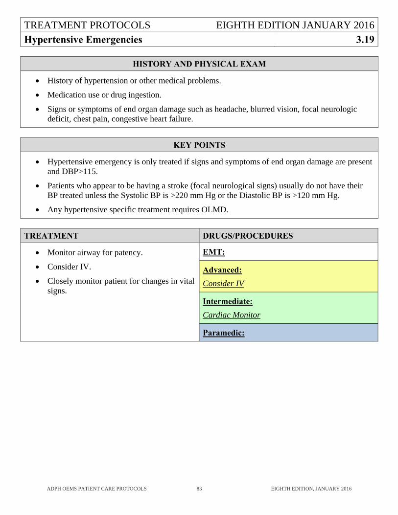
TREATMENT PROTOCOLS EIGHTH EDITION JANUARY 2016
Hypertensive Emergencies 3.19
ADPH OEMS PATIENT CARE PROTOCOLS 83 EIGHTH EDITION, JANUARY 2016
HISTORY AND PHYSICAL EXAM
History of hypertension or other medical problems.
Medication use or drug ingestion.
Signs or symptoms of end organ damage such as headache, blurred vision, focal neurologic deficit, chest pain, congestive heart failure.
KEY POINTS
Hypertensive emergency is only treated if signs and symptoms of end organ damage are present and DBP>115.
Patients who appear to be having a stroke (focal neurological signs) usually do not have their BP treated unless the Systolic BP is >220 mm Hg or the Diastolic BP is >120 mm Hg.
Any hypertensive specific treatment requires OLMD.
TREATMENT DRUGS/PROCEDURES
Monitor airway for patency.
Consider IV.
Closely monitor patient for changes in vital signs.
EMT:
Advanced:
Consider IV
Intermediate:
Cardiac Monitor
Paramedic:
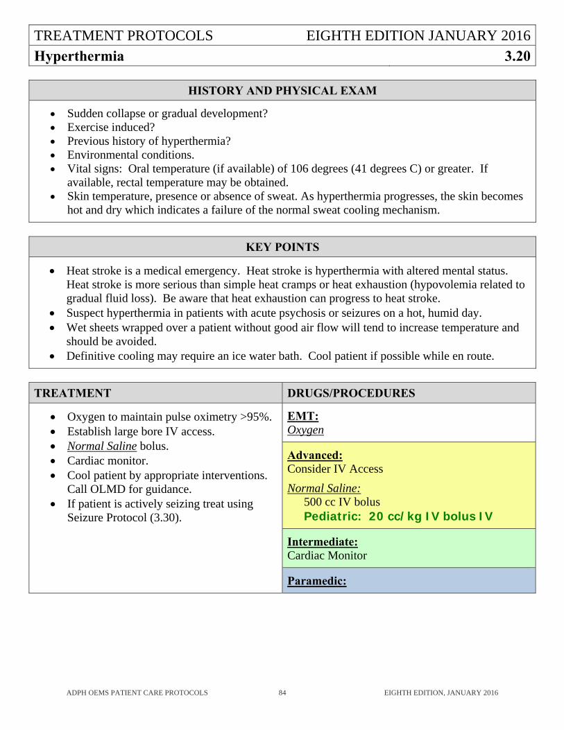
TREATMENT PROTOCOLS EIGHTH EDITION JANUARY 2016
Hyperthermia 3.20
ADPH OEMS PATIENT CARE PROTOCOLS 84 EIGHTH EDITION, JANUARY 2016
HISTORY AND PHYSICAL EXAM
Sudden collapse or gradual development? Exercise induced? Previous history of hyperthermia? Environmental conditions. Vital signs: Oral temperature (if available) of 106 degrees (41 degrees C) or greater. If
available, rectal temperature may be obtained. Skin temperature, presence or absence of sweat. As hyperthermia progresses, the skin becomes
hot and dry which indicates a failure of the normal sweat cooling mechanism.
KEY POINTS
Heat stroke is a medical emergency. Heat stroke is hyperthermia with altered mental status. Heat stroke is more serious than simple heat cramps or heat exhaustion (hypovolemia related to gradual fluid loss). Be aware that heat exhaustion can progress to heat stroke.
Suspect hyperthermia in patients with acute psychosis or seizures on a hot, humid day. Wet sheets wrapped over a patient without good air flow will tend to increase temperature and
should be avoided. Definitive cooling may require an ice water bath. Cool patient if possible while en route.
TREATMENT DRUGS/PROCEDURES
Oxygen to maintain pulse oximetry >95%. Establish large bore IV access. Normal Saline bolus. Cardiac monitor. Cool patient by appropriate interventions.
Call OLMD for guidance. If patient is actively seizing treat using
Seizure Protocol (3.30).
EMT: Oxygen
Advanced: Consider IV Access
Normal Saline: 500 cc IV bolus Pediatric: 20 cc/kg IV bolus IV
Intermediate: Cardiac Monitor
Paramedic:
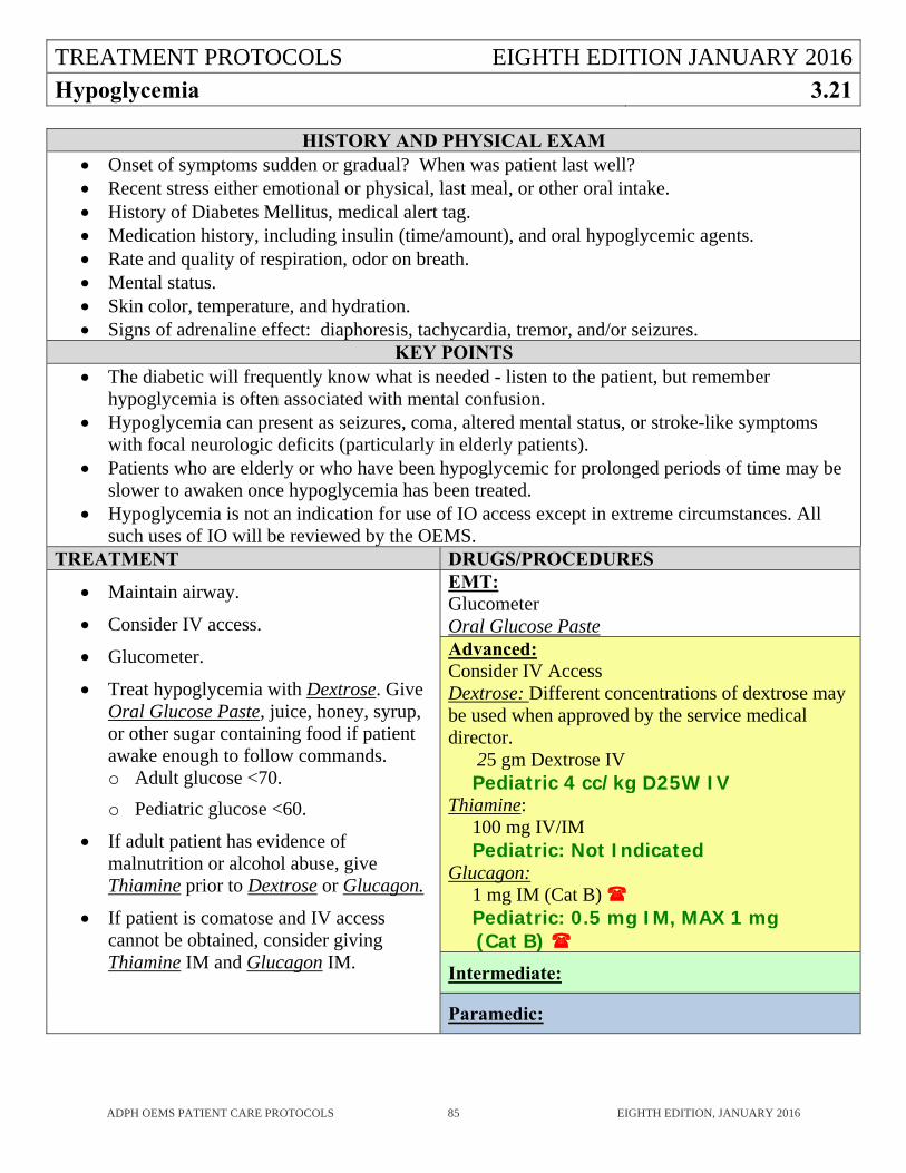
TREATMENT PROTOCOLS EIGHTH EDITION JANUARY 2016
Hypoglycemia 3.21
ADPH OEMS PATIENT CARE PROTOCOLS 85 EIGHTH EDITION, JANUARY 2016
HISTORY AND PHYSICAL EXAM Onset of symptoms sudden or gradual? When was patient last well? Recent stress either emotional or physical, last meal, or other oral intake. History of Diabetes Mellitus, medical alert tag. Medication history, including insulin (time/amount), and oral hypoglycemic agents. Rate and quality of respiration, odor on breath. Mental status. Skin color, temperature, and hydration. Signs of adrenaline effect: diaphoresis, tachycardia, tremor, and/or seizures.
KEY POINTS The diabetic will frequently know what is needed - listen to the patient, but remember
hypoglycemia is often associated with mental confusion. Hypoglycemia can present as seizures, coma, altered mental status, or stroke-like symptoms
with focal neurologic deficits (particularly in elderly patients). Patients who are elderly or who have been hypoglycemic for prolonged periods of time may be
slower to awaken once hypoglycemia has been treated. Hypoglycemia is not an indication for use of IO access except in extreme circumstances. All
such uses of IO will be reviewed by the OEMS. TREATMENT DRUGS/PROCEDURES
Maintain airway.
Consider IV access.
Glucometer.
Treat hypoglycemia with Dextrose. Give Oral Glucose Paste, juice, honey, syrup, or other sugar containing food if patient awake enough to follow commands. o Adult glucose <70.
o Pediatric glucose <60.
If adult patient has evidence of malnutrition or alcohol abuse, give Thiamine prior to Dextrose or Glucagon.
If patient is comatose and IV access cannot be obtained, consider giving Thiamine IM and Glucagon IM.
EMT: Glucometer Oral Glucose Paste Advanced: Consider IV Access Dextrose: Different concentrations of dextrose may be used when approved by the service medical director. 25 gm Dextrose IV Pediatric 4 cc/kg D25W IV Thiamine: 100 mg IV/IM Pediatric: Not Indicated Glucagon: 1 mg IM (Cat B) Pediatric: 0.5 mg IM, MAX 1 mg (Cat B)
Intermediate:
Paramedic:
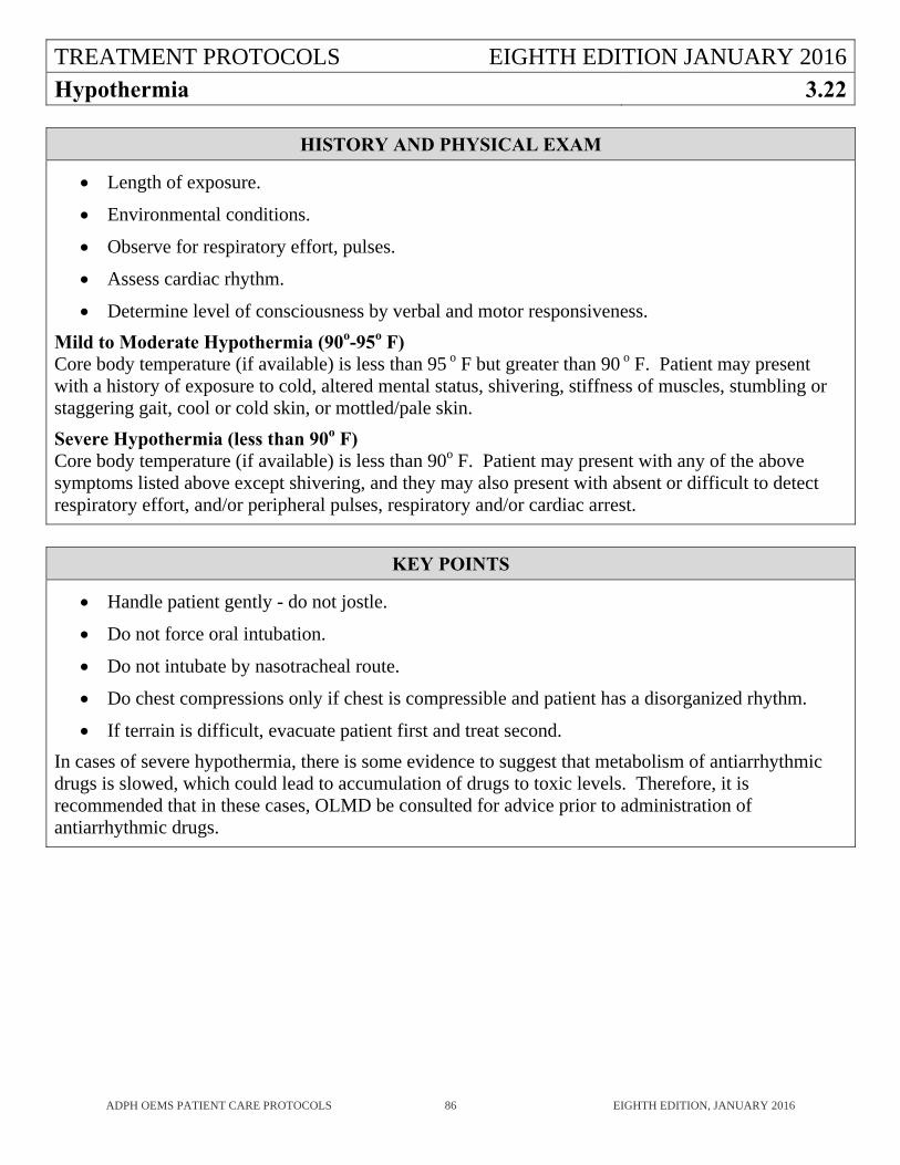
TREATMENT PROTOCOLS EIGHTH EDITION JANUARY 2016
Hypothermia 3.22
ADPH OEMS PATIENT CARE PROTOCOLS 86 EIGHTH EDITION, JANUARY 2016
HISTORY AND PHYSICAL EXAM
Length of exposure.
Environmental conditions.
Observe for respiratory effort, pulses.
Assess cardiac rhythm.
Determine level of consciousness by verbal and motor responsiveness.
Mild to Moderate Hypothermia (90o-95o F) Core body temperature (if available) is less than 95 o F but greater than 90 o F. Patient may present with a history of exposure to cold, altered mental status, shivering, stiffness of muscles, stumbling or staggering gait, cool or cold skin, or mottled/pale skin.
Severe Hypothermia (less than 90o F) Core body temperature (if available) is less than 90o F. Patient may present with any of the above symptoms listed above except shivering, and they may also present with absent or difficult to detect respiratory effort, and/or peripheral pulses, respiratory and/or cardiac arrest.
KEY POINTS
Handle patient gently - do not jostle.
Do not force oral intubation.
Do not intubate by nasotracheal route.
Do chest compressions only if chest is compressible and patient has a disorganized rhythm.
If terrain is difficult, evacuate patient first and treat second.
In cases of severe hypothermia, there is some evidence to suggest that metabolism of antiarrhythmic drugs is slowed, which could lead to accumulation of drugs to toxic levels. Therefore, it is recommended that in these cases, OLMD be consulted for advice prior to administration of antiarrhythmic drugs.
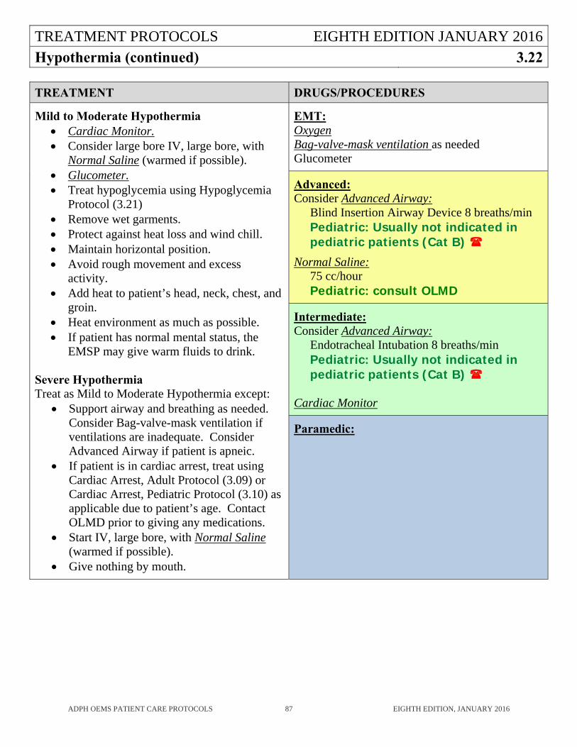
TREATMENT PROTOCOLS EIGHTH EDITION JANUARY 2016
Hypothermia (continued) 3.22
ADPH OEMS PATIENT CARE PROTOCOLS 87 EIGHTH EDITION, JANUARY 2016
TREATMENT DRUGS/PROCEDURES
Mild to Moderate Hypothermia Cardiac Monitor. Consider large bore IV, large bore, with
Normal Saline (warmed if possible). Glucometer. Treat hypoglycemia using Hypoglycemia
Protocol (3.21) Remove wet garments. Protect against heat loss and wind chill. Maintain horizontal position. Avoid rough movement and excess
activity. Add heat to patient’s head, neck, chest, and
groin. Heat environment as much as possible. If patient has normal mental status, the
EMSP may give warm fluids to drink. Severe Hypothermia Treat as Mild to Moderate Hypothermia except:
Support airway and breathing as needed. Consider Bag-valve-mask ventilation if ventilations are inadequate. Consider Advanced Airway if patient is apneic.
If patient is in cardiac arrest, treat using Cardiac Arrest, Adult Protocol (3.09) or Cardiac Arrest, Pediatric Protocol (3.10) as applicable due to patient’s age. Contact OLMD prior to giving any medications.
Start IV, large bore, with Normal Saline (warmed if possible).
Give nothing by mouth.
EMT: Oxygen Bag-valve-mask ventilation as needed Glucometer
Advanced: Consider Advanced Airway:
Blind Insertion Airway Device 8 breaths/min Pediatric: Usually not indicated in pediatric patients (Cat B)
Normal Saline: 75 cc/hour Pediatric: consult OLMD
Intermediate: Consider Advanced Airway:
Endotracheal Intubation 8 breaths/min Pediatric: Usually not indicated in pediatric patients (Cat B)
Cardiac Monitor
Paramedic:
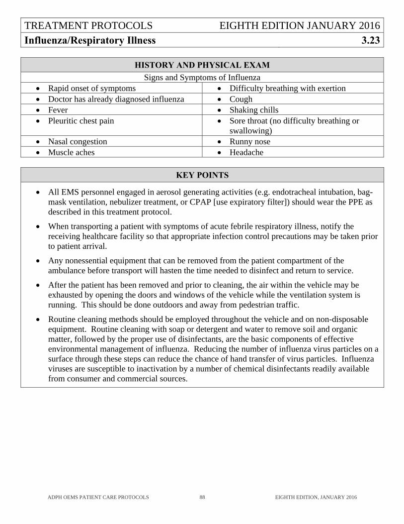
TREATMENT PROTOCOLS EIGHTH EDITION JANUARY 2016
Influenza/Respiratory Illness 3.23
ADPH OEMS PATIENT CARE PROTOCOLS 88 EIGHTH EDITION, JANUARY 2016
HISTORY AND PHYSICAL EXAM
Signs and Symptoms of Influenza Rapid onset of symptoms Difficulty breathing with exertion Doctor has already diagnosed influenza Cough Fever Shaking chills Pleuritic chest pain Sore throat (no difficulty breathing or
swallowing) Nasal congestion Runny nose Muscle aches Headache
KEY POINTS
All EMS personnel engaged in aerosol generating activities (e.g. endotracheal intubation, bag-mask ventilation, nebulizer treatment, or CPAP [use expiratory filter]) should wear the PPE as described in this treatment protocol.
When transporting a patient with symptoms of acute febrile respiratory illness, notify the receiving healthcare facility so that appropriate infection control precautions may be taken prior to patient arrival.
Any nonessential equipment that can be removed from the patient compartment of the ambulance before transport will hasten the time needed to disinfect and return to service.
After the patient has been removed and prior to cleaning, the air within the vehicle may be exhausted by opening the doors and windows of the vehicle while the ventilation system is running. This should be done outdoors and away from pedestrian traffic.
Routine cleaning methods should be employed throughout the vehicle and on non-disposable equipment. Routine cleaning with soap or detergent and water to remove soil and organic matter, followed by the proper use of disinfectants, are the basic components of effective environmental management of influenza. Reducing the number of influenza virus particles on a surface through these steps can reduce the chance of hand transfer of virus particles. Influenza viruses are susceptible to inactivation by a number of chemical disinfectants readily available from consumer and commercial sources.
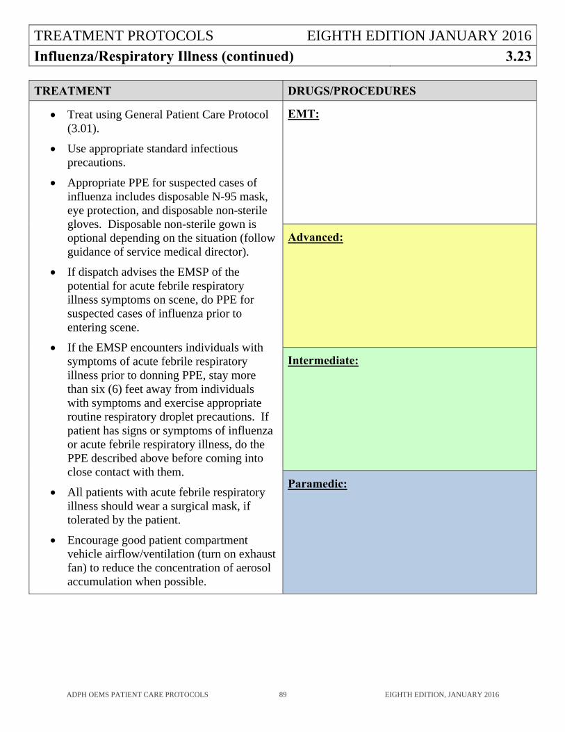
TREATMENT PROTOCOLS EIGHTH EDITION JANUARY 2016
Influenza/Respiratory Illness (continued) 3.23
ADPH OEMS PATIENT CARE PROTOCOLS 89 EIGHTH EDITION, JANUARY 2016
TREATMENT DRUGS/PROCEDURES
Treat using General Patient Care Protocol (3.01).
Use appropriate standard infectious precautions.
Appropriate PPE for suspected cases of influenza includes disposable N-95 mask, eye protection, and disposable non-sterile gloves. Disposable non-sterile gown is optional depending on the situation (follow guidance of service medical director).
If dispatch advises the EMSP of the potential for acute febrile respiratory illness symptoms on scene, do PPE for suspected cases of influenza prior to entering scene.
If the EMSP encounters individuals with symptoms of acute febrile respiratory illness prior to donning PPE, stay more than six (6) feet away from individuals with symptoms and exercise appropriate routine respiratory droplet precautions. If patient has signs or symptoms of influenza or acute febrile respiratory illness, do the PPE described above before coming into close contact with them.
All patients with acute febrile respiratory illness should wear a surgical mask, if tolerated by the patient.
Encourage good patient compartment vehicle airflow/ventilation (turn on exhaust fan) to reduce the concentration of aerosol accumulation when possible.
EMT:
Advanced:
Intermediate:
Paramedic:
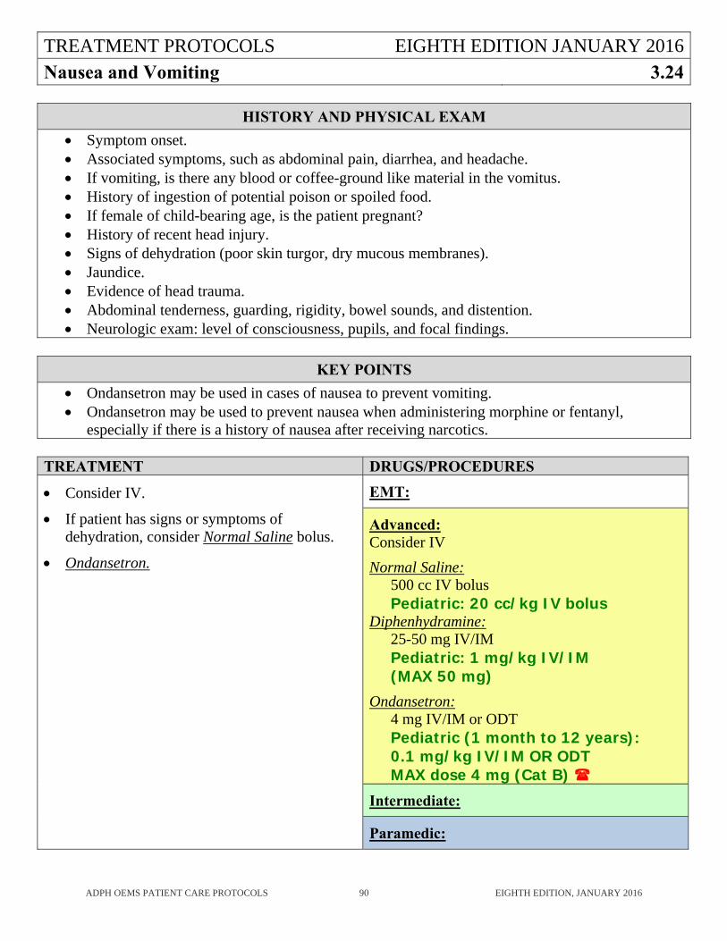
TREATMENT PROTOCOLS EIGHTH EDITION JANUARY 2016
Nausea and Vomiting 3.24
ADPH OEMS PATIENT CARE PROTOCOLS 90 EIGHTH EDITION, JANUARY 2016
HISTORY AND PHYSICAL EXAM
Symptom onset. Associated symptoms, such as abdominal pain, diarrhea, and headache. If vomiting, is there any blood or coffee-ground like material in the vomitus. History of ingestion of potential poison or spoiled food. If female of child-bearing age, is the patient pregnant? History of recent head injury. Signs of dehydration (poor skin turgor, dry mucous membranes). Jaundice. Evidence of head trauma. Abdominal tenderness, guarding, rigidity, bowel sounds, and distention. Neurologic exam: level of consciousness, pupils, and focal findings.
KEY POINTS
Ondansetron may be used in cases of nausea to prevent vomiting. Ondansetron may be used to prevent nausea when administering morphine or fentanyl,
especially if there is a history of nausea after receiving narcotics.
TREATMENT DRUGS/PROCEDURES
Consider IV.
If patient has signs or symptoms of dehydration, consider Normal Saline bolus.
Ondansetron.
EMT:
Advanced: Consider IV
Normal Saline: 500 cc IV bolus Pediatric: 20 cc/kg IV bolus
Diphenhydramine: 25-50 mg IV/IM Pediatric: 1 mg/kg IV/IM (MAX 50 mg)
Ondansetron: 4 mg IV/IM or ODT Pediatric (1 month to 12 years): 0.1 mg/kg IV/IM OR ODT MAX dose 4 mg (Cat B)
Intermediate:
Paramedic:
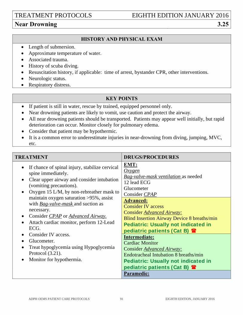
TREATMENT PROTOCOLS EIGHTH EDITION JANUARY 2016
Near Drowning 3.25
ADPH OEMS PATIENT CARE PROTOCOLS 91 EIGHTH EDITION, JANUARY 2016
HISTORY AND PHYSICAL EXAM
Length of submersion. Approximate temperature of water. Associated trauma. History of scuba diving. Resuscitation history, if applicable: time of arrest, bystander CPR, other interventions. Neurologic status. Respiratory distress.
KEY POINTS
If patient is still in water, rescue by trained, equipped personnel only. Near drowning patients are likely to vomit, use caution and protect the airway. All near drowning patients should be transported. Patients may appear well initially, but rapid
deterioration can occur. Monitor closely for pulmonary edema. Consider that patient may be hypothermic. It is a common error to underestimate injuries in near-drowning from diving, jumping, MVC,
etc.
TREATMENT DRUGS/PROCEDURES
If chance of spinal injury, stabilize cervical spine immediately.
Clear upper airway and consider intubation (vomiting precautions).
Oxygen 15 L/M, by non-rebreather mask to maintain oxygen saturation >95%, assist with Bag-valve-mask and suction as necessary.
Consider CPAP or Advanced Airway. Attach cardiac monitor, perform 12-Lead
ECG. Consider IV access. Glucometer. Treat hypoglycemia using Hypoglycemia
Protocol (3.21). Monitor for hypothermia.
EMT: Oxygen Bag-valve-mask ventilation as needed 12 lead ECG Glucometer Consider CPAP Advanced: Consider IV access Consider Advanced Airway: Blind Insertion Airway Device 8 breaths/min Pediatric: Usually not indicated in pediatric patients (Cat B) Intermediate: Cardiac Monitor Consider Advanced Airway: Endotracheal Intubation 8 breaths/min Pediatric: Usually not indicated in pediatric patients (Cat B) Paramedic:
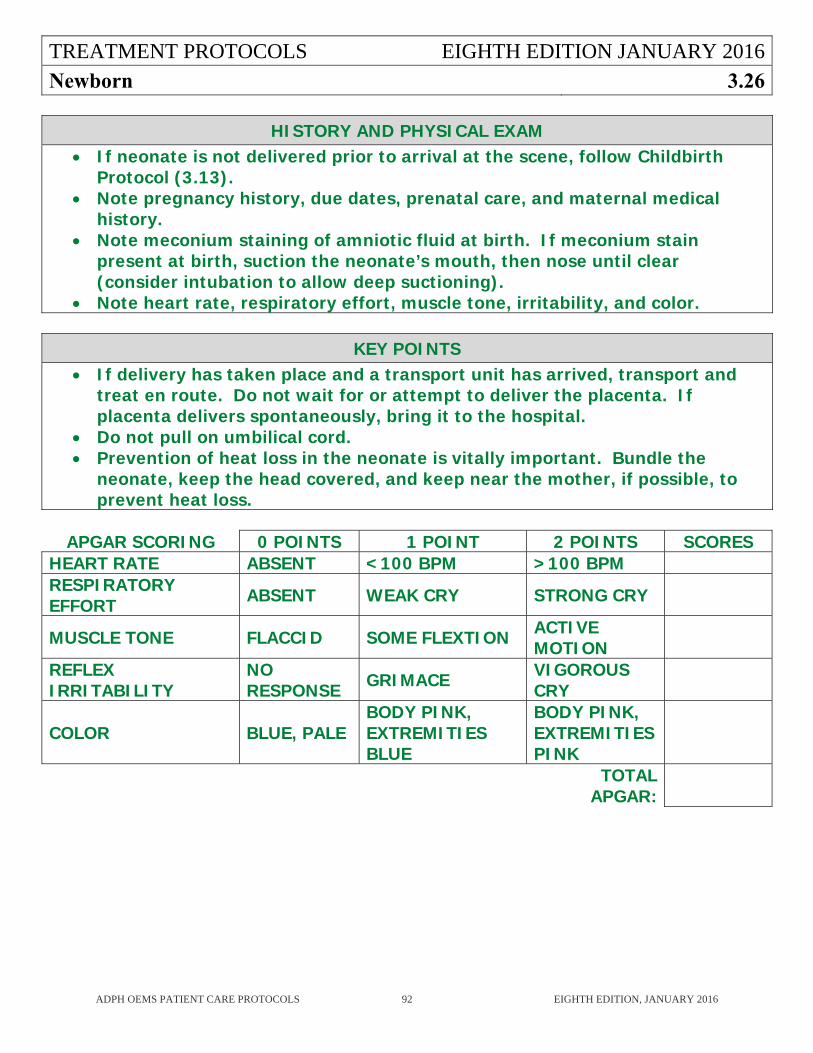
TREATMENT PROTOCOLS EIGHTH EDITION JANUARY 2016
Newborn 3.26
ADPH OEMS PATIENT CARE PROTOCOLS 92 EIGHTH EDITION, JANUARY 2016
HISTORY AND PHYSICAL EXAM If neonate is not delivered prior to arrival at the scene, follow Childbirth
Protocol (3.13). Note pregnancy history, due dates, prenatal care, and maternal medical
history. Note meconium staining of amniotic fluid at birth. If meconium stain
present at birth, suction the neonate’s mouth, then nose until clear (consider intubation to allow deep suctioning).
Note heart rate, respiratory effort, muscle tone, irritability, and color.
KEY POINTS If delivery has taken place and a transport unit has arrived, transport and
treat en route. Do not wait for or attempt to deliver the placenta. If placenta delivers spontaneously, bring it to the hospital.
Do not pull on umbilical cord. Prevention of heat loss in the neonate is vitally important. Bundle the
neonate, keep the head covered, and keep near the mother, if possible, to prevent heat loss.
APGAR SCORING 0 POINTS 1 POINT 2 POINTS SCORESHEART RATE ABSENT <100 BPM >100 BPM RESPIRATORY EFFORT ABSENT WEAK CRY STRONG CRY
MUSCLE TONE FLACCID SOME FLEXTION ACTIVE MOTION
REFLEX IRRITABILITY
NO RESPONSE GRIMACE VIGOROUS
CRY
COLOR BLUE, PALE BODY PINK, EXTREMITIES BLUE
BODY PINK, EXTREMITIES PINK
TOTAL APGAR:
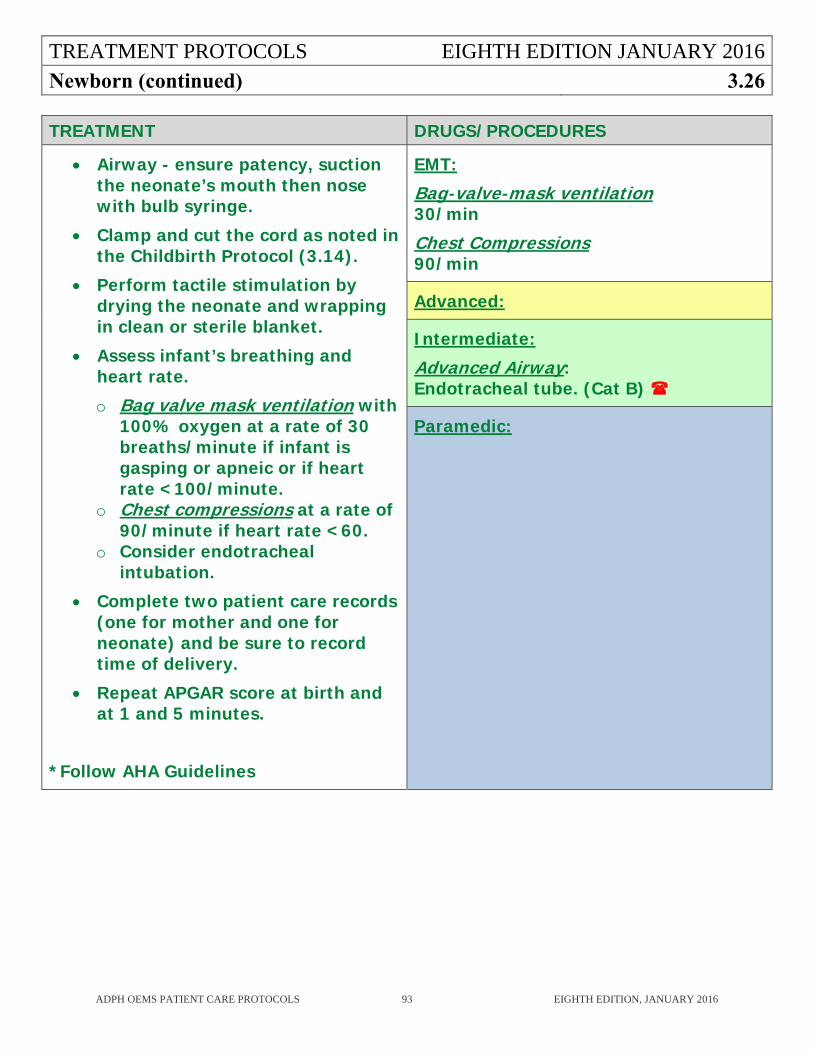
TREATMENT PROTOCOLS EIGHTH EDITION JANUARY 2016
Newborn (continued) 3.26
ADPH OEMS PATIENT CARE PROTOCOLS 93 EIGHTH EDITION, JANUARY 2016
TREATMENT DRUGS/PROCEDURES
Airway - ensure patency, suction the neonate’s mouth then nose with bulb syringe.
Clamp and cut the cord as noted in the Childbirth Protocol (3.14).
Perform tactile stimulation by drying the neonate and wrapping in clean or sterile blanket.
Assess infant’s breathing and heart rate.
o Bag valve mask ventilation with 100% oxygen at a rate of 30 breaths/minute if infant is gasping or apneic or if heart rate <100/minute.
o Chest compressions at a rate of 90/minute if heart rate <60.
o Consider endotracheal intubation.
Complete two patient care records (one for mother and one for neonate) and be sure to record time of delivery.
Repeat APGAR score at birth and at 1 and 5 minutes.
*Follow AHA Guidelines
EMT:
Bag-valve-mask ventilation 30/min
Chest Compressions 90/min
Advanced:
Intermediate:
Advanced Airway: Endotracheal tube. (Cat B)
Paramedic:
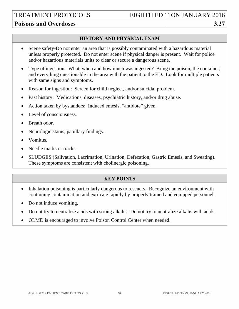
TREATMENT PROTOCOLS EIGHTH EDITION JANUARY 2016
Poisons and Overdoses 3.27
ADPH OEMS PATIENT CARE PROTOCOLS 94 EIGHTH EDITION, JANUARY 2016
HISTORY AND PHYSICAL EXAM
Scene safety-Do not enter an area that is possibly contaminated with a hazardous material unless properly protected. Do not enter scene if physical danger is present. Wait for police and/or hazardous materials units to clear or secure a dangerous scene.
Type of ingestion: What, when and how much was ingested? Bring the poison, the container, and everything questionable in the area with the patient to the ED. Look for multiple patients with same signs and symptoms.
Reason for ingestion: Screen for child neglect, and/or suicidal problem.
Past history: Medications, diseases, psychiatric history, and/or drug abuse.
Action taken by bystanders: Induced emesis, “antidote” given.
Level of consciousness.
Breath odor.
Neurologic status, papillary findings.
Vomitus.
Needle marks or tracks.
SLUDGES (Salivation, Lacrimation, Urination, Defecation, Gastric Emesis, and Sweating). These symptoms are consistent with cholinergic poisoning.
KEY POINTS
Inhalation poisoning is particularly dangerous to rescuers. Recognize an environment with continuing contamination and extricate rapidly by properly trained and equipped personnel.
Do not induce vomiting.
Do not try to neutralize acids with strong alkalis. Do not try to neutralize alkalis with acids.
OLMD is encouraged to involve Poison Control Center when needed.

TREATMENT PROTOCOLS EIGHTH EDITION JANUARY 2016
Poisons and Overdoses (continued) 3.27
ADPH OEMS PATIENT CARE PROTOCOLS 95 EIGHTH EDITION, JANUARY 2016
TREATMENT DRUGS/PROCEDURES
EXTERNAL / INHALATION POISONING
Follow local Hazardous Material Protocol.
Protect medical personnel.
Remove the patient from contaminated area or remove contaminant from the patient.
Remove contaminated clothing.
Flush contaminated skin and eyes with copious amounts of water.
Oxygen to maintain pulse oximetry >95%. If suspicion of Carbon Monoxide poisoning, remember pulse oximetry is unreliable.
Cardiac Monitor.
Consider IV Access.
If cholinergic or organophosphate poisoning, administer Atropine
EMT:
Advanced:
Consider IV Access
Intermediate:
Cardiac Monitor
Paramedic:
Atropine:
2 mg IV/IM every 5 minutes; titrate to effect. (Cat B)
Pediatrics: 0.02 mg/kg IV/IM MIN dose 0.1 mg, MAX single dose is 0.5 mg. (Cat B)
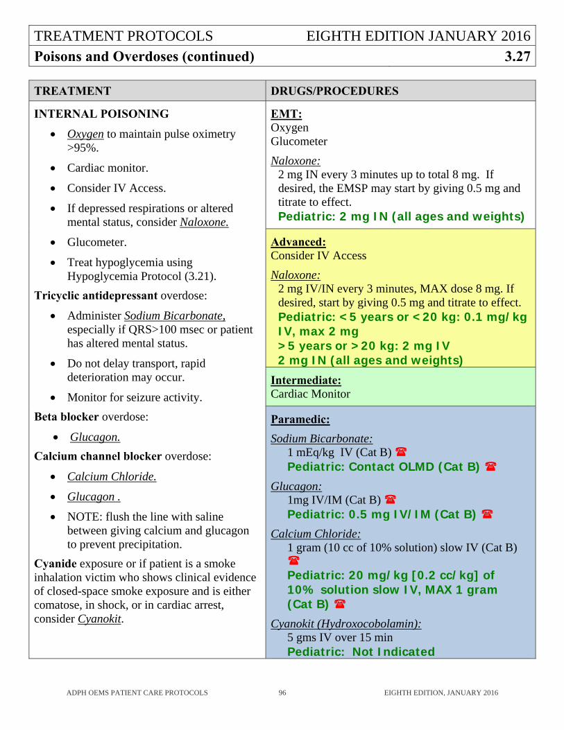
TREATMENT PROTOCOLS EIGHTH EDITION JANUARY 2016Poisons and Overdoses (continued) 3.27
ADPH OEMS PATIENT CARE PROTOCOLS 96 EIGHTH EDITION, JANUARY 2016
TREATMENT DRUGS/PROCEDURES
INTERNAL POISONING
Oxygen to maintain pulse oximetry >95%.
Cardiac monitor.
Consider IV Access.
If depressed respirations or altered mental status, consider Naloxone.
Glucometer.
Treat hypoglycemia using Hypoglycemia Protocol (3.21).
Tricyclic antidepressant overdose:
Administer Sodium Bicarbonate, especially if QRS>100 msec or patient has altered mental status.
Do not delay transport, rapid deterioration may occur.
Monitor for seizure activity.
Beta blocker overdose:
Glucagon.
Calcium channel blocker overdose:
Calcium Chloride.
Glucagon .
NOTE: flush the line with saline between giving calcium and glucagon to prevent precipitation.
Cyanide exposure or if patient is a smoke inhalation victim who shows clinical evidence of closed-space smoke exposure and is either comatose, in shock, or in cardiac arrest, consider Cyanokit.
EMT: Oxygen Glucometer
Naloxone: 2 mg IN every 3 minutes up to total 8 mg. If desired, the EMSP may start by giving 0.5 mg and titrate to effect. Pediatric: 2 mg IN (all ages and weights)
Advanced: Consider IV Access
Naloxone: 2 mg IV/IN every 3 minutes, MAX dose 8 mg. If desired, start by giving 0.5 mg and titrate to effect. Pediatric: <5 years or <20 kg: 0.1 mg/kg IV, max 2 mg >5 years or >20 kg: 2 mg IV 2 mg IN (all ages and weights)
Intermediate: Cardiac Monitor
Paramedic:
Sodium Bicarbonate: 1 mEq/kg IV (Cat B) Pediatric: Contact OLMD (Cat B)
Glucagon: 1mg IV/IM (Cat B) Pediatric: 0.5 mg IV/IM (Cat B)
Calcium Chloride: 1 gram (10 cc of 10% solution) slow IV (Cat B) Pediatric: 20 mg/kg [0.2 cc/kg] of 10% solution slow IV, MAX 1 gram (Cat B)
Cyanokit (Hydroxocobolamin): 5 gms IV over 15 min Pediatric: Not Indicated
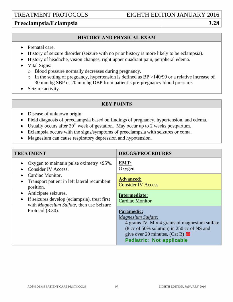
TREATMENT PROTOCOLS EIGHTH EDITION JANUARY 2016
Preeclampsia/Eclampsia 3.28
ADPH OEMS PATIENT CARE PROTOCOLS 97 EIGHTH EDITION, JANUARY 2016
HISTORY AND PHYSICAL EXAM
Prenatal care. History of seizure disorder (seizure with no prior history is more likely to be eclampsia). History of headache, vision changes, right upper quadrant pain, peripheral edema. Vital Signs:
o Blood pressure normally decreases during pregnancy. o In the setting of pregnancy, hypertension is defined as BP >140/90 or a relative increase of
30 mm hg SBP or 20 mm hg DBP from patient’s pre-pregnancy blood pressure. Seizure activity.
KEY POINTS
Disease of unknown origin. Field diagnosis of preeclampsia based on findings of pregnancy, hypertension, and edema. Usually occurs after 20th week of gestation. May occur up to 2 weeks postpartum. Eclampsia occurs with the signs/symptoms of preeclampsia with seizures or coma. Magnesium can cause respiratory depression and hypotension.
TREATMENT DRUGS/PROCEDURES
Oxygen to maintain pulse oximetry >95%. Consider IV Access. Cardiac Monitor. Transport patient in left lateral recumbent
position. Anticipate seizures. If seizures develop (eclampsia), treat first
with Magnesium Sulfate, then use Seizure Protocol (3.30).
EMT: Oxygen
Advanced: Consider IV Access
Intermediate: Cardiac Monitor
Paramedic: Magnesium Sulfate:
4 grams IV. Mix 4 grams of magnesium sulfate (8 cc of 50% solution) in 250 cc of NS and give over 20 minutes. (Cat B) Pediatric: Not applicable
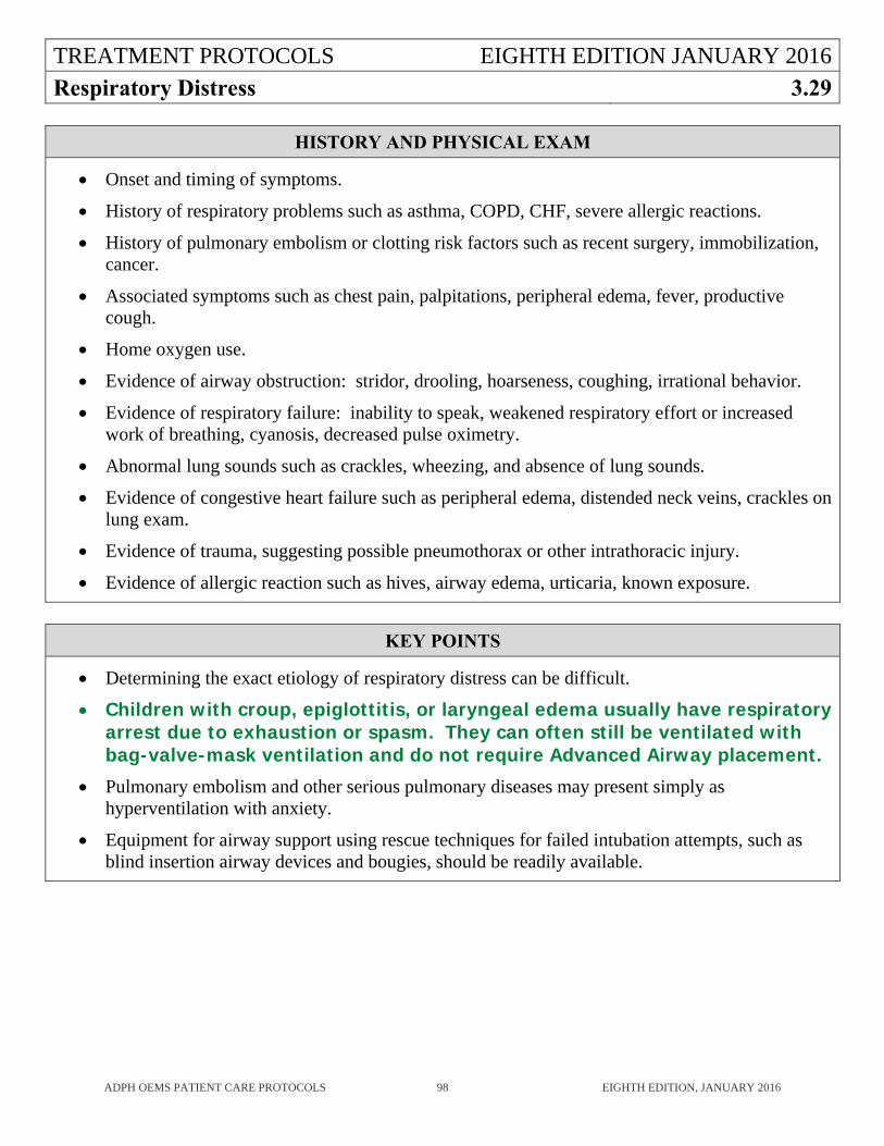
TREATMENT PROTOCOLS EIGHTH EDITION JANUARY 2016
Respiratory Distress 3.29
ADPH OEMS PATIENT CARE PROTOCOLS 98 EIGHTH EDITION, JANUARY 2016
HISTORY AND PHYSICAL EXAM
Onset and timing of symptoms.
History of respiratory problems such as asthma, COPD, CHF, severe allergic reactions.
History of pulmonary embolism or clotting risk factors such as recent surgery, immobilization, cancer.
Associated symptoms such as chest pain, palpitations, peripheral edema, fever, productive cough.
Home oxygen use.
Evidence of airway obstruction: stridor, drooling, hoarseness, coughing, irrational behavior.
Evidence of respiratory failure: inability to speak, weakened respiratory effort or increased work of breathing, cyanosis, decreased pulse oximetry.
Abnormal lung sounds such as crackles, wheezing, and absence of lung sounds.
Evidence of congestive heart failure such as peripheral edema, distended neck veins, crackles on lung exam.
Evidence of trauma, suggesting possible pneumothorax or other intrathoracic injury.
Evidence of allergic reaction such as hives, airway edema, urticaria, known exposure.
KEY POINTS
Determining the exact etiology of respiratory distress can be difficult.
Children with croup, epiglottitis, or laryngeal edema usually have respiratory arrest due to exhaustion or spasm. They can often still be ventilated with bag-valve-mask ventilation and do not require Advanced Airway placement.
Pulmonary embolism and other serious pulmonary diseases may present simply as hyperventilation with anxiety.
Equipment for airway support using rescue techniques for failed intubation attempts, such as blind insertion airway devices and bougies, should be readily available.
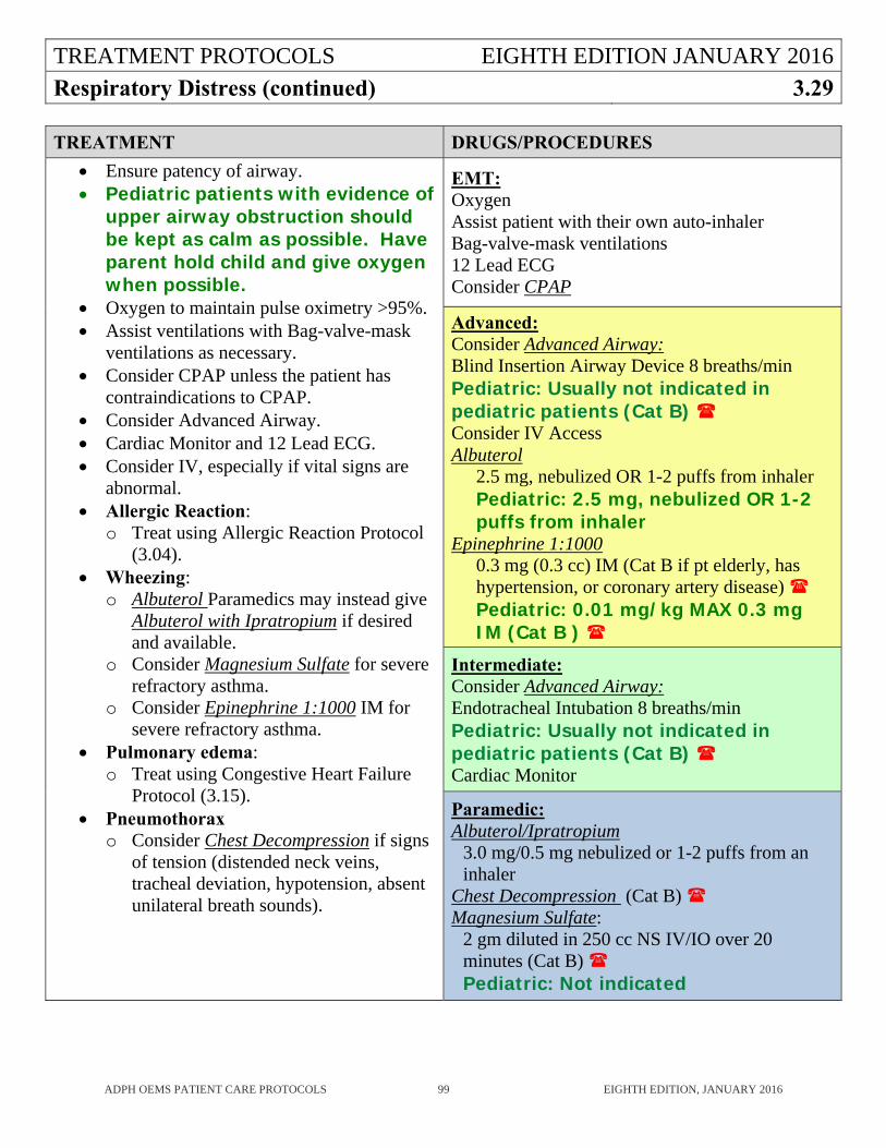
TREATMENT PROTOCOLS EIGHTH EDITION JANUARY 2016
Respiratory Distress (continued) 3.29
ADPH OEMS PATIENT CARE PROTOCOLS 99 EIGHTH EDITION, JANUARY 2016
TREATMENT DRUGS/PROCEDURES
Ensure patency of airway. Pediatric patients with evidence of
upper airway obstruction should be kept as calm as possible. Have parent hold child and give oxygen when possible.
Oxygen to maintain pulse oximetry >95%. Assist ventilations with Bag-valve-mask
ventilations as necessary. Consider CPAP unless the patient has
contraindications to CPAP. Consider Advanced Airway. Cardiac Monitor and 12 Lead ECG. Consider IV, especially if vital signs are
abnormal. Allergic Reaction:
o Treat using Allergic Reaction Protocol (3.04).
Wheezing: o Albuterol Paramedics may instead give
Albuterol with Ipratropium if desired and available.
o Consider Magnesium Sulfate for severe refractory asthma.
o Consider Epinephrine 1:1000 IM for severe refractory asthma.
Pulmonary edema: o Treat using Congestive Heart Failure
Protocol (3.15). Pneumothorax
o Consider Chest Decompression if signs of tension (distended neck veins, tracheal deviation, hypotension, absent unilateral breath sounds).
EMT: Oxygen Assist patient with their own auto-inhaler Bag-valve-mask ventilations 12 Lead ECG Consider CPAP
Advanced: Consider Advanced Airway: Blind Insertion Airway Device 8 breaths/min Pediatric: Usually not indicated in pediatric patients (Cat B) Consider IV Access Albuterol
2.5 mg, nebulized OR 1-2 puffs from inhaler Pediatric: 2.5 mg, nebulized OR 1-2 puffs from inhaler
Epinephrine 1:1000 0.3 mg (0.3 cc) IM (Cat B if pt elderly, has hypertension, or coronary artery disease) Pediatric: 0.01 mg/kg MAX 0.3 mg IM (Cat B )
Intermediate: Consider Advanced Airway: Endotracheal Intubation 8 breaths/min Pediatric: Usually not indicated in pediatric patients (Cat B) Cardiac Monitor
Paramedic: Albuterol/Ipratropium
3.0 mg/0.5 mg nebulized or 1-2 puffs from an inhaler
Chest Decompression (Cat B) Magnesium Sulfate:
2 gm diluted in 250 cc NS IV/IO over 20 minutes (Cat B) Pediatric: Not indicated
TREATMENT PROTOCOLS EIGHTH EDITION JANUARY 2016
Seizure 3.30
ADPH OEMS PATIENT CARE PROTOCOLS 100 EIGHTH EDITION, JANUARY 2016
HISTORY AND PHYSICAL EXAM
Onset and duration. History and description of seizures. Medications and compliance. History or evidence of trauma, particularly head trauma. History of diabetes, headache, recreational drug or alcohol use, pregnancy. Level of consciousness. Ongoing seizure activity. Incontinence. Focal neurologic signs.
KEY POINTS
Don’t force things into seizing patient’s mouths. Seizures may be caused by arrythmias, particularly in patients over 50. Seizure activity may be caused by cerebral hypoxia from cardiac arrest, always check a pulse
when seizures terminate. Seizures in pediatric patients are commonly febrile seizures and are usually benign and short
lived. Pregnant women who seize may be eclamptic, treat using the Preeclampsia/Eclampsia Protocol
(3.28).
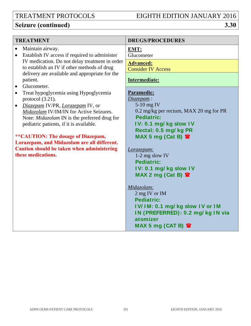
TREATMENT PROTOCOLS EIGHTH EDITION JANUARY 2016
Seizure (continued) 3.30
ADPH OEMS PATIENT CARE PROTOCOLS 101 EIGHTH EDITION, JANUARY 2016
TREATMENT DRUGS/PROCEDURES
Maintain airway. Establish IV access if required to administer
IV medication. Do not delay treatment in order to establish an IV if other methods of drug delivery are available and appropriate for the patient.
Glucometer. Treat hypoglycemia using Hypoglycemia
protocol (3.21). Diazepam IV/PR, Lorazepam IV, or
Midazolam IV/IM/IN for Active Seizures. Note: Midazolam IN is the preferred drug for pediatric patients, if it is available.
**CAUTION: The dosage of Diazepam, Lorazepam, and Midazolam are all different. Caution should be taken when administering these medications.
EMT: Glucometer
Advanced: Consider IV Access
Intermediate:
Paramedic: Diazepam :
5-10 mg IV 0.2 mg/kg per rectum, MAX 20 mg for PR
Pediatric: IV: 0.1 mg/kg slow IV Rectal: 0.5 mg/kg PR MAX 5 mg (Cat B)
Lorazepam:
1-2 mg slow IV Pediatric: IV: 0.1 mg/kg slow IV MAX 2 mg (Cat B)
Midazolam: 2 mg IV or IM Pediatric: IV/IM: 0.1 mg/kg slow IV or IM IN (PREFERRED): 0.2 mg/kg IN via atomizer MAX 5 mg (CAT B)
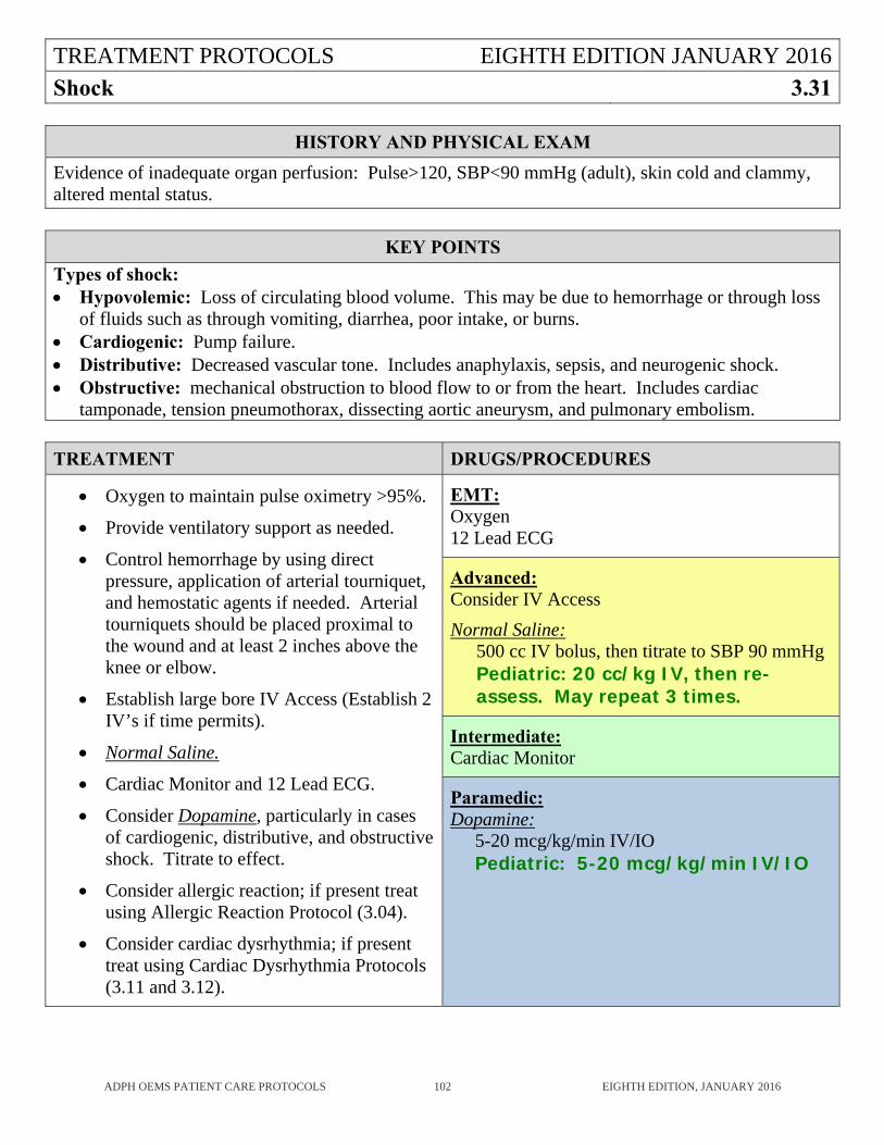
TREATMENT PROTOCOLS EIGHTH EDITION JANUARY 2016
Shock 3.31
ADPH OEMS PATIENT CARE PROTOCOLS 102 EIGHTH EDITION, JANUARY 2016
HISTORY AND PHYSICAL EXAM
Evidence of inadequate organ perfusion: Pulse>120, SBP<90 mmHg (adult), skin cold and clammy, altered mental status.
KEY POINTS
Types of shock: Hypovolemic: Loss of circulating blood volume. This may be due to hemorrhage or through loss
of fluids such as through vomiting, diarrhea, poor intake, or burns. Cardiogenic: Pump failure. Distributive: Decreased vascular tone. Includes anaphylaxis, sepsis, and neurogenic shock. Obstructive: mechanical obstruction to blood flow to or from the heart. Includes cardiac
tamponade, tension pneumothorax, dissecting aortic aneurysm, and pulmonary embolism.
TREATMENT DRUGS/PROCEDURES
Oxygen to maintain pulse oximetry >95%.
Provide ventilatory support as needed.
Control hemorrhage by using direct pressure, application of arterial tourniquet, and hemostatic agents if needed. Arterial tourniquets should be placed proximal to the wound and at least 2 inches above the knee or elbow.
Establish large bore IV Access (Establish 2 IV’s if time permits).
Normal Saline.
Cardiac Monitor and 12 Lead ECG.
Consider Dopamine, particularly in cases of cardiogenic, distributive, and obstructive shock. Titrate to effect.
Consider allergic reaction; if present treat using Allergic Reaction Protocol (3.04).
Consider cardiac dysrhythmia; if present treat using Cardiac Dysrhythmia Protocols (3.11 and 3.12).
EMT: Oxygen 12 Lead ECG
Advanced: Consider IV Access
Normal Saline: 500 cc IV bolus, then titrate to SBP 90 mmHg Pediatric: 20 cc/kg IV, then re-assess. May repeat 3 times.
Intermediate: Cardiac Monitor
Paramedic: Dopamine:
5-20 mcg/kg/min IV/IO Pediatric: 5-20 mcg/kg/min IV/IO
TREATMENT PROTOCOLS EIGHTH EDITION JANUARY 2016
Spinal Injury 3.32
ADPH OEMS PATIENT CARE PROTOCOLS 103 EIGHTH EDITION, JANUARY 2016
HISTORY AND PHYSICAL EXAM
Mechanism of Injury: Elements that should increase suspicion for spine injury and prompt screening for spinal injury include:
Axial Loading (diving). Blunt trauma to head or neck. Motor Vehicle Crash. Fall> 3 feet or an adult falling from standing height. Any violent mechanism of injury with high energy transfer. History of arthritis of spine.
Patient Reliability: Assessment for spinal injury can only be utilized if the patient is alert, calm, cooperative, and not intoxicated. If there is a communication barrier, the patient cannot be properly assessed and based on mechanism and any complaint of injury the patient should receive SMR.
Distracting Injury: Any painful injury might distract the patient from the pain of a spine injury. Both medical as well as traumatic causes for pain can be considered a distracting injury. If the patient has an injury or illness that seems to be causing enough pain to provide a distraction, the spine cannot be cleared clinically.
Neurologic Evaluation: A patient who is reliable and has no distracting injury should be examined for any neurologic deficits. Perform the following assessments bilaterally in the upper and lower extremities. Responses should be symmetrical. Any abnormalities should prompt SMR.
Motor: • Have the patient spread the fingers of his or her hand and resist as the EMSP tries to squeeze
them together. There should be some resistance against the squeeze. • Ask the patient to hold his or her hand out with the palm facing down. While supporting the
wrist, ask the patient to resist while the EMSP pushes down on the dorsal surface of the hand or fingers. The patient should be able to provide some resistance.
• Place a hand on the bottom of the patient’s foot at the great toe. Ask the patient to push down against resistance. The patient should be able to apply pressure to the EMSP’s hand.
• Move a hand to the top of the foot and ask the patient to pull his or her toe towards nose against the resistance. The patient should be able to apply pressure to the EMSP’s hand.
Sensory: • Assess for the ability to distinguish soft and sharp sensation in each hand and foot. Use a sharp
object and a soft object. A corner of a gauze pad and a pencil may be used. • Alternately apply the soft and then the sharp object to each extremity. Do not let the patient
know which one was used. Ask the patient whether the sensation is soft or sharp. Repeat soft and sharp in all extremities.
• The patient should be able to distinguish soft from sharp.
TREATMENT PROTOCOLS EIGHTH EDITION JANUARY 2016
Spinal Injury (continued) 3.32
ADPH OEMS PATIENT CARE PROTOCOLS 104 EIGHTH EDITION, JANUARY 2016
HISTORY AND PHYSICAL EXAM (continued)
Complaints of Pain or Examination Tenderness: Palpate the entire spine. Any complaint of pain or tenderness to palpation along any part of the
spine should be considered an indication that the patient requires spinal precautions. Ask the patient about sensations of numbness, tingling, shooting pain, or motor weakness in any
extremity. Any positive response requires full SMR. Some components of the sensory examination are subjective. When in doubt, apply SMR.
Documentation: In any case where there is head and/or facial injury, or a mechanism of injury suggesting the possibility of a cervical spine injury, clear and concise documentation is absolutely essential. In the cases where the decision not to provide SMR is made, documentation must include the following information:
• The examination was performed on a reliable patient. • The patient denies having any spinal pain. • The patient denies having any extremity weakness or loss of movement. • The patient denies having any tingling or feeling of pins and needles in the extremities. • There is no pain on palpation of the spine. • Motor function is intact in all of the extremities. • Sensation is intact in all extremities.
KEY POINTS
Full SMR as an automatic response to trauma may not always be in the patient’s best interest. Patients packaged on hard SMR devices may develop complications or problems due to laying on a hard spineboard. These complications or problems could potentially be avoided if a spinal assessment tool is utilized to reduce the number of patients unnecessarily placed in SMR.
Patients with penetrating trauma to the head, neck, or torso and no evidence of spinal injury do not require full SMR.
Use of a backboard for stabilization of some other injury than the spine, or to move the patient does not mean that SMR is indicated. If a backboard device is used only to move the patient, it should be removed as soon as practical.
When implementing SMR, do not secure the head to the backboard before securing the body because this can cause torsion on the neck.
SMR with a cervical collar and a vacuum mattress is a recommended technique. A vacuum mattress, when available, is preferred for all but short transports.
TREATMENT PROTOCOLS EIGHTH EDITION JANUARY 2016
Spinal Injury (continued) 3.32
ADPH OEMS PATIENT CARE PROTOCOLS 105 EIGHTH EDITION, JANUARY 2016
KEY POINTS (continued)
Vomiting should be expected in head injury patients. Therefore, the patient should be securely strapped to a long board to enable board and patient to be turned as a unit. EMSPs should be aware that additional help may be necessary during transport to turn patient and manage airway while maintaining cervical spine integrity.
Chin straps that could compromise the airway should be removed as the patient is secured to the long board.
Most adults require 1 to 1 ½ inches of firm padding behind the head to assume standard neutral anatomic position, and some additional padding behind the neck is necessary for full support. Most children require padding under the shoulders to maintain neutral spinal alignment.
A rigid cervical collar, continuous manual in-line support during rapid extrication onto a long spine board, and rapid transport should be substituted for more time consuming methods in the severely traumatized patient requiring immediate life saving intervention.
Airway problems, respiratory difficulty, and shock are common in the traumatized patient. Alternate techniques for performing airway procedures should be used in spinal injured patients. To maintain proper control of the cervical spine during endotracheal intubation, in-line stabilization must be performed by two EMSPs.
If any motion restriction techniques cause an increase in pain or neurologic deficit, the patient should be stabilized in position found or position of greatest comfort.
Geriatric patients (over 55) should raise a higher index of suspicion for the EMSP due to physiologic aging changes. The EMSP’s awareness of the need to provide for cervical spine motion restriction should be more acute in these patients.
Patients with one or more acutely paralyzed limbs in the setting of acute trauma should be entered into the Alabama Trauma System where applicable.
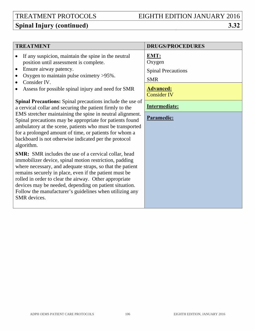
TREATMENT PROTOCOLS EIGHTH EDITION JANUARY 2016
Spinal Injury (continued) 3.32
ADPH OEMS PATIENT CARE PROTOCOLS 106 EIGHTH EDITION, JANUARY 2016
TREATMENT DRUGS/PROCEDURES
If any suspicion, maintain the spine in the neutral position until assessment is complete.
Ensure airway patency. Oxygen to maintain pulse oximetry >95%. Consider IV. Assess for possible spinal injury and need for SMR Spinal Precautions: Spinal precautions include the use of a cervical collar and securing the patient firmly to the EMS stretcher maintaining the spine in neutral alignment. Spinal precautions may be appropriate for patients found ambulatory at the scene, patients who must be transported for a prolonged amount of time, or patients for whom a backboard is not otherwise indicated per the protocol algorithm.
SMR: SMR includes the use of a cervical collar, head immobilizer device, spinal motion restriction, padding where necessary, and adequate straps, so that the patient remains securely in place, even if the patient must be rolled in order to clear the airway. Other appropriate devices may be needed, depending on patient situation. Follow the manufacturer’s guidelines when utilizing any SMR devices.
EMT: Oxygen
Spinal Precautions
SMR
Advanced: Consider IV
Intermediate:
Paramedic:
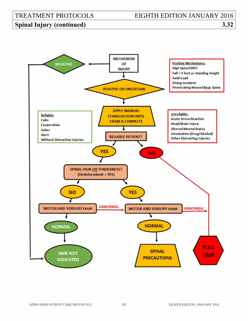
TREATMENT PROTOCOLS EIGHTH EDITION JANUARY 2016
Spinal Injury (continued) 3.32
ADPH OEMS PATIENT CARE PROTOCOLS 107 EIGHTH EDITION, JANUARY 2016
TREATMENT PROTOCOLS EIGHTH EDITION JANUARY 2016
Stroke 3.33
ADPH OEMS PATIENT CARE PROTOCOLS 108 EIGHTH EDITION, JANUARY 2016
HISTORY AND PHYSICAL EXAM
Last time patient was seen normal. Existing previous neurologic deficit. Stroke risk factors (hypertension, diabetes, heart disease, smoking, dysrhythmias, blood thinner
use, or previous stroke). Has the patient had any recent similar events? Level of consciousness: Alert, Responds to Voice, Responds to Pain, Unconscious.
Perform FAST stroke scale (Face, Arm, Speech, Time): 1. Face: Assess for facial droop: have the patient show teeth or smile.
Normal – both sides of face move equally. Abnormal – one side of face does not move as well as the other side.
2. Arm: Assess for arm drift: have the patient close eyes and hold both arms straight out; with palms up, for 10 seconds. Normal – both arms move the same or both arms do not move at all. Abnormal – one arm does not move or one arm drifts down compared to the other.
3. Speech: Assess for abnormal speech: have the patient say “you can’t teach an old dog new tricks.” Normal – patient uses correct words with no slurring. Abnormal – patient slurs words, uses the wrong words, or is unable to speak.
4. Time: If any of above are positive, attempt to determine the time of symptom onset (clock time).
If any component of the FAST stroke scale is abnormal, the patient is very likely to be suffering from an acute stroke.
TREATMENT PROTOCOLS EIGHTH EDITION JANUARY 2016
Stroke (continued) 3.33
ADPH OEMS PATIENT CARE PROTOCOLS 109 EIGHTH EDITION, JANUARY 2016
KEY POINTS
This protocol is for patients who have an acute episode of neurologic deficit without any evidence of trauma. If the patient has altered mental status, treat using the Altered Mental Status Protocol (3.05).
Determining the exact time of symptom onset is critical since administration of thrombolytic drugs for the treatment of stroke is time-dependent.
High blood pressure during an acute stroke may be compensatory, do not attempt to lower it without consulting OLMD.
Intravenous glucose may aggravate the effects of ischemia upon brain tissue. Do not administer glucose unless hypoglycemia is documented.
If in a region with a stroke system, notify the ATCC and transport the patient to the appropriate ready stroke center.
When possible, bring a knowledgeable friend or family member with the patient to assist with providing the patient’s history at the hospital. If it is not possible to for that person to accompany the patient, attempt to obtain a cell phone number for someone who can provide the receiving hospital with patient history and details of the event.
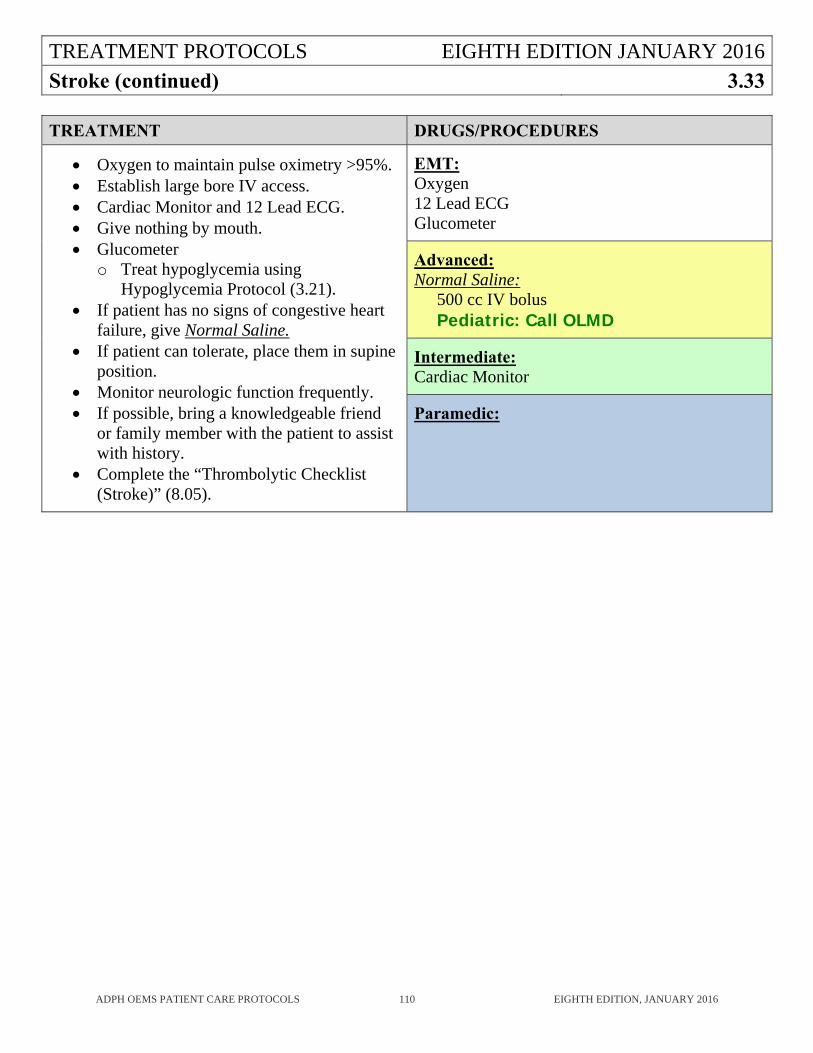
TREATMENT PROTOCOLS EIGHTH EDITION JANUARY 2016
Stroke (continued) 3.33
ADPH OEMS PATIENT CARE PROTOCOLS 110 EIGHTH EDITION, JANUARY 2016
TREATMENT DRUGS/PROCEDURES
Oxygen to maintain pulse oximetry >95%. Establish large bore IV access. Cardiac Monitor and 12 Lead ECG. Give nothing by mouth. Glucometer
o Treat hypoglycemia using Hypoglycemia Protocol (3.21).
If patient has no signs of congestive heart failure, give Normal Saline.
If patient can tolerate, place them in supine position.
Monitor neurologic function frequently. If possible, bring a knowledgeable friend
or family member with the patient to assist with history.
Complete the “Thrombolytic Checklist (Stroke)” (8.05).
EMT: Oxygen 12 Lead ECG Glucometer
Advanced: Normal Saline:
500 cc IV bolus Pediatric: Call OLMD
Intermediate: Cardiac Monitor
Paramedic:
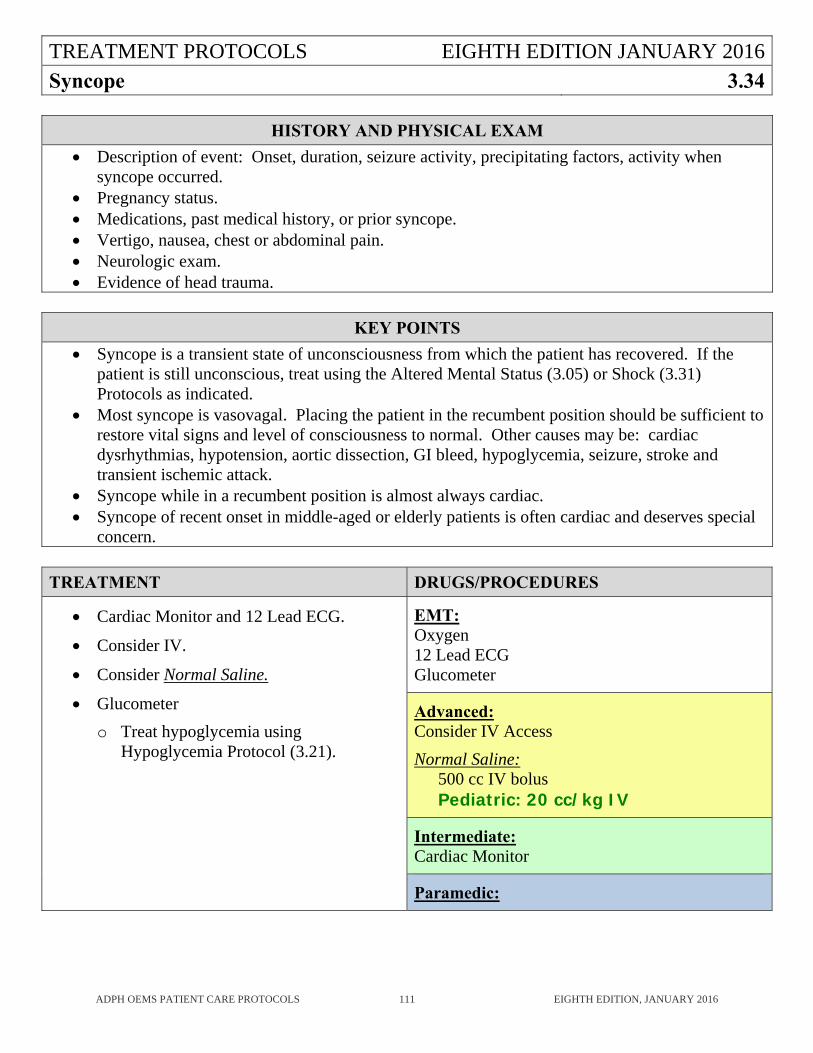
TREATMENT PROTOCOLS EIGHTH EDITION JANUARY 2016
Syncope 3.34
ADPH OEMS PATIENT CARE PROTOCOLS 111 EIGHTH EDITION, JANUARY 2016
HISTORY AND PHYSICAL EXAM
Description of event: Onset, duration, seizure activity, precipitating factors, activity when syncope occurred.
Pregnancy status. Medications, past medical history, or prior syncope. Vertigo, nausea, chest or abdominal pain. Neurologic exam. Evidence of head trauma.
KEY POINTS
Syncope is a transient state of unconsciousness from which the patient has recovered. If the patient is still unconscious, treat using the Altered Mental Status (3.05) or Shock (3.31) Protocols as indicated.
Most syncope is vasovagal. Placing the patient in the recumbent position should be sufficient to restore vital signs and level of consciousness to normal. Other causes may be: cardiac dysrhythmias, hypotension, aortic dissection, GI bleed, hypoglycemia, seizure, stroke and transient ischemic attack.
Syncope while in a recumbent position is almost always cardiac. Syncope of recent onset in middle-aged or elderly patients is often cardiac and deserves special
concern.
TREATMENT DRUGS/PROCEDURES
Cardiac Monitor and 12 Lead ECG.
Consider IV.
Consider Normal Saline.
Glucometer
o Treat hypoglycemia using Hypoglycemia Protocol (3.21).
EMT: Oxygen 12 Lead ECG Glucometer
Advanced: Consider IV Access
Normal Saline: 500 cc IV bolus Pediatric: 20 cc/kg IV
Intermediate: Cardiac Monitor
Paramedic:
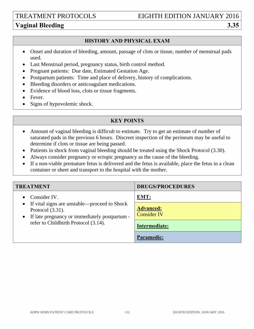
TREATMENT PROTOCOLS EIGHTH EDITION JANUARY 2016
Vaginal Bleeding 3.35
ADPH OEMS PATIENT CARE PROTOCOLS 112 EIGHTH EDITION, JANUARY 2016
HISTORY AND PHYSICAL EXAM
Onset and duration of bleeding, amount, passage of clots or tissue, number of menstrual pads used.
Last Menstrual period, pregnancy status, birth control method. Pregnant patients: Due date, Estimated Gestation Age. Postpartum patients: Time and place of delivery, history of complications. Bleeding disorders or anticoagulant medications. Evidence of blood loss, clots or tissue fragments. Fever. Signs of hypovolemic shock.
KEY POINTS
Amount of vaginal bleeding is difficult to estimate. Try to get an estimate of number of saturated pads in the previous 6 hours. Discreet inspection of the perineum may be useful to determine if clots or tissue are being passed.
Patients in shock from vaginal bleeding should be treated using the Shock Protocol (3.30). Always consider pregnancy or ectopic pregnancy as the cause of the bleeding. If a non-viable premature fetus is delivered and the fetus is available, place the fetus in a clean
container or sheet and transport to the hospital with the mother.
TREATMENT DRUGS/PROCEDURES
Consider IV. If vital signs are unstable—proceed to Shock
Protocol (3.31). If late pregnancy or immediately postpartum -
refer to Childbirth Protocol (3.14).
EMT:
Advanced: Consider IV
Intermediate:
Paramedic:

ADPH EMS PROTOCOLS EIGHTH EDITION
PROCEDURES 4
ADPH OEMS PATIENT CARE PROTOCOLS 113 EIGHTH EDITION, JANUARY 2016
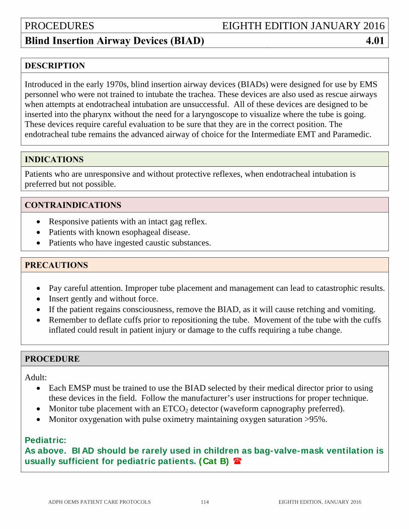
PROCEDURES EIGHTH EDITION JANUARY 2016
Blind Insertion Airway Devices (BIAD) 4.01
ADPH OEMS PATIENT CARE PROTOCOLS 114 EIGHTH EDITION, JANUARY 2016
DESCRIPTION
Introduced in the early 1970s, blind insertion airway devices (BIADs) were designed for use by EMS personnel who were not trained to intubate the trachea. These devices are also used as rescue airways when attempts at endotracheal intubation are unsuccessful. All of these devices are designed to be inserted into the pharynx without the need for a laryngoscope to visualize where the tube is going. These devices require careful evaluation to be sure that they are in the correct position. The endotracheal tube remains the advanced airway of choice for the Intermediate EMT and Paramedic.
INDICATIONS
Patients who are unresponsive and without protective reflexes, when endotracheal intubation is preferred but not possible.
CONTRAINDICATIONS
Responsive patients with an intact gag reflex. Patients with known esophageal disease. Patients who have ingested caustic substances.
PRECAUTIONS
Pay careful attention. Improper tube placement and management can lead to catastrophic results. Insert gently and without force. If the patient regains consciousness, remove the BIAD, as it will cause retching and vomiting. Remember to deflate cuffs prior to repositioning the tube. Movement of the tube with the cuffs
inflated could result in patient injury or damage to the cuffs requiring a tube change.
PROCEDURE
Adult: Each EMSP must be trained to use the BIAD selected by their medical director prior to using
these devices in the field. Follow the manufacturer’s user instructions for proper technique. Monitor tube placement with an ETCO2 detector (waveform capnography preferred). Monitor oxygenation with pulse oximetry maintaining oxygen saturation >95%.
Pediatric: As above. BIAD should be rarely used in children as bag-valve-mask ventilation is usually sufficient for pediatric patients. (Cat B)
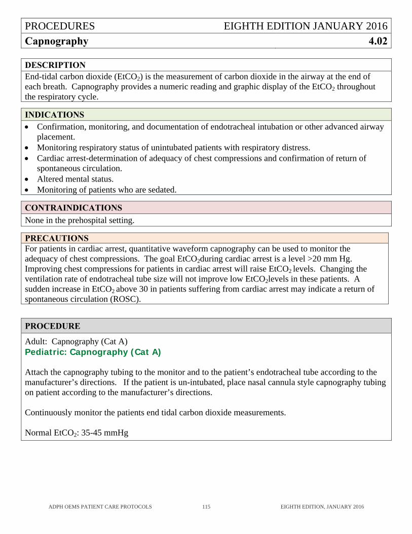
PROCEDURES EIGHTH EDITION JANUARY 2016
Capnography 4.02
ADPH OEMS PATIENT CARE PROTOCOLS 115 EIGHTH EDITION, JANUARY 2016
DESCRIPTION End-tidal carbon dioxide (EtCO2) is the measurement of carbon dioxide in the airway at the end of each breath. Capnography provides a numeric reading and graphic display of the EtCO2 throughout the respiratory cycle.
INDICATIONS Confirmation, monitoring, and documentation of endotracheal intubation or other advanced airway
placement. Monitoring respiratory status of unintubated patients with respiratory distress. Cardiac arrest-determination of adequacy of chest compressions and confirmation of return of
spontaneous circulation. Altered mental status. Monitoring of patients who are sedated.
CONTRAINDICATIONS
None in the prehospital setting.
PRECAUTIONS For patients in cardiac arrest, quantitative waveform capnography can be used to monitor the adequacy of chest compressions. The goal EtCO2during cardiac arrest is a level >20 mm Hg. Improving chest compressions for patients in cardiac arrest will raise EtCO2 levels. Changing the ventilation rate of endotracheal tube size will not improve low EtCO2levels in these patients. A sudden increase in EtCO2 above 30 in patients suffering from cardiac arrest may indicate a return of spontaneous circulation (ROSC).
PROCEDURE
Adult: Capnography (Cat A) Pediatric: Capnography (Cat A) Attach the capnography tubing to the monitor and to the patient’s endotracheal tube according to the manufacturer’s directions. If the patient is un-intubated, place nasal cannula style capnography tubing on patient according to the manufacturer’s directions. Continuously monitor the patients end tidal carbon dioxide measurements. Normal EtCO2: 35-45 mmHg
PROCEDURES EIGHTH EDITION JANUARY 2016
Cardioversion (Synchronized) 4.03
ADPH OEMS PATIENT CARE PROTOCOLS 116 EIGHTH EDITION, JANUARY 2016
DESCRIPTION
Synchronized cardioversion is indicated for patients with tachydysrhythmias associated with cardiopulmonary instability.
INDICATIONS
Tachycardia with serious signs and symptoms related to the tachycardia leading to hemodynamic instability. These include: altered mental status, ongoing chest pain, hypotension, or other signs of shock.
Ventricular rate >150 beats/min for adults. Ventricular rate >220 for infants <1 year. Ventricular rate >180 for children 1-8 years.
CONTRAINDICATIONS
None in the prehospital setting.
PRECAUTIONS
Consider sedation premedication when possible with benzodiazepine or narcotic. Have available airway management equipment. Establish IV access prior to cardioversion attempt.
PROCEDURE
Attach cardiac monitor to patient using paddles or adhesive pads. Press the synchronization button on the defibrillator. Select appropriate energy level:
Atrial fibrillation 120-200 J , (Cat B) Stable Monomorphic Ventricular Tachycardia 100 J (Cat B) Supraventricular Tachycardia (SVT), Atrial Flutter: 50-100 J (Cat B)
For each energy setting, increase in stepwise fashion per manufacturer’s recommendation.
Pediatric: 1st dose 0.5-1 J/kg 2nd dose 2 J/kg (Cat B)
Announce “Charging Defibrillator”. Push the charge button on the defibrillator. When fully charged, announce “I am going to shock on three”, “1, 2, 3, All Clear”. After confirming all personnel are clear, push the shock delivery button on the defibrillator. Re-assess the patient and his/her heart rhythm.
PROCEDURES EIGHTH EDITION JANUARY 2016
Chest Decompression 4.04
ADPH OEMS PATIENT CARE PROTOCOLS 117 EIGHTH EDITION, JANUARY 2016
DESCRIPTION
The emergency procedure of introducing a large-bore catheter/needle into the pleural space of the chest to provide temporary relief for the patient suffering from a symptomatic tension pneumothorax.
INDICATIONS
PRIMARY- ABSOLUTE REQUIREMENTS A patient who is assessed to have a life-threatening tension pneumothorax manifested by:
Absent breath sounds on one side. Profound shock with a systolic blood pressure of 80 mmHg or less in an adult. Criteria for
profound shock in a child must be determined by OLMD. A patient with a flail chest severe enough to require endotracheal intubation for persistent
hypoxia should have a precautionary needle decompression on the side of the injury.
SECONDARY INDICATIONS (Suggestive but not sufficient without the above) Distended neck veins (may not be present if there is severe hemorrhage). Tracheal shift away from the affected side (late and rare). Altered mental status (almost always present). Increased airway resistance to ventilation, especially if intubated. Tympany (hyperresonance) to percussion on the affected side. Subcutaneous air in an intubated patient.
CONTRAINDICATIONS
Patient has only simple pneumothorax (loss of breath sounds on one side but normal systolic blood pressure is not an indication for decompression).
Patient with a symptomatic tension pneumothorax that can be relieved by the removal of an occlusive dressing from an open chest wound.
PRECAUTIONS
Catheter becomes kinked or pulls out allowing tension pneumothorax to reoccur. Intercostal vascular or nerve injury from incorrect placement. Pneumothorax or hemothorax. Direct injury to the lung. Pericardial/cardiac injury from incorrect placement or catheter that is too long. Infection.
PROCEDURES EIGHTH EDITION JANUARY 2016
Chest Decompression (continued) 4.04
ADPH OEMS PATIENT CARE PROTOCOLS 118 EIGHTH EDITION, JANUARY 2016
PROCEDURE
Traumatic Cardiac Arrest: (Cat A)
Tension pneumothorax without cardiac arrest: (Cat B)
Provide 100% oxygen and assist ventilations as needed.
Identify the second or third intercostal space on the anterior chest at the midclavicular line on the same side as the tension pneumothorax. This may be done by feeling for the “angle of Louis,” the bump located on the sternum about a quarter of the way from the suprasternal notch. Follow the interspace just below this bump to the midclavicular line to insert the catheter.
Prepare the area with an antiseptic.
Remove the plastic cap from a 2.5 inch (6.0 to 7.0 cm) large-bore (12 or 14 gauge) over-the-needle catheter. Insert the needle into the skin over the superior border of the third rib, midclavicular line, and direct it into the intercostal space at a 90-degree angle to the rib. Direction of the bevel is irrelevant to successful results. As the needle enters the pleural space, there will be a “pop.” If a tension pneumothorax is present, there will be a hiss of air as the pneumothorax is decompressed. Advance the catheter all the way to the hub and remove the needle. The catheter hub must be stabilized to the chest with tape.
Attach a one-way valve (such as an Asherman Chest Seal), if possible.
Leave the catheter in place until it is replaced by a chest tube at the hospital. Monitor the catheter to be sure it remains patent.
A copy of the patient care report, the Chest Decompression Report (Form 8.01) and the name of the receiving physician must be submitted to the Regional EMS Agency and the Office of EMS (OEMS) for review within 10 working days.
If desired, the EMSP can call the ATCC for OLMD from a trauma center.
PROCEDURES EIGHTH EDITION JANUARY 2016
Continuous Positive Airway Pressure (CPAP) 4.05
ADPH OEMS PATIENT CARE PROTOCOLS 119 EIGHTH EDITION, JANUARY 2016
DESCRIPTION
Continuous Positive Airway Pressure (CPAP) has been shown to rapidly improve vital signs, gas exchange, and the work of breathing. It also decreases the sense of dyspnea, and decreases the need for endotracheal intubation in the patients who suffer from shortness of breath from congestive heart failure and/or acute pulmonary edema. CPAP is also shown to improve dyspnea associated with pneumonia, asthma, bronchitis, and emphysema. CPAP improves hemodynamics of patients with chronic obstructive pulmonary disease (COPD), by reducing preload and afterload. When desired and approved by the service medical director, BiPAP may be substituted for CPAP.
INDICATIONS
Respiratory Distress in patients who: Are awake, oriented, and able to follow commands. Have the ability to maintain an open airway (GCS>10). Have a respiratory rate greater than 25 breaths per minute with a pulse oximetry reading of
<95%. Have a systolic blood pressure above 90 mmHg. Are using accessory muscles during respirations. Are over 12 years of age and are able to be fitted with the CPAP mask.
CONTRAINDICATIONS
Pneumothorax. Respiratory arrest or agonal respirations. Unconsciousness. Shock associated with cardiac insufficiency. Penetrating chest trauma. Persistent nausea/vomiting. Facial abnormalities. Active upper GI bleeding or history of recent gastric surgery. Children under 12 years of age.
PRECAUTIONS
Monitor patient for gastric distension which may lead to vomiting. Use nitroglycerine tablets to avoid nitroglycerine spray from being dispersed on patient / EMS
crew. If CPAP is suddenly discontinued, patients may become apneic. Monitor closely. Advise receiving hospital as soon as possible so they can prepare for patient’s arrival.
PROCEDURES EIGHTH EDITION JANUARY 2016Continuous Positive Airway Pressure (CPAP) (continued)
4.05
ADPH OEMS PATIENT CARE PROTOCOLS 120 EIGHTH EDITION, JANUARY 2016
PROCEDURE
Make sure the patient does not have a pneumothorax. Confirm breath sounds in ALL lung fields.
Place patient in a sitting position.
Attach cardiac monitor and pulse oximetry. Assess vital signs and pulse oximetry at least every
5 mins.
If BP <90 mmHg systolic, contact OLMD prior to beginning CPAP. OLMD may override this contraindication.
Use maximum 10cmH20 pressure.
Explain the procedure to the patient.
o Example: “You are going to feel some pressure from the mask, but this will help you breathe easier.”
o Place delivery device over mouth and nose, and set oxygen flow at 15 l/m with no pressure. Ask the patient to hold the mask in place.
o Instruct patient to breathe through his/her nose slowly, and exhale through their mouth as long as possible (count slowly and aloud to four, and then instruct to inhale slowly). It is better not to strap the mask in place but to continue to have the patient hold the mask in place (with assistance). This makes it easier to recognize if the patient is tiring, or if the patient’s level of consciousness is decreasing.
Check for air leaks, and correct if necessary. Then begin to advance the O2 pressure with the
device. Do not adjust the device beyond the pressure required to begin to see positive changes in the patient’s condition, such as improving pulse oximetry, decreased level of anxiety, decreased work of breathing, and improved heart rate.
Treatment should be given continuously throughout transport to ED. Continue to coach patient to keep mask in place and re-adjust as needed. If respiratory status or level of consciousness deteriorates, remove device, and consider bag
valve mask ventilation and/or placement of advanced airway. Documentation on the patient care record should include:
o CPAP level – (5 or 10cm H2O). o FiO2 – (100%). o Pulse oximetry minimum every 5 minutes. o Vital sign minimum every 5 minutes. o Response to treatment. o Any adverse reaction.
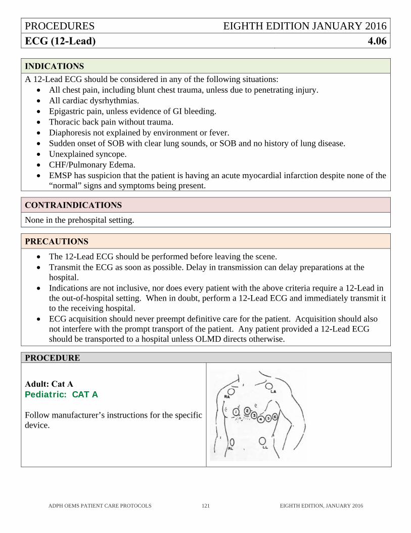
PROCEDURES EIGHTH EDITION JANUARY 2016
ECG (12-Lead) 4.06
ADPH OEMS PATIENT CARE PROTOCOLS 121 EIGHTH EDITION, JANUARY 2016
INDICATIONS
A 12-Lead ECG should be considered in any of the following situations: All chest pain, including blunt chest trauma, unless due to penetrating injury. All cardiac dysrhythmias. Epigastric pain, unless evidence of GI bleeding. Thoracic back pain without trauma. Diaphoresis not explained by environment or fever. Sudden onset of SOB with clear lung sounds, or SOB and no history of lung disease. Unexplained syncope. CHF/Pulmonary Edema. EMSP has suspicion that the patient is having an acute myocardial infarction despite none of the
“normal” signs and symptoms being present.
CONTRAINDICATIONS
None in the prehospital setting.
PRECAUTIONS
The 12-Lead ECG should be performed before leaving the scene. Transmit the ECG as soon as possible. Delay in transmission can delay preparations at the
hospital. Indications are not inclusive, nor does every patient with the above criteria require a 12-Lead in
the out-of-hospital setting. When in doubt, perform a 12-Lead ECG and immediately transmit it to the receiving hospital.
ECG acquisition should never preempt definitive care for the patient. Acquisition should also not interfere with the prompt transport of the patient. Any patient provided a 12-Lead ECG should be transported to a hospital unless OLMD directs otherwise.
PROCEDURE
Adult: Cat A Pediatric: CAT A Follow manufacturer’s instructions for the specific device.
PROCEDURES EIGHTH EDITION JANUARY 2016
Endotracheal Intubation (Oral) 4.07
ADPH OEMS PATIENT CARE PROTOCOLS 122 EIGHTH EDITION, JANUARY 2016
DESCRIPTION
Use of bag valve mask ventilation is not considered sufficient to provide and maintain a protected airway in adult patients except for limited time periods prior to intubation, or during medication administration in the altered mental status protocol. Adult patients who are unconscious, do not have a gag reflex, and need positive pressure ventilation should be intubated by the endotracheal route as soon as indicated.
INDICATIONS
Cardiac arrest. Inability of a conscious patient to ventilate adequately. Inability of the patient to protect the airway. Inability of the EMSP to ventilate the unconscious patient with conventional methods.
CONTRAINDICATIONS
Responsive patients with an intact gag reflex.
PRECAUTIONS
Adequate ventilation and oxygenation must be provided between attempts. Improper tube placement and management can lead to catastrophic results. Pay careful attention
to confirm endotracheal tube placement. If the patient regains consciousness, remove the endotracheal tube as it will cause retching and
vomiting. When the patient is moved after intubation, it is essential to verify that the tube position remains
correct in the new patient position. Remember to deflate cuff prior to repositioning the tube. Movement of the tube with the cuff
inflated could result in patient injury or damage to the cuff, requiring a tube change. Transportation should not be delayed for multiple attempted intubations. Children are almost always best ventilated with a BVM. It is rare to need to
intubate a child. Use of the bougie to facilitate intubation is contraindicated in children.
PROCEDURES EIGHTH EDITION JANUARY 2016
Endotracheal Intubation (Oral) (continued) 4.07
ADPH OEMS PATIENT CARE PROTOCOLS 123 EIGHTH EDITION, JANUARY 2016
PROCEDURE
Adult: Endotracheal Intubation (Oral) (CAT A)
Pediatric: Endotracheal Intubation (Oral) (Cat B)
1. Ventilation by Bag Valve Mask should precede any attempt at intubation.
2. The maximum interruption of ventilation for endotracheal intubation should be 30 seconds.
3. Insert the endotracheal tube using the correct technique for that device.
4. For difficult adult orotracheal intubations where cords are not visible or where the angle is such that it is very difficult to get the tube through the cords, a bougie can be very helpful. Insert the bougie through the cords and then slip the tube over the bougie and slide it down through the cords. Then remove the bougie and verify tube placement.
5. Verification of proper tube placement must immediately be confirmed with auscultation. The abdomen should be auscultated first and then the chest checked for equal bilateral breath sounds and rise.
6. After verification of tube placement using auscultation, verification of tube placement using capnography (qualitative colorimetric or quantitative waveform capnography) should be performed. Use of one or the other is mandatory. Esophageal Detection Device (EDD- suction bulb or syringe) may also be used, but should not replace capnography.
7. Continuously monitor tube placement with waveform capnography if available.
8. Monitor oxygenation with pulse oximeter. Maintain oxygen saturation reading >95%.
9. Ventilate at the appropriate rate as indicated in the protocols.
10. Consider nasogastric tube placement.
PROCEDURES EIGHTH EDITION JANUARY 2016
Endotracheal Intubation (Nasal) 4.08
ADPH OEMS PATIENT CARE PROTOCOLS 124 EIGHTH EDITION, JANUARY 2016
DESCRIPTION
Nasotracheal intubation may be used as an alternative to orotracheal intubation in rare circumstances. It is a very difficult procedure because it must be done without viewing the pharynx and vocal cords. To be successful, the EMSP must be able to appreciate the intensity of the breath sounds of spontaneously breathing patients.
INDICATIONS
The nasotracheal route of endotracheal intubation may be indicated when ventilatory assistance is needed, but the EMSP cannot ventilate successfully with a bag-valve-mask and cannot open the adult patient’s mouth because of clenched jaws.
CONTRAINDICATIONS
Apnea. Suspected epiglottitis. Age less than 12 years. Major facial trauma. Suspected anterior basilar skull fracture (Raccoon Eyes). Foreign bodies or polyps in the nares. Recent nasal surgery. Epistaxis or history of frequent epistaxis. Patients taking warfarin or other anticoagulants. Patients with known clotting disorders.
PRECAUTIONS
Adequate ventilation and oxygenation must be provided between attempts. Improper tube placement can lead to catastrophic results. Pay careful attention to confirm
endotracheal tube placement. When the patient is moved after intubation, it is essential to verify that the tube position remains
correct in the new patient position. Quantitative capnography is the best method to monitor placement of the tube. Remember to deflate cuff prior to repositioning the tube. Movement of the tube with the cuff
inflated could result in patient injury or damage to the cuff, requiring a tube change. Transportation should not be delayed for multiple attempted intubations.
PROCEDURES EIGHTH EDITION JANUARY 2016
Endotracheal Intubation (Nasal) (continued) 4.08
ADPH OEMS PATIENT CARE PROTOCOLS 125 EIGHTH EDITION, JANUARY 2016
PROCEDURE
Adult: Endotracheal Intubation (Nasal) (Cat A)
Pediatric: Endotracheal Intubation (Nasal) Not Indicated. If in doubt, contact OLMD. (Cat B)
1. Ventilation by Bag Valve Mask should precede any attempt at intubation.
2. The maximum interruption of ventilation for endotracheal intubation should be 30 seconds.
3. Insert the endotracheal tube using the correct technique for that device.
4. Verification of proper tube placement must immediately be confirmed with auscultation. The abdomen should be auscultated first and then the chest checked for equal bilateral breath sounds and rise.
5. After verification of tube placement using auscultation, verification of tube placement using capnography (qualitative colorimetric or quantitative waveform capnography) should be performed. Use of one or the other is mandatory. Esophageal Detection Device (EDD- suction bulb or syringe) may also be used, but should not replace capnography.
6. Continuously monitor tube placement with waveform capnography if available.
7. Monitor oxygenation with pulse oximeter. Maintain oxygen saturation reading >95%.
8. Ventilate at the appropriate rate as indicated in the protocols.
PROCEDURES EIGHTH EDITION JANUARY 2016
External Pacing 4.09
ADPH OEMS PATIENT CARE PROTOCOLS 126 EIGHTH EDITION, JANUARY 2016
DESCRIPTION
External pacing is the technique of electrical cardiac pacing accomplished by using skin electrodes to pass repetitive electrical impulses through the thorax.
INDICATIONS
Bradycardia (heart rate <60) with evidence of inadequate perfusion such as hypotension BP <90, altered mental status, unresponsive to atropine therapy or unable to establish IV/IO access, pulmonary edema, chest pain, or dyspnea.
CONTRAINDICATIONS
External pacing should not be used in the following settings: Asystole or PEA. Patients <14 years of age. Patients meeting death in the field criteria. Patients with signs of penetrating or blunt trauma.
PRECAUTIONS
May be painful for awake patients.
PROCEDURE
Adult: External Pacing (Cat B) Pediatric: External Pacing (Cat B) 1. Attach pacemaker leads and verify the monitor is displaying a cardiac rhythm. 2. Attach pacing electrodes to anterior and posterior chest just to the left of the sternum and spinal
column, respectively. 3. Begin pacing with a heart rate of 80 beats per minute at minimum current output. Increase current
output until pulse rate captures to match rhythm strip. Use the minimum current output to maintain capture.
4. Assess for capture by observing the monitor and the patient for changes in mental status, pulse, or blood pressure.
5. If the patient complains of pain during pacing despite reduced current output, consider Morphine Sulfate.
Morphine Sulfate: 4 mg IV initial dose, titrate to pain relief in 2 mg doses, every 3-5 minutes, 10 mg MAX. If pain not relieved after 10 mg, call OLMD for further doses. (Cat B)
6. In the event of electrical capture and no pulses, use Cardiac Arrest (PEA) Protocol (3.09). 7. If there is no response to Atropine and /or pacing, or if a change in pacing rate is desired, consult
OLMD for further orders.
PROCEDURES EIGHTH EDITION JANUARY 2016
Hemostatic Agents 4.10
ADPH OEMS PATIENT CARE PROTOCOLS 127 EIGHTH EDITION, JANUARY 2016
DESCRIPTION
Hemostatic agents can be used to control exsanguinating hemorrhage when use of direct pressure and tourniquets fail.
INDICATIONS
Exsanguinating hemorrhage that cannot be controlled by direct pressure or by tourniquet. This is most likely to involve wounds of axilla, groin, neck, face, or scalp.
CONTRAINDICATIONS
Minor bleeding. Bleeding that can be controlled by direct pressure. Bleeding that can be controlled by application of a tourniquet. Open abdominal or chest wounds may be treated with hemostatic agent impregnated gauze, but not
with powdered hemostatic agents.
PRECAUTIONS
None.
PROCEDURE
Adult: Hemostatic Agents (Cat A)
Pediatric: Hemostatic Agents (Cat A)
1. Each service must be trained to use the hemostatic agent selected by their medical director. (See Hemostatic Agents 6.03.)
2. Follow the manufacturer’s user instructions for proper technique.
3. Pack the wound with the chosen hemostatic agent.
4. Apply direct pressure over the wound for a minimum of 3 minutes or until bleeding stops.
5. Apply pressure dressing over wound and hemostatic agent.
6. Advise receiving hospital personnel of use of hemostatic agent.
PROCEDURES EIGHTH EDITION JANUARY 2016
Intraosseous Therapy 4.11
ADPH OEMS PATIENT CARE PROTOCOLS 128 EIGHTH EDITION, JANUARY 2016
DESCRIPTION
An alternative technique for establishing intravenous access in critical patients when peripheral intravenous access is unobtainable or too time consuming, and the patient’s outcome will be compromised if no intravascular access is obtained prior to hospital arrival.
INDICATIONS
A life-threatening condition exists such as cardiac arrest or shock with systolic blood pressure less than 90 mmHg and a peripheral IV cannot, or is unlikely to be established. For the critical patient, the EMSP should consider IO when two IV attempts have been made or 90 seconds have been spent trying to find a vein. Inability to locate an appropriate vein site is equivalent to an attempt. It is not necessary to actually penetrate the skin with a needle.
CONTRAINDICATIONS
Cellulitis or infection overlying the site. Fracture in the same bone or a proximal vascular injury. Severe pelvic trauma. A previous intraosseous attempt in the same bone.
PRECAUTIONS
Incorrect placement may lead to: sub-periosteal infusion, extravasation due to prior attempt in same bone, or through-and-through puncture of the bone.
Complications include plugging of needle with bone or marrow, growth plate damage, osteomyelitis, compression to popliteal vessels, or the tibial nerve due to extravasation, pulmonary embolism, fracture of the tibia.
This procedure should not delay transport time. This procedure should not be used for a precautionary IV. Hypoglycemia is not an indication for IO except in extreme circumstances. All use of IO for this
will be reviewed by the Office of EMS.
PROCEDURES EIGHTH EDITION JANUARY 2016
Intraosseous Therapy (continued) 4.11
ADPH OEMS PATIENT CARE PROTOCOLS 129 EIGHTH EDITION, JANUARY 2016
PROCEDURE
Cardiac Arrest or Shock (BP<90mmHg): Adult (Cat A) Pediatric (Cat A) All other applications: Adult (Cat B) Pediatric (Cat B) The Medical Director for the EMS service will approve the IO device(s) to be used by that service. (See Intraosseous Needle Insertion Devices 6.04). EMSP must complete training on their approved IO device prior to using it in the field. The proximal tibia and the proximal humerus are the only authorized sites for all devices except the FAST-1, which is a sternal device only for use in adults. FAST-1 is not approved for pediatric patients. 1. Palpate the landmarks on the tibia (or sternum for FAST-1) and note the entry point. 2. Prepare the surface with antiseptic and dry with a sterile gauze pad. 3. Insert the device using the correct procedure, insertion point, and special precautions for that
device. 4. If extravasation should occur, further attempts at the site and extremity should be avoided. 5. IO fluid administration causes pain for conscious patients and is related to intramedullary pressure.
If the adult patient is conscious, slowly administer one or two cc (20 - 40 mg) of 2% Lidocaine through the IO (utilizing the Lidocaine pre-filled syringe) prior to the initial fluid bolus.
6. Although gravity drainage may suffice, pressurized infusions (blood pump, syringes and stopcock,
or pressure bag) may be needed during resuscitation.
7. When the EMSP leaves the patient with the receiving hospital, be sure that they know how to remove the device and have any special equipment needed to accomplish this.
PROCEDURES EIGHTH EDITION JANUARY 2016
Intravenous Therapy 4.12
ADPH OEMS PATIENT CARE PROTOCOLS 130 EIGHTH EDITION, JANUARY 2016
DESCRIPTION Intravenous therapy is the introduction of fluids and other substances into the venous side of the circulatory system to replace blood loss due to hemorrhage, for electrolyte or fluid replacement, and for introduction of medications into the vascular system.
INDICATIONS
Any time a medication or Normal Saline solution is administered as a continuous infusion.
CONTRAINDICATIONS
None in the prehospital setting.
PRECAUTIONS
At the time of transfer of care from one agency to another, the patient care report should include that amount of solution already infused and the rate at which the solution is infusing.
All infusions and patient response should be closely monitored and documented. An extension set should be at least 10” long, contain one or more injection sites, a slide clam p,
and have a volume capacity of not less than 6.0ml. A “small-bore” extension set should be 3” long and have a volume capacity of 0.15ml. In addition to regular IV line(s), every trauma patient should have extension tubing attached
between the IV hub and the solution bag.
PROCEDURE Adult: Intravenous Therapy (Cat A) Pediatric: Intravenous Therapy (Cat A) 1. IV access.
a. Establish intravenous access and prepare Normal Saline. b. Connect an extension set between the IV hub and the solution bag and tubing. c. All IVs will be started using macrodrips, unless otherwise indicated.
2. IV access with and IV lock. a. Establish IV access. b. Connect a “small-bore” extension set between the IV hub and male adapter plug. c. After placement, the line should be flushed with normal saline. d. If the IV lock system is used for the administration of medication, the line must be flushed after
each administration. 3. IV Infusions.
a. Connect the IV tubing to the IV lock and the solution bag. b. Open the flow control and begin infusing the solution at the appropriate rate. c. An infusion pump may be used, if available and desired. Use according to manufacturer’s
directions.
PROCEDURES EIGHTH EDITION JANUARY 2016
Patient Restraint 4.13
ADPH OEMS PATIENT CARE PROTOCOLS 131 EIGHTH EDITION, JANUARY 2016
DESCRIPTION
Patient restraints should be utilized only when necessary and in those situations where the patient is exhibiting behavior that the EMSP believes presents a danger to the patient and/or others. This procedure is not to be used on patients specifically refusing treatment unless they are placed under a police hold or OLMD has given orders to restrain the patient. This procedure also applies to patients being treated under implied consent. Agitated, anxious, and uncooperative patients can often be calmed adequately by eliminating threatening or forced activities, and by backing off. Attempt to gain the confidence of the patient.
INDICATIONS
To restrain patients when necessary for the protection of the EMSP and/or the patient.
CONTRAINDICATIONS
None in the prehospital setting.
PRECAUTIONS
Physical restraint MUST be used any time a potentially violent or unstable patient (head injury, patient under the influence of intoxicants, or altered mental status for ANY other reason) is transported by air ambulance, even if the patient is sedated.
PROCEDURE
Adult: Patient Restraint (Cat B) Pediatric: Patient Restraint (Cat B) 1. Ensure sufficient manpower is present to control the patient while restraining him/her. If law
enforcement or additional manpower is needed, call for it prior to attempting restraint procedures. The EMSP should not endanger himself/herself or the rest of the ambulance crew.
2. Use the minimum physical restraint required to accomplish necessary patient care and ensure safe transportation. Avoid placing restraints in such a way as to preclude evaluation of the patient’s medical status (airway, breathing, and circulation). Consider whether placement of restraints will interfere with necessary patient care activities or will cause further harm.
3. Patient should be transported face up. A Reeves Sleeve® could be useful. 4. Secure ALL extremities. Try to restrain lower extremities first at both ankles and the knees. Next,
restrain the patient’s arms at his/her sides using a restraint on each wrist. 5. If necessary, utilize cervical spine precautions to control violent head or body movements. 6. Place padding under patient’s head and wherever else needed to prevent the patient from further
harming himself/herself, or restricting circulation. 7. Document airway, breathing, and circulatory status of patient every 15 minutes, including
circulatory status of restrained extremities. 8. Document the need for physical restraint to prevent possible harm to the patient or others.
PROCEDURES EIGHTH EDITION JANUARY 2016
Rectal Diazepam Administration 4.14
ADPH OEMS PATIENT CARE PROTOCOLS 132 EIGHTH EDITION, JANUARY 2016
DESCRIPTION
The placement of a soft, flexible catheter into the rectum for the purpose of administering diazepam.
INDICATIONS
Treatment of seizures in pediatric or adult patients when IV access is unobtainable.
CONTRAINDICATIONS
Known rectal abnormality.
PRECAUTIONS
IV administration of diazepam is the preferred route. Do not force the catheter in. Catheter should advance with little or no resistance. There is a risk of respiratory depression with the administration of diazepam, therefore the patient
should be monitored closely for signs of respiratory depression. This procedure may be performed en route to the hospital. It should not delay transport. The condition of the rectal mucosa, the presence of fecal matter, and the metabolic status of the
patient may affect absorption. It is recommended that the EMSP prepare all of his/her equipment in advance of the procedure.
PROCEDURE
Adult: Rectal Administration of Diazepam Pediatric: Rectal Administration of Diazepam (Cat B) 1. Diazepam solution can be deposited into the rectal lumen using an IV catheter that is 4-6 cm
(2”) in length. 2. Remove the flexible catheter from an IV needle that is the appropriate length and attach to
the syringe containing the diazepam. 3. If available, medication delivery systems designed specifically for rectal medication
insertion may be used instead. 4. Advance the flexible catheter 4-6 cm into the rectum and administer the medication. 5. Flush the catheter with 3 cc of air after administering the diazepam. Flushing with fluid will
dilute the medication. 6. Hold the buttocks together for 1-2 minutes to prevent any leakage of medication.
PROCEDURES EIGHTH EDITION JANUARY 2016
Transportation of Pediatric Patients 4.15
ADPH OEMS PATIENT CARE PROTOCOLS 133 EIGHTH EDITION, JANUARY 2016
DESCRIPTION
Appropriate restraint of children is critical to prevent a serious or potentially fatal injury of pediatric patients being transported on ambulances. The greatest potential for injury occurs when an unrestrained child becomes a projectile object upon a sudden stop or crash.
INDICATIONS Any time a child is transported in a prehospital vehicle.
CONTRAINDICATIONS None.
PRECAUTIONS It is recognized that in certain cases there may be more children to be
transported than there are restraint devices available in which to place them. If the ambulance is equipped with passenger side airbags, children under the
age of 12 years should not be transported in the passenger seat. These guidelines may not be consistent with the official instructions for use of
a child restraint in a passenger vehicle. These guidelines assume that the ambulance is equipped with a cot and
fastener system that has been successfully tested under vehicle crash conditions.
PROCEDURES EIGHTH EDITION JANUARY 2016
Transportation of Pediatric Patients (continued) 4.15
ADPH OEMS PATIENT CARE PROTOCOLS 134 EIGHTH EDITION, JANUARY 2016
PROCEDURE
Pediatric: Transportation of Pediatric Patients (Cat A) 1. Whenever possible, all pediatric patients should be safely and appropriately
restrained during transport. Safe and appropriate transport does not include having a child held by another person who is riding or strapped to the gurney.
2. Available child restraint systems should be used for all pediatric patients. These systems should include those specifically produced for secure transport on an ambulance stretcher that includes an integrated five-point harness. A child’s own car seat, appropriately secured to the stretcher, often proves to be an excellent source of a restraint system.
3. Children who are not patients should not routinely be transported in the ambulance. There may be extenuating circumstances that require such transport. In those cases, the child should be placed in an appropriate child restraint seat, in the appropriate position, in either the passenger area or patient area of the ambulance.
a. <1 year old and <20 lbs.: rear facing infant seat. b. <4 years old and <40 lbs.: forward facing toddler seat. c. 4-8 years old and 40 lbs.: booster seats with lap/shoulder belt. d. <12 years old: back seat, restrained.
While it is not recommended using a child’s own car seat for transportation post accident, such may be better than no restraint during transport. In addition, it is recognized that the very nature of emergency circumstances may require some compromises of best practices. If a child is found in a child restraint that is still visually intact, it may be better to move the child in that restraint to the ambulance for transport than to transfer the child to a different restraint. If there is a question, this should be discussed with the OLMD.
ADPH EMS PROTOCOLS EIGHTH EDITION
MEDICATIONS 5
ADPH OEMS PATIENT CARE PROTOCOLS 135 EIGHTH EDITION, JANUARY 2016
This section is provided for reference information only. Medications may be administered only as defined by treatment protocol unless online medical direction orders a deviation.
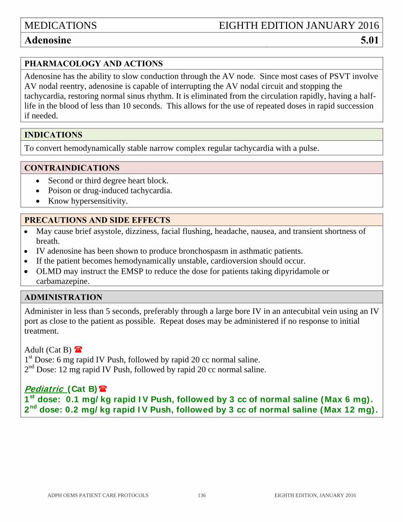
MEDICATIONS EIGHTH EDITION JANUARY 2016
Adenosine 5.01
ADPH OEMS PATIENT CARE PROTOCOLS 136 EIGHTH EDITION, JANUARY 2016
PHARMACOLOGY AND ACTIONS
Adenosine has the ability to slow conduction through the AV node. Since most cases of PSVT involve AV nodal reentry, adenosine is capable of interrupting the AV nodal circuit and stopping the tachycardia, restoring normal sinus rhythm. It is eliminated from the circulation rapidly, having a half-life in the blood of less than 10 seconds. This allows for the use of repeated doses in rapid succession if needed.
INDICATIONS
To convert hemodynamically stable narrow complex regular tachycardia with a pulse.
CONTRAINDICATIONS
Second or third degree heart block. Poison or drug-induced tachycardia. Know hypersensitivity.
PRECAUTIONS AND SIDE EFFECTS May cause brief asystole, dizziness, facial flushing, headache, nausea, and transient shortness of
breath. IV adenosine has been shown to produce bronchospasm in asthmatic patients. If the patient becomes hemodynamically unstable, cardioversion should occur. OLMD may instruct the EMSP to reduce the dose for patients taking dipyridamole or
carbamazepine.
ADMINISTRATION
Administer in less than 5 seconds, preferably through a large bore IV in an antecubital vein using an IV port as close to the patient as possible. Repeat doses may be administered if no response to initial treatment. Adult (Cat B) 1st Dose: 6 mg rapid IV Push, followed by rapid 20 cc normal saline. 2nd Dose: 12 mg rapid IV Push, followed by rapid 20 cc normal saline. Pediatric (Cat B) 1st dose: 0.1 mg/kg rapid IV Push, followed by 3 cc of normal saline (Max 6 mg). 2nd dose: 0.2 mg/kg rapid IV Push, followed by 3 cc of normal saline (Max 12 mg).
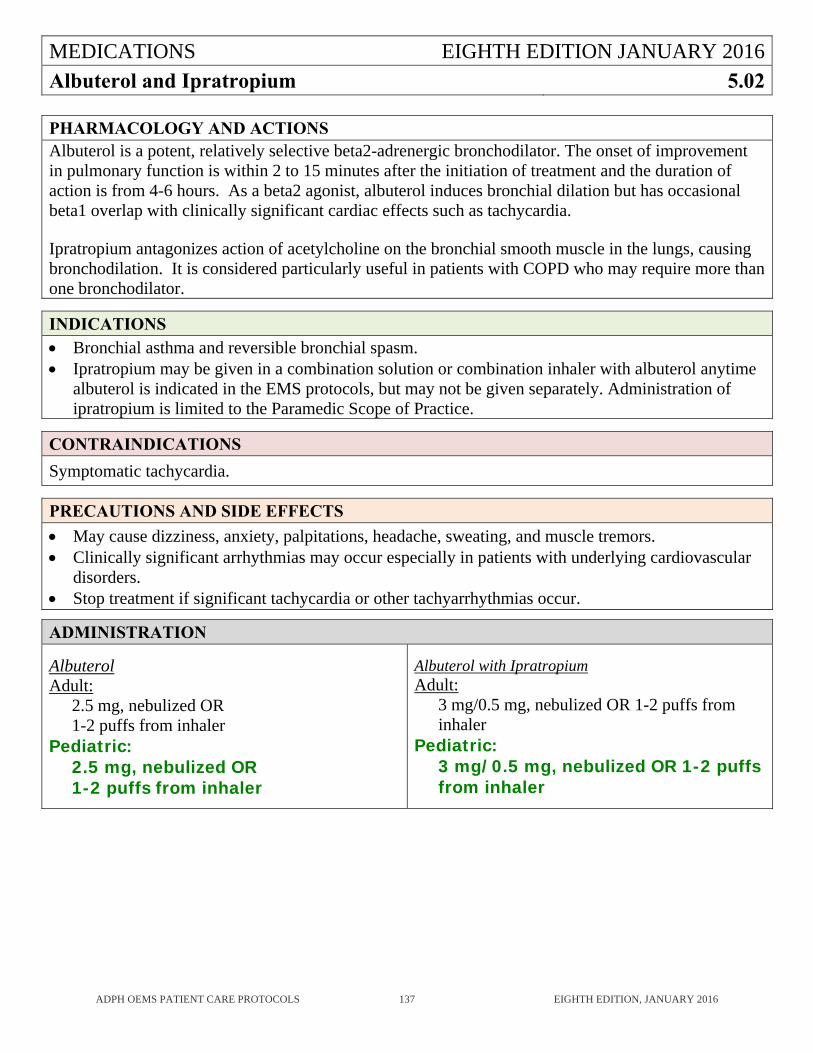
MEDICATIONS EIGHTH EDITION JANUARY 2016
Albuterol and Ipratropium 5.02
ADPH OEMS PATIENT CARE PROTOCOLS 137 EIGHTH EDITION, JANUARY 2016
PHARMACOLOGY AND ACTIONS Albuterol is a potent, relatively selective beta2-adrenergic bronchodilator. The onset of improvement in pulmonary function is within 2 to 15 minutes after the initiation of treatment and the duration of action is from 4-6 hours. As a beta2 agonist, albuterol induces bronchial dilation but has occasional beta1 overlap with clinically significant cardiac effects such as tachycardia. Ipratropium antagonizes action of acetylcholine on the bronchial smooth muscle in the lungs, causing bronchodilation. It is considered particularly useful in patients with COPD who may require more than one bronchodilator.
INDICATIONS Bronchial asthma and reversible bronchial spasm. Ipratropium may be given in a combination solution or combination inhaler with albuterol anytime
albuterol is indicated in the EMS protocols, but may not be given separately. Administration of ipratropium is limited to the Paramedic Scope of Practice.
CONTRAINDICATIONS
Symptomatic tachycardia.
PRECAUTIONS AND SIDE EFFECTS
May cause dizziness, anxiety, palpitations, headache, sweating, and muscle tremors. Clinically significant arrhythmias may occur especially in patients with underlying cardiovascular
disorders. Stop treatment if significant tachycardia or other tachyarrhythmias occur.
ADMINISTRATION
Albuterol Adult:
2.5 mg, nebulized OR 1-2 puffs from inhaler
Pediatric: 2.5 mg, nebulized OR 1-2 puffs from inhaler
Albuterol with Ipratropium Adult:
3 mg/0.5 mg, nebulized OR 1-2 puffs from inhaler
Pediatric: 3 mg/0.5 mg, nebulized OR 1-2 puffs from inhaler
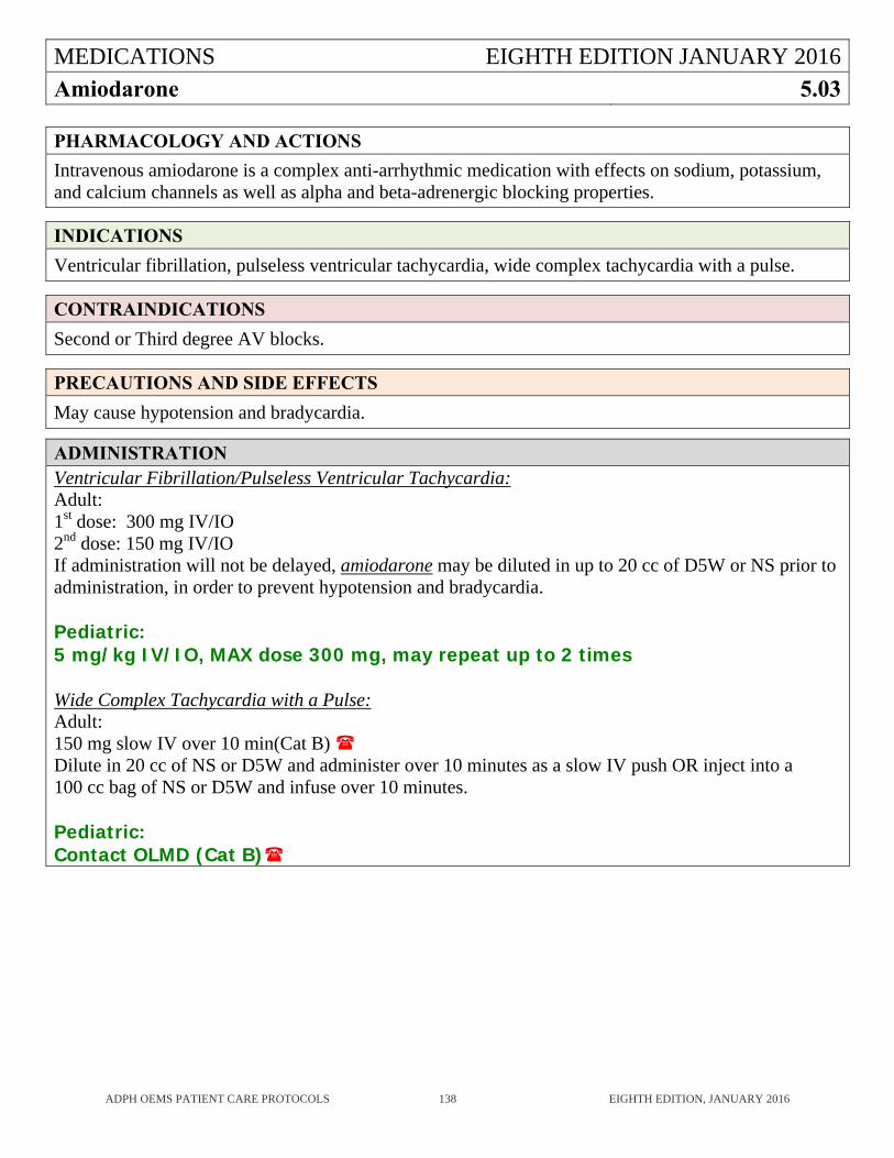
MEDICATIONS EIGHTH EDITION JANUARY 2016
Amiodarone 5.03
ADPH OEMS PATIENT CARE PROTOCOLS 138 EIGHTH EDITION, JANUARY 2016
PHARMACOLOGY AND ACTIONS
Intravenous amiodarone is a complex anti-arrhythmic medication with effects on sodium, potassium, and calcium channels as well as alpha and beta-adrenergic blocking properties.
INDICATIONS
Ventricular fibrillation, pulseless ventricular tachycardia, wide complex tachycardia with a pulse.
CONTRAINDICATIONS
Second or Third degree AV blocks.
PRECAUTIONS AND SIDE EFFECTS
May cause hypotension and bradycardia.
ADMINISTRATION Ventricular Fibrillation/Pulseless Ventricular Tachycardia: Adult: 1st dose: 300 mg IV/IO 2nd dose: 150 mg IV/IO If administration will not be delayed, amiodarone may be diluted in up to 20 cc of D5W or NS prior to administration, in order to prevent hypotension and bradycardia. Pediatric: 5 mg/kg IV/IO, MAX dose 300 mg, may repeat up to 2 times Wide Complex Tachycardia with a Pulse: Adult: 150 mg slow IV over 10 min(Cat B) Dilute in 20 cc of NS or D5W and administer over 10 minutes as a slow IV push OR inject into a 100 cc bag of NS or D5W and infuse over 10 minutes. Pediatric: Contact OLMD (Cat B)
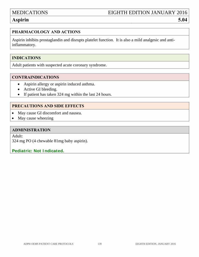
MEDICATIONS EIGHTH EDITION JANUARY 2016
Aspirin 5.04
ADPH OEMS PATIENT CARE PROTOCOLS 139 EIGHTH EDITION, JANUARY 2016
PHARMACOLOGY AND ACTIONS
Aspirin inhibits prostaglandin and disrupts platelet function. It is also a mild analgesic and anti-inflammatory.
INDICATIONS
Adult patients with suspected acute coronary syndrome.
CONTRAINDICATIONS
Aspirin allergy or aspirin induced asthma. Active GI bleeding. If patient has taken 324 mg within the last 24 hours.
PRECAUTIONS AND SIDE EFFECTS
May cause GI discomfort and nausea. May cause wheezing
ADMINISTRATION
Adult: 324 mg PO (4 chewable 81mg baby aspirin). Pediatric: Not Indicated.
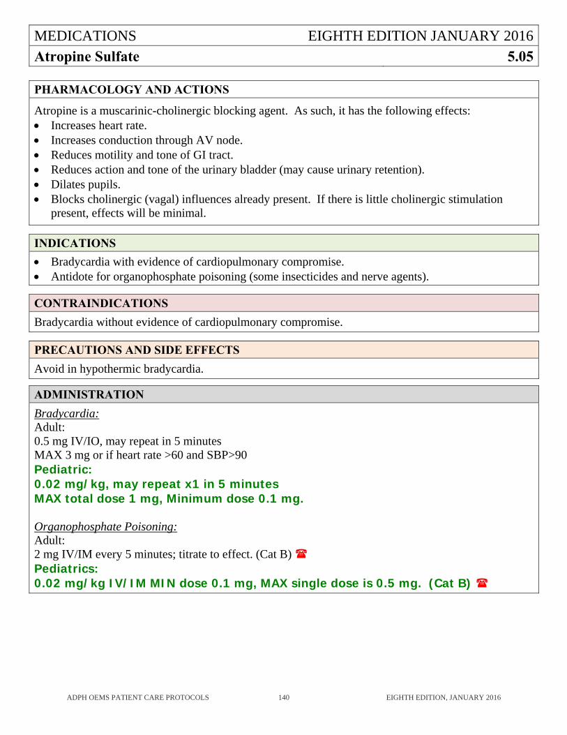
MEDICATIONS EIGHTH EDITION JANUARY 2016
Atropine Sulfate 5.05
ADPH OEMS PATIENT CARE PROTOCOLS 140 EIGHTH EDITION, JANUARY 2016
PHARMACOLOGY AND ACTIONS
Atropine is a muscarinic-cholinergic blocking agent. As such, it has the following effects: Increases heart rate. Increases conduction through AV node. Reduces motility and tone of GI tract. Reduces action and tone of the urinary bladder (may cause urinary retention). Dilates pupils. Blocks cholinergic (vagal) influences already present. If there is little cholinergic stimulation
present, effects will be minimal.
INDICATIONS
Bradycardia with evidence of cardiopulmonary compromise. Antidote for organophosphate poisoning (some insecticides and nerve agents).
CONTRAINDICATIONS
Bradycardia without evidence of cardiopulmonary compromise.
PRECAUTIONS AND SIDE EFFECTS
Avoid in hypothermic bradycardia.
ADMINISTRATION
Bradycardia: Adult: 0.5 mg IV/IO, may repeat in 5 minutes MAX 3 mg or if heart rate >60 and SBP>90 Pediatric: 0.02 mg/kg, may repeat x1 in 5 minutes MAX total dose 1 mg, Minimum dose 0.1 mg. Organophosphate Poisoning: Adult: 2 mg IV/IM every 5 minutes; titrate to effect. (Cat B) Pediatrics: 0.02 mg/kg IV/IM MIN dose 0.1 mg, MAX single dose is 0.5 mg. (Cat B)
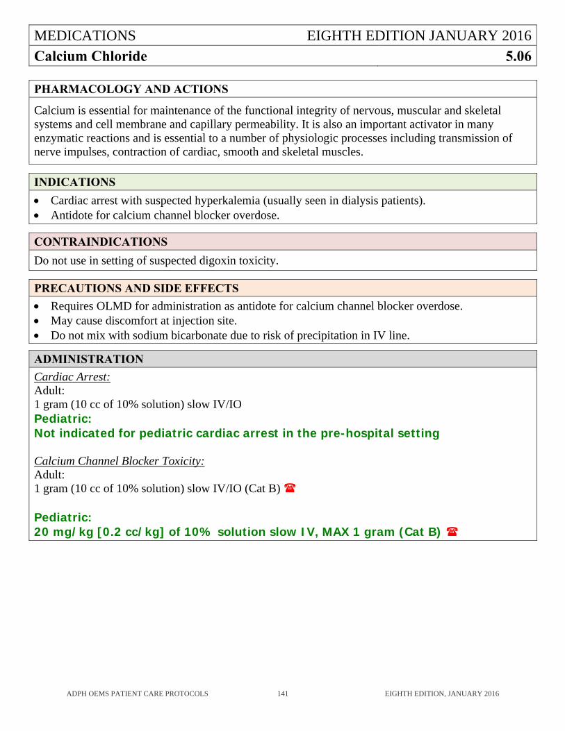
MEDICATIONS EIGHTH EDITION JANUARY 2016
Calcium Chloride 5.06
ADPH OEMS PATIENT CARE PROTOCOLS 141 EIGHTH EDITION, JANUARY 2016
PHARMACOLOGY AND ACTIONS
Calcium is essential for maintenance of the functional integrity of nervous, muscular and skeletal systems and cell membrane and capillary permeability. It is also an important activator in many enzymatic reactions and is essential to a number of physiologic processes including transmission of nerve impulses, contraction of cardiac, smooth and skeletal muscles.
INDICATIONS
Cardiac arrest with suspected hyperkalemia (usually seen in dialysis patients). Antidote for calcium channel blocker overdose.
CONTRAINDICATIONS
Do not use in setting of suspected digoxin toxicity.
PRECAUTIONS AND SIDE EFFECTS
Requires OLMD for administration as antidote for calcium channel blocker overdose. May cause discomfort at injection site. Do not mix with sodium bicarbonate due to risk of precipitation in IV line.
ADMINISTRATION
Cardiac Arrest: Adult: 1 gram (10 cc of 10% solution) slow IV/IO Pediatric: Not indicated for pediatric cardiac arrest in the pre-hospital setting Calcium Channel Blocker Toxicity: Adult: 1 gram (10 cc of 10% solution) slow IV/IO (Cat B)
Pediatric: 20 mg/kg [0.2 cc/kg] of 10% solution slow IV, MAX 1 gram (Cat B)
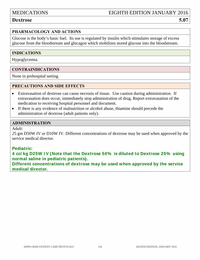
MEDICATIONS EIGHTH EDITION JANUARY 2016
Dextrose 5.07
ADPH OEMS PATIENT CARE PROTOCOLS 142 EIGHTH EDITION, JANUARY 2016
PHARMACOLOGY AND ACTIONS
Glucose is the body’s basic fuel. Its use is regulated by insulin which stimulates storage of excess glucose from the bloodstream and glucagon which mobilizes stored glucose into the bloodstream.
INDICATIONS
Hypoglycemia.
CONTRAINDICATIONS
None in prehospital setting.
PRECAUTIONS AND SIDE EFFECTS
Extravasation of dextrose can cause necrosis of tissue. Use caution during administration. If extravasation does occur, immediately stop administration of drug. Report extravasation of the medication to receiving hospital personnel and document.
If there is any evidence of malnutrition or alcohol abuse, thiamine should precede the administration of dextrose (adult patients only).
ADMINISTRATION Adult: 25 gm D50W IV or D10W IV. Different concentrations of dextrose may be used when approved by the service medical director. Pediatric: 4 cc/kg D25W IV (Note that the Dextrose 50% is diluted to Dextrose 25% using normal saline in pediatric patients). Different concentrations of dextrose may be used when approved by the service medical director.
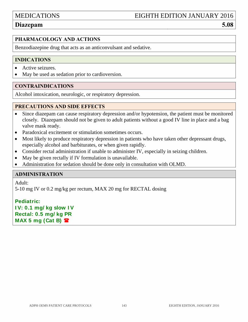
MEDICATIONS EIGHTH EDITION JANUARY 2016
Diazepam 5.08
ADPH OEMS PATIENT CARE PROTOCOLS 143 EIGHTH EDITION, JANUARY 2016
PHARMACOLOGY AND ACTIONS
Benzodiazepine drug that acts as an anticonvulsant and sedative.
INDICATIONS
Active seizures. May be used as sedation prior to cardioversion.
CONTRAINDICATIONS
Alcohol intoxication, neurologic, or respiratory depression.
PRECAUTIONS AND SIDE EFFECTS Since diazepam can cause respiratory depression and/or hypotension, the patient must be monitored
closely. Diazepam should not be given to adult patients without a good IV line in place and a bag valve mask ready.
Paradoxical excitement or stimulation sometimes occurs. Most likely to produce respiratory depression in patients who have taken other depressant drugs,
especially alcohol and barbiturates, or when given rapidly. Consider rectal administration if unable to administer IV, especially in seizing children. May be given rectally if IV formulation is unavailable. Administration for sedation should be done only in consultation with OLMD.
ADMINISTRATION
Adult: 5-10 mg IV or 0.2 mg/kg per rectum, MAX 20 mg for RECTAL dosing Pediatric: IV: 0.1 mg/kg slow IV Rectal: 0.5 mg/kg PR MAX 5 mg (Cat B)
MEDICATIONS EIGHTH EDITION JANUARY 2016
Diphenhydramine 5.09
ADPH OEMS PATIENT CARE PROTOCOLS 144 EIGHTH EDITION, JANUARY 2016
PHARMACOLOGY AND ACTIONS
An antihistamine which blocks action of histamines released from cells during an allergic reaction; also has anticholinergic properties which makes it useful to treat or prevent acute dystonic reactions to antipsychotic drugs. These reactions include: oculogyric crisis, acute torticollis, and facial grimacing.
INDICATIONS Treatment of allergic reactions. Treatment or prevention of acute dystonic reactions to antipsychotic drugs. Nausea or vomiting
CONTRAINDICATIONS Known hypersensitivity. Newborns. Nursing mothers (relative contraindication).
PRECAUTIONS AND SIDE EFFECTS
Usually causes sedation, however it may paradoxically cause excitation in children. May have additive sedation effect with alcohol or other CNS depressants. May cause hypotension when given IV.
ADMINISTRATION
Adult: 25-50 mg IV/IM Pediatric: 1 mg/kg IV/IM (MAX 50 mg) Diphenhydramine is Cat A for Allergic Reaction, Cat B for Altered Mental Status in both pediatric and adult patients.
MEDICATIONS EIGHTH EDITION JANUARY 2016
Dopamine 5.10
ADPH OEMS PATIENT CARE PROTOCOLS 145 EIGHTH EDITION, JANUARY 2016
PHARMACOLOGY AND ACTIONS
Chemical precursor of nor-epinephrine which occurs naturally in man and which has both alpha and beta-receptor and dopaminergic stimulating actions. Its actions differ with dosage given: 1-5 mcg/kg/min - dilates renal and mesenteric blood vessels (no effect on heart rate or blood
pressure). 2-10 mcg/kg/min - beta effects on heart which usually increases cardiac output without greatly
increasing heart rate or blood pressure. 10-20 mcg/kg/min - alpha peripheral effects cause peripheral vasoconstriction and increased blood
pressure. 20-40 mcg/kg/min - alpha effects reverse dilatation or renal and mesenteric vessels with resultant
decreased flow.
INDICATIONS
Treatment of refractory shock, particularly cardiogenic, distributive, or obstructive.
CONTRAINDICATIONS
Hypovolemic shock, especially with hypotension.
PRECAUTIONS AND SIDE EFFECTS
May induce tachyarrhythmias, in which case infusion should be decreased or stopped. High doses (10 mcg/kg) may cause peripheral vasoconstriction . Should not be added to sodium bicarbonate or other alkaline solutions since dopamine will be
inactivated in alkaline solutions. Consider hypovolemia and treat this with appropriate fluids before administration of dopamine. Dopamine is best administered by an infusion pump to accurately regulate rate. It may be
hazardous when used in the field without an infusion pump. Monitor closely.
ADMINISTRATION 5-20 mcg/kg/min IV/IO Mix 800 mg dopamine in 500 cc Normal Saline to produce concentration of 1600 mcg/ml. Start infusion rate 2-5 mcg/kg/min. Gradually increase by 5 mcg/kg/min until desired effect is achieved. Use microdrip chamber only. See dosage chart on next page. Pediatric: 5-20 mcg/kg/min IV/IO Mix 200 mg in 500 cc Normal Saline to produce concentration of 400 mcg/ml. Start infusion rate 2-5 mcg/kg/min. Gradually increase by 5 mcg/kg/min until desired effect is achieved. Use microdrip chamber only.
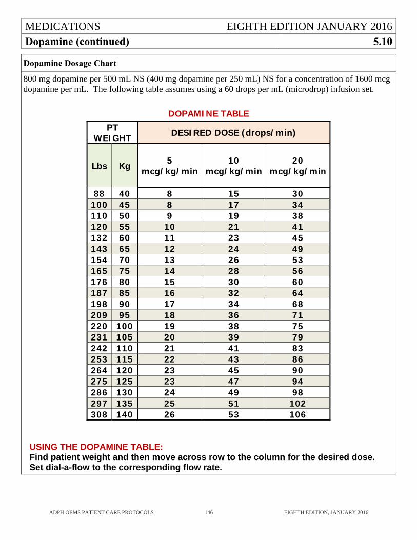
MEDICATIONS EIGHTH EDITION JANUARY 2016
Dopamine (continued) 5.10
ADPH OEMS PATIENT CARE PROTOCOLS 146 EIGHTH EDITION, JANUARY 2016
Dopamine Dosage Chart
800 mg dopamine per 500 mL NS (400 mg dopamine per 250 mL) NS for a concentration of 1600 mcg dopamine per mL. The following table assumes using a 60 drops per mL (microdrop) infusion set. DOPAMINE TABLE
PT WEIGHT DESIRED DOSE (drops/min)
Lbs Kg 5 mcg/kg/min
10 mcg/kg/min
20
mcg/kg/min
88 40 8 15 30 100 45 8 17 34 110 50 9 19 38 120 55 10 21 41 132 60 11 23 45 143 65 12 24 49 154 70 13 26 53 165 75 14 28 56 176 80 15 30 60 187 85 16 32 64 198 90 17 34 68 209 95 18 36 71 220 100 19 38 75 231 105 20 39 79 242 110 21 41 83 253 115 22 43 86 264 120 23 45 90 275 125 23 47 94 286 130 24 49 98 297 135 25 51 102 308 140 26 53 106
USING THE DOPAMINE TABLE: Find patient weight and then move across row to the column for the desired dose. Set dial-a-flow to the corresponding flow rate.
MEDICATIONS EIGHTH EDITION JANUARY 2016
Epinephrine 5.11
ADPH OEMS PATIENT CARE PROTOCOLS 147 EIGHTH EDITION, JANUARY 2016
PHARMACOLOGY AND ACTIONS
Catecholamine with alpha and beta effects which increases heart rate and blood pressure. Potent bronchodilator.
INDICATIONS Cardiac Arrest. Pediatric Bradycardia with Cardiopulmonary Compromise. Moderate and Severe Allergic Reactions. Severe Refractory Wheezing.
CONTRAINDICATIONS
Uncontrolled hypertension is a relative contraindication.
PRECAUTIONS AND SIDE EFFECTS
Epinephrine increases cardiac work and can precipitate angina, myocardial infarction or major dysrhythmias in an individual with ischemic heart disease. Extreme caution should be used when treating patients with allergic reaction or wheezing with Epinephrine due to the cardiac side effects.
Wheezing in the elderly is most commonly a sign of conditions which do not require Epinephrine such as pneumonia, pulmonary embolism, or pulmonary edema.
Other side effects include anxiety and tremor. Use extreme caution to prevent accidentally giving Epinephrine 1:1000 intravenously which could
lead to accidental overdose of Epinephrine.
ADMINISTRATION Moderate allergic reaction and severe refractory wheezing: Epinephrine 1:1000
0.3 mg (0.3cc) IM (Cat A) If pt is age 65 or older, has history of heart disease, or uncontrolled hypertension contact OLMD prior to administration (Cat B) Pediatric: 0.01 mg/kg MAX 0.3 mg IM
Major allergic reaction: Epinephrine 1:10,000
0.3 mg (3 cc) IV repeat every 5 minutes as needed. (Cat B) Pediatric: 0.01 mg/kg (0.1 cc/kg) MAX 0.3 mg (3 cc) IV, repeat every 5 minutes as needed. (Cat B)
Cardiac arrest: 1 mg IV/IO every 3-5 min 0.01 mg/kg (0.1 cc/kg) 1:10,000 IV/IO every 3-5 min.
Pediatric bradycardia with evidence of cardiopulmonary compromise: 0.01 mg/kg (0.1 cc/kg) 1:10,000 IV/IO every 3-5 min. If given ET: 0.1 mg/kg (1:1,000)
MEDICATIONS EIGHTH EDITION JANUARY 2016
Fentanyl 5.12
ADPH OEMS PATIENT CARE PROTOCOLS 148 EIGHTH EDITION, JANUARY 2016
INDICATIONS
Severe pain of any etiology.
CONTRAINDICATIONS
Known allergy to Fentanyl. Oxygen saturation less than 90% or significant respiratory depression.
PRECAUTIONS AND SIDE EFFECTS
Fentanyl causes neurologic and respiratory depression. Respiratory depression may be worse in patients with underlying lung disease or concomitant use of other depressant drugs such as benzodiazepines or alcohol. Respiratory support must be available when administering Fentanyl.
Fentanyl can be reversed with naloxone. Check and document vital signs and patient response after each dose. Fentanyl may cause nausea and/or vomiting. Consider simultaneous administration of Ondansetron
whenever administering Fentanyl. When Fentanyl is given to treat pain, the goal is reduction of pain not total elimination of pain.
ADMINISTRATION
Pain Management: Adult: 1 mcg/kg slow IV push/IM/IN, 50 mcg MAX. May repeat once. If pain not relieved after second dose, call OLMD for further doses. (Cat B) Pediatric: 1 mcg/kg slow IV push/IN, 50 mcg MAX (Cat B)
MEDICATIONS EIGHTH EDITION JANUARY 2016
Furosemide 5.13
ADPH OEMS PATIENT CARE PROTOCOLS 149 EIGHTH EDITION, JANUARY 2016
PHARMACOLOGY AND ACTIONS
Potent diuretic which acts primarily by inhibiting sodium re-absorption in the kidney. When given IV, it also causes immediate rapid venous dilation, which probably accounts for its positive effect in pulmonary edema. Peak effect: 30-60 min after IV administration, duration 2 hours.
INDICATIONS
Acute pulmonary edema, such as that seen with congestive heart failure.
CONTRAINDICATIONS
None in prehospital setting.
PRECAUTIONS AND SIDE EFFECTS
Monitor closely; can lead to profound diuresis with resultant shock and electrolyte depletion. A. Hypovolemia, hypotension, hyponatremia, and hypokalemia are the main toxic effects. The hypokalemia induced is of concern in digitalized patients and particularly those who have digitalis toxicity.
ADMINISTRATION
40 mg IV (Cat B) Pediatric: Call OLMD (Cat B)
MEDICATIONS EIGHTH EDITION JANUARY 2016
Glucagon 5.14
ADPH OEMS PATIENT CARE PROTOCOLS 150 EIGHTH EDITION, JANUARY 2016
PHARMACOLOGY AND ACTIONS
Increases serum glucose by releasing glycogen stores from the liver. Will not work if patient is malnourished.
Counteracts effects of Beta Blocker or Calcium Channel Blocker overdose.
INDICATIONS
Symptomatic Hypoglycemic states (Glucometer reading <70 adults or <60 in children), when an IV cannot be established.
The unconscious patient, when a glucometer reading cannot be obtained and an IV cannot be established.
Known Beta Blocker or Calcium Channel Blocker overdose with hypotension.
CONTRAINDICATIONS
Glucagon is not a first line medication and is to be used ONLY when the EMSP is unable to start an IV on a patient who has symptomatic hypoglycemia (altered mental status).
PRECAUTIONS AND SIDE EFFECTS
May cause tachycardia because of catecholamine release. May cause nausea and vomiting. Only the diluent supplied by the manufacturer should be used to mix the glucagon. Thiamine (IM) should precede the administration of glucagon in any adult patient when there is
evidence of alcoholism or malnutrition.
ADMINISTRATION
Adult: 1mg IV/IM (Cat B) Pediatric: 0.5 mg IV/IM (Cat B)
MEDICATIONS EIGHTH EDITION JANUARY 2016
Haloperidol 5.15
ADPH OEMS PATIENT CARE PROTOCOLS 151 EIGHTH EDITION, JANUARY 2016
PHARMACOLOGY AND ACTIONS
Antipsychotic drug that acts as a dopamine antagonist. When given IM, time to peak drug concentration is 20 minutes; duration of action is several hours.
INDICATIONS
Altered Mental Status when patient is combative and potential for harm to patient and/or personnel is present.
CONTRAINDICATIONS
Known hypersensitivity to haloperidol. Patients with known reversible cause of altered mental status. QT prolongation or history of torsades de pointes.
PRECAUTIONS AND SIDE EFFECTS
Give with diphenhydramine to prevent extrapyramidal symptoms which are a common side effect. Use caution when treating elderly patients who may require smaller doses to achieve therapeutic
effect. Haloperidol has been associated with cardiac arrest in patients with prolonged QT intervals.
Patients who receive haloperidol should be closely monitored for cardiac arrhythmia, particularly when the medication is given IV.
May cause neuroleptic malignant syndrome.
ADMINISTRATION
Adult: 5 mg IM. May repeat every 15 minutes up to total 20 mg as needed for agitation (Cat B) Pediatric: 0.1 mg/kg IM (Cat B) (MAX dose 5 mg)
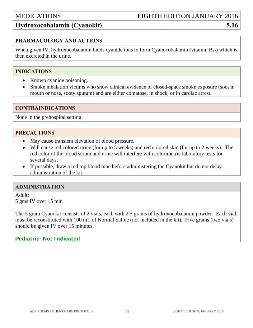
MEDICATIONS EIGHTH EDITION JANUARY 2016
Hydroxocobalamin (Cyanokit) 5.16
ADPH OEMS PATIENT CARE PROTOCOLS 152 EIGHTH EDITION, JANUARY 2016
PHARMACOLOGY AND ACTIONS
When given IV, hydroxocobalamin binds cyanide ions to form Cyanocobalamin (vitamin B12) which is then excreted in the urine.
INDICATIONS
Known cyanide poisoning. Smoke inhalation victims who show clinical evidence of closed-space smoke exposure (soot in
mouth or nose, sooty sputum) and are either comatose, in shock, or in cardiac arrest.
CONTRAINDICATIONS
None in the prehospital setting.
PRECAUTIONS
May cause transient elevation of blood pressure. Will cause red colored urine (for up to 5 weeks) and red colored skin (for up to 2 weeks). The
red color of the blood serum and urine will interfere with colorimetric laboratory tests for several days.
If possible, draw a red top blood tube before administering the Cyanokit but do not delay administration of the kit.
ADMINISTRATION
Adult: 5 gms IV over 15 min The 5 gram Cyanokit consists of 2 vials, each with 2.5 grams of hydroxocobalamin powder. Each vial must be reconstituted with 100 mL of Normal Saline (not included in the kit). Five grams (two vials) should be given IV over 15 minutes. Pediatric: Not Indicated
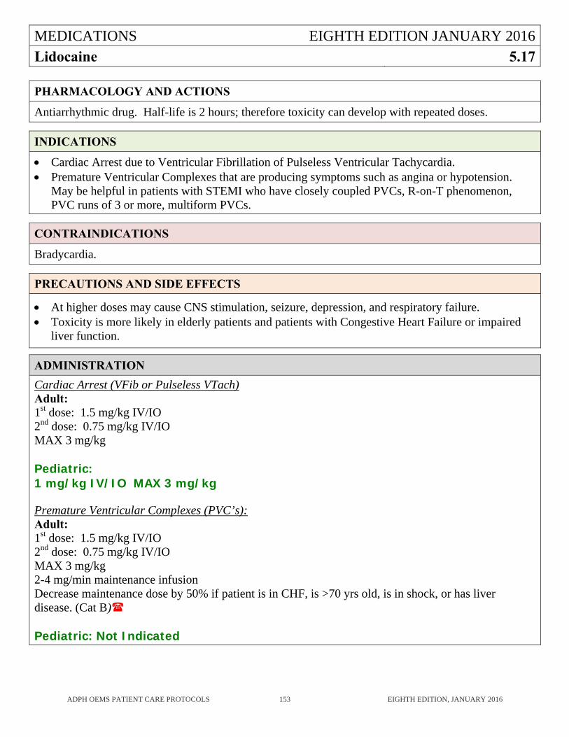
MEDICATIONS EIGHTH EDITION JANUARY 2016
Lidocaine 5.17
ADPH OEMS PATIENT CARE PROTOCOLS 153 EIGHTH EDITION, JANUARY 2016
PHARMACOLOGY AND ACTIONS
Antiarrhythmic drug. Half-life is 2 hours; therefore toxicity can develop with repeated doses.
INDICATIONS
Cardiac Arrest due to Ventricular Fibrillation of Pulseless Ventricular Tachycardia. Premature Ventricular Complexes that are producing symptoms such as angina or hypotension.
May be helpful in patients with STEMI who have closely coupled PVCs, R-on-T phenomenon, PVC runs of 3 or more, multiform PVCs.
CONTRAINDICATIONS
Bradycardia.
PRECAUTIONS AND SIDE EFFECTS
At higher doses may cause CNS stimulation, seizure, depression, and respiratory failure. Toxicity is more likely in elderly patients and patients with Congestive Heart Failure or impaired
liver function.
ADMINISTRATION
Cardiac Arrest (VFib or Pulseless VTach) Adult: 1st dose: 1.5 mg/kg IV/IO 2nd dose: 0.75 mg/kg IV/IO MAX 3 mg/kg Pediatric: 1 mg/kg IV/IO MAX 3 mg/kg Premature Ventricular Complexes (PVC’s): Adult: 1st dose: 1.5 mg/kg IV/IO 2nd dose: 0.75 mg/kg IV/IO MAX 3 mg/kg 2-4 mg/min maintenance infusion Decrease maintenance dose by 50% if patient is in CHF, is >70 yrs old, is in shock, or has liver disease. (Cat B)
Pediatric: Not Indicated
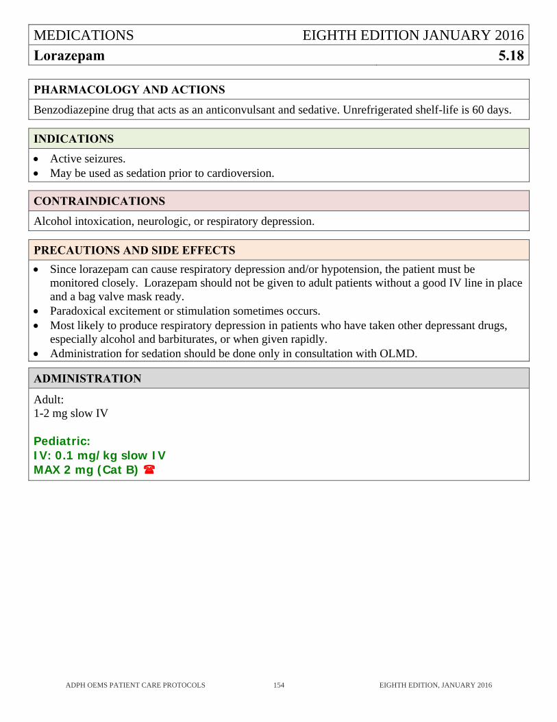
MEDICATIONS EIGHTH EDITION JANUARY 2016
Lorazepam 5.18
ADPH OEMS PATIENT CARE PROTOCOLS 154 EIGHTH EDITION, JANUARY 2016
PHARMACOLOGY AND ACTIONS
Benzodiazepine drug that acts as an anticonvulsant and sedative. Unrefrigerated shelf-life is 60 days.
INDICATIONS
Active seizures. May be used as sedation prior to cardioversion.
CONTRAINDICATIONS
Alcohol intoxication, neurologic, or respiratory depression.
PRECAUTIONS AND SIDE EFFECTS
Since lorazepam can cause respiratory depression and/or hypotension, the patient must be monitored closely. Lorazepam should not be given to adult patients without a good IV line in place and a bag valve mask ready.
Paradoxical excitement or stimulation sometimes occurs. Most likely to produce respiratory depression in patients who have taken other depressant drugs,
especially alcohol and barbiturates, or when given rapidly. Administration for sedation should be done only in consultation with OLMD.
ADMINISTRATION
Adult: 1-2 mg slow IV Pediatric: IV: 0.1 mg/kg slow IV MAX 2 mg (Cat B)
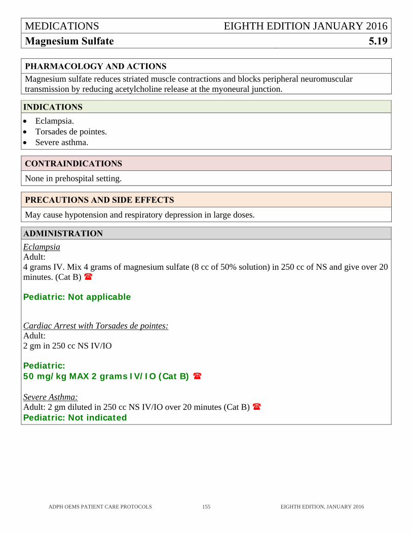
MEDICATIONS EIGHTH EDITION JANUARY 2016
Magnesium Sulfate 5.19
ADPH OEMS PATIENT CARE PROTOCOLS 155 EIGHTH EDITION, JANUARY 2016
PHARMACOLOGY AND ACTIONS
Magnesium sulfate reduces striated muscle contractions and blocks peripheral neuromuscular transmission by reducing acetylcholine release at the myoneural junction.
INDICATIONS
Eclampsia. Torsades de pointes. Severe asthma.
CONTRAINDICATIONS
None in prehospital setting.
PRECAUTIONS AND SIDE EFFECTS
May cause hypotension and respiratory depression in large doses.
ADMINISTRATION
Eclampsia Adult: 4 grams IV. Mix 4 grams of magnesium sulfate (8 cc of 50% solution) in 250 cc of NS and give over 20 minutes. (Cat B) Pediatric: Not applicable Cardiac Arrest with Torsades de pointes: Adult: 2 gm in 250 cc NS IV/IO
Pediatric: 50 mg/kg MAX 2 grams IV/IO (Cat B) Severe Asthma: Adult: 2 gm diluted in 250 cc NS IV/IO over 20 minutes (Cat B) Pediatric: Not indicated
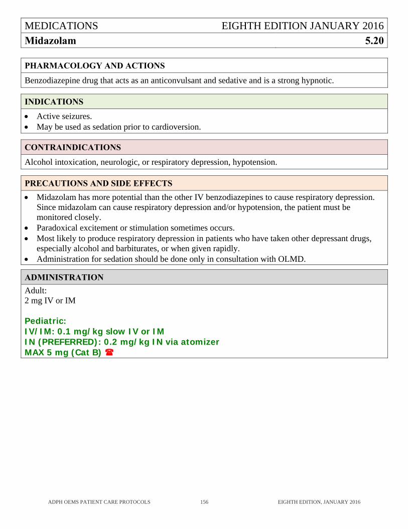
MEDICATIONS EIGHTH EDITION JANUARY 2016
Midazolam 5.20
ADPH OEMS PATIENT CARE PROTOCOLS 156 EIGHTH EDITION, JANUARY 2016
PHARMACOLOGY AND ACTIONS
Benzodiazepine drug that acts as an anticonvulsant and sedative and is a strong hypnotic.
INDICATIONS
Active seizures. May be used as sedation prior to cardioversion.
CONTRAINDICATIONS
Alcohol intoxication, neurologic, or respiratory depression, hypotension.
PRECAUTIONS AND SIDE EFFECTS
Midazolam has more potential than the other IV benzodiazepines to cause respiratory depression. Since midazolam can cause respiratory depression and/or hypotension, the patient must be monitored closely.
Paradoxical excitement or stimulation sometimes occurs. Most likely to produce respiratory depression in patients who have taken other depressant drugs,
especially alcohol and barbiturates, or when given rapidly. Administration for sedation should be done only in consultation with OLMD.
ADMINISTRATION
Adult: 2 mg IV or IM Pediatric: IV/IM: 0.1 mg/kg slow IV or IM IN (PREFERRED): 0.2 mg/kg IN via atomizer MAX 5 mg (Cat B)
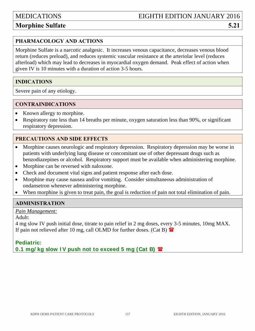
MEDICATIONS EIGHTH EDITION JANUARY 2016
Morphine Sulfate 5.21
ADPH OEMS PATIENT CARE PROTOCOLS 157 EIGHTH EDITION, JANUARY 2016
PHARMACOLOGY AND ACTIONS
Morphine Sulfate is a narcotic analgesic. It increases venous capacitance, decreases venous blood return (reduces preload), and reduces systemic vascular resistance at the arteriolar level (reduces afterload) which may lead to decreases in myocardial oxygen demand. Peak effect of action when given IV is 10 minutes with a duration of action 3-5 hours.
INDICATIONS
Severe pain of any etiology.
CONTRAINDICATIONS
Known allergy to morphine. Respiratory rate less than 14 breaths per minute, oxygen saturation less than 90%, or significant
respiratory depression.
PRECAUTIONS AND SIDE EFFECTS
Morphine causes neurologic and respiratory depression. Respiratory depression may be worse in patients with underlying lung disease or concomitant use of other depressant drugs such as benzodiazepines or alcohol. Respiratory support must be available when administering morphine.
Morphine can be reversed with naloxone. Check and document vital signs and patient response after each dose. Morphine may cause nausea and/or vomiting. Consider simultaneous administration of
ondansetron whenever administering morphine. When morphine is given to treat pain, the goal is reduction of pain not total elimination of pain.
ADMINISTRATION
Pain Management: Adult: 4 mg slow IV push initial dose, titrate to pain relief in 2 mg doses, every 3-5 minutes, 10mg MAX. If pain not relieved after 10 mg, call OLMD for further doses. (Cat B) Pediatric: 0.1 mg/kg slow IV push not to exceed 5 mg (Cat B)
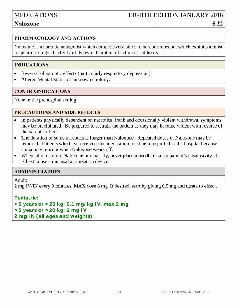
MEDICATIONS EIGHTH EDITION JANUARY 2016
Naloxone 5.22
ADPH OEMS PATIENT CARE PROTOCOLS 158 EIGHTH EDITION, JANUARY 2016
PHARMACOLOGY AND ACTIONS
Naloxone is a narcotic antagonist which competitively binds to narcotic sites but which exhibits almost no pharmacological activity of its own. Duration of action is 1-4 hours.
INDICATIONS
Reversal of narcotic effects (particularly respiratory depression). Altered Mental Status of unknown etiology.
CONTRAINDICATIONS
None in the prehospital setting.
PRECAUTIONS AND SIDE EFFECTS
In patients physically dependent on narcotics, frank and occasionally violent withdrawal symptoms may be precipitated. Be prepared to restrain the patient as they may become violent with reverse of the narcotic effect.
The duration of some narcotics is longer than Naloxone. Repeated doses of Naloxone may be required. Patients who have received this medication must be transported to the hospital because coma may reoccur when Naloxone wears off.
When administering Naloxone intranasally, never place a needle inside a patient’s nasal cavity. It is best to use a mucosal atomization device.
ADMINISTRATION
Adult: 2 mg IV/IN every 3 minutes, MAX dose 8 mg. If desired, start by giving 0.5 mg and titrate to effect. Pediatric: <5 years or <20 kg: 0.1 mg/kg IV, max 2 mg >5 years or >20 kg: 2 mg IV 2 mg IN (all ages and weights)
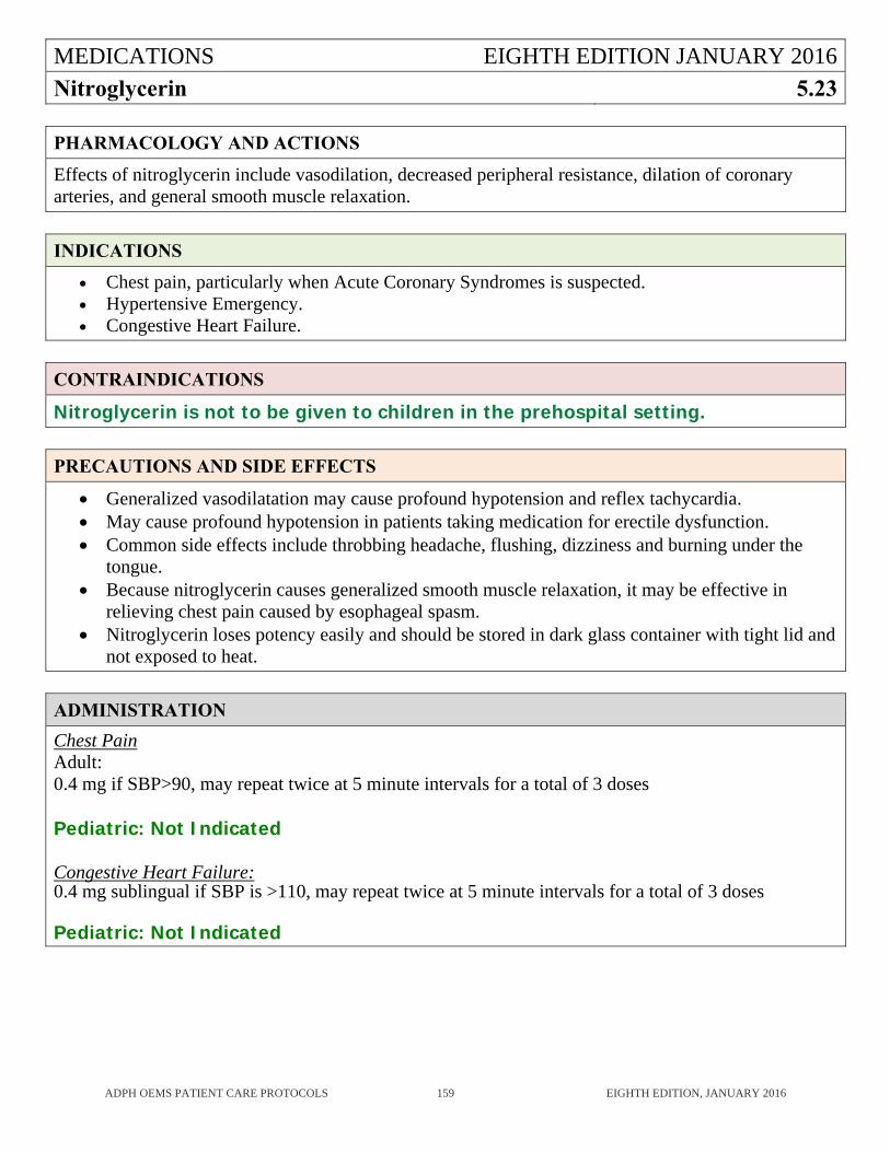
MEDICATIONS EIGHTH EDITION JANUARY 2016
Nitroglycerin 5.23
ADPH OEMS PATIENT CARE PROTOCOLS 159 EIGHTH EDITION, JANUARY 2016
PHARMACOLOGY AND ACTIONS
Effects of nitroglycerin include vasodilation, decreased peripheral resistance, dilation of coronary arteries, and general smooth muscle relaxation.
INDICATIONS
Chest pain, particularly when Acute Coronary Syndromes is suspected. Hypertensive Emergency. Congestive Heart Failure.
CONTRAINDICATIONS
Nitroglycerin is not to be given to children in the prehospital setting.
PRECAUTIONS AND SIDE EFFECTS
Generalized vasodilatation may cause profound hypotension and reflex tachycardia. May cause profound hypotension in patients taking medication for erectile dysfunction. Common side effects include throbbing headache, flushing, dizziness and burning under the
tongue. Because nitroglycerin causes generalized smooth muscle relaxation, it may be effective in
relieving chest pain caused by esophageal spasm. Nitroglycerin loses potency easily and should be stored in dark glass container with tight lid and
not exposed to heat.
ADMINISTRATION
Chest Pain Adult: 0.4 mg if SBP>90, may repeat twice at 5 minute intervals for a total of 3 doses Pediatric: Not Indicated Congestive Heart Failure: 0.4 mg sublingual if SBP is >110, may repeat twice at 5 minute intervals for a total of 3 doses Pediatric: Not Indicated
MEDICATIONS EIGHTH EDITION JANUARY 2016
Nitrous Oxide 5.24
ADPH OEMS PATIENT CARE PROTOCOLS 160 EIGHTH EDITION, JANUARY 2016
PHARMACOLOGY AND ACTIONS
Nitrous Oxide is a blended mixture of 50% nitrous oxide and 50% oxygen which has potent analgesic effects. The high concentration of oxygen delivered with the nitrous oxide will increase the amount of oxygen in the blood.
INDICATIONS
Severe pain.
CONTRAINDICATIONS
Patients who cannot comprehend verbal instructions. Patients with altered mental status. Patients with suspected pneumothorax. Patients with abdominal pain. Patients who have COPD where the high oxygen concentration may depress ventilatory effort.
PRECAUTIONS AND SIDE EFFECTS
It is essential that Nitrous Oxide be self-administered. May cause nausea and vomiting.
ADMINISTRATION
Adult : Self-administer until the pain is significantly relieved or until patient drops the mask. (Cat B) Pediatric: Consult with OLMD. (Cat B)
MEDICATIONS EIGHTH EDITION JANUARY 2016
Normal Saline 5.25
ADPH OEMS PATIENT CARE PROTOCOLS 161 EIGHTH EDITION, JANUARY 2016
PHARMACOLOGY AND ACTIONS
0.9% Saline solution.
INDICATIONS
Normal Saline is indicated for replacement of fluid volume losses such as in trauma, burns, dehydration, or shock, and is the only IV fluid authorized by these protocols. Where IVs are used to maintain venous access, a heparin or saline lock may be substituted. They must be properly maintained to prevent occlusion.
CONTRAINDICATIONS
None in the prehospital setting.
PRECAUTIONS AND SIDE EFFECTS
In patients in which fluid overload is a problem, Normal Saline may be used with a microdrip, and this microdrip may be used to administer prehospital medications; also consider the use of a saline lock.
ADMINISTRATION
Adult: Rate and amount to be given varies with the specific protocol. Pediatric: Rate and amount to be given varies with the specific protocol.
MEDICATIONS EIGHTH EDITION JANUARY 2016
Ondansetron 5.26
ADPH OEMS PATIENT CARE PROTOCOLS 162 EIGHTH EDITION, JANUARY 2016
PHARMACOLOGY AND ACTIONS
Ondansetron acts as an antiemetic by selectively antagonizing serotonin 5-HT3.
INDICATIONS
Nausea or vomiting.
CONTRAINDICATIONS
Allergy to Ondansetron. Age less than one month.
PRECAUTIONS AND SIDE EFFECTS
Can rarely cause extrapyramidal symptoms.
ADMINISTRATION
Adult: 4 mg IV/IM or ODT (Orally Dissolving Tablet) Pediatric (1 month to 12 years): 0.1mg/kg IV/IM or ODT MAX dose 4 mg (Cat B)
MEDICATIONS EIGHTH EDITION JANUARY 2016
Oxygen 5.27
ADPH OEMS PATIENT CARE PROTOCOLS 163 EIGHTH EDITION, JANUARY 2016
PHARMACOLOGY AND ACTIONS
Oxygen added to the inspired air raises the amount of oxygen in the blood and, therefore, the amount delivered to the tissues.
INDICATIONS
Hypoxia or respiratory distress from any cause. Acute chest pain in which Acute Coronary Syndrome. Shock (decreased oxygenation of tissues) from any cause. Major trauma. Carbon monoxide poisoning.
CONTRAINDICATIONS
None in prehospital setting.
PRECAUTIONS AND SIDE EFFECTS
If the patient is not breathing adequately, the EMSP must assist their ventilations. Provision of oxygen alone is not enough.
A small percentage of patients with chronic lung disease breathe because they are hypoxic. Administration of oxygen may abolish their respiratory drive. Do not withhold oxygen because of this possibility, however, be prepared to assist ventilations. Monitor oxygen saturation with a pulse oximeter and, if available, monitor ventilations using capnography. Use just enough oxygen to maintain pulse oximeter reading of >95%.
Restlessness may be an important sign of hypoxia.
ADMINISTRATION
Method Flow Rate O2% Inspired Air
Room Air 21%
Nasal Cannula (prongs) 1 L/min 24%
2 L/min 28%
Face Mask 6 L/min 44%
Oxygen Reservoir (mask) 10-12 L/min 90%
Bag-Valve Mask with 100% valve and reservoir
High glow regulated to inflate reservoir at proper rate
90%+
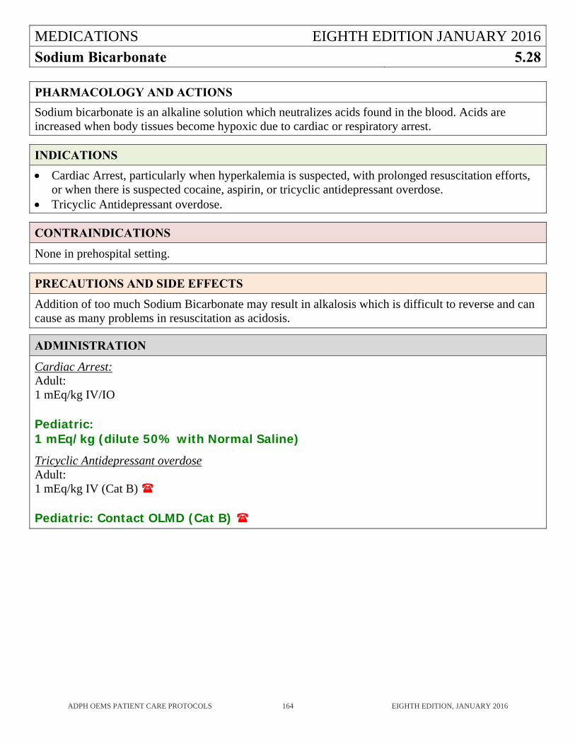
MEDICATIONS EIGHTH EDITION JANUARY 2016
Sodium Bicarbonate 5.28
ADPH OEMS PATIENT CARE PROTOCOLS 164 EIGHTH EDITION, JANUARY 2016
PHARMACOLOGY AND ACTIONS
Sodium bicarbonate is an alkaline solution which neutralizes acids found in the blood. Acids are increased when body tissues become hypoxic due to cardiac or respiratory arrest.
INDICATIONS
Cardiac Arrest, particularly when hyperkalemia is suspected, with prolonged resuscitation efforts, or when there is suspected cocaine, aspirin, or tricyclic antidepressant overdose.
Tricyclic Antidepressant overdose.
CONTRAINDICATIONS
None in prehospital setting.
PRECAUTIONS AND SIDE EFFECTS
Addition of too much Sodium Bicarbonate may result in alkalosis which is difficult to reverse and can cause as many problems in resuscitation as acidosis.
ADMINISTRATION
Cardiac Arrest: Adult: 1 mEq/kg IV/IO Pediatric: 1 mEq/kg (dilute 50% with Normal Saline) Tricyclic Antidepressant overdose Adult: 1 mEq/kg IV (Cat B) Pediatric: Contact OLMD (Cat B)
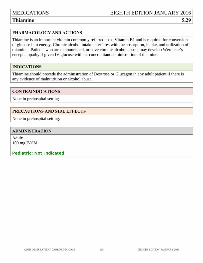
MEDICATIONS EIGHTH EDITION JANUARY 2016
Thiamine 5.29
ADPH OEMS PATIENT CARE PROTOCOLS 165 EIGHTH EDITION, JANUARY 2016
PHARMACOLOGY AND ACTIONS
Thiamine is an important vitamin commonly referred to as Vitamin B1 and is required for conversion of glucose into energy. Chronic alcohol intake interferes with the absorption, intake, and utilization of thiamine. Patients who are malnourished, or have chronic alcohol abuse, may develop Wernicke’s encephalopathy if given IV glucose without concomitant administration of thiamine.
INDICATIONS
Thiamine should precede the administration of Dextrose or Glucagon in any adult patient if there is any evidence of malnutrition or alcohol abuse.
CONTRAINDICATIONS
None in prehospital setting.
PRECAUTIONS AND SIDE EFFECTS
None in prehospital setting.
ADMINISTRATION
Adult: 100 mg IV/IM Pediatric: Not Indicated
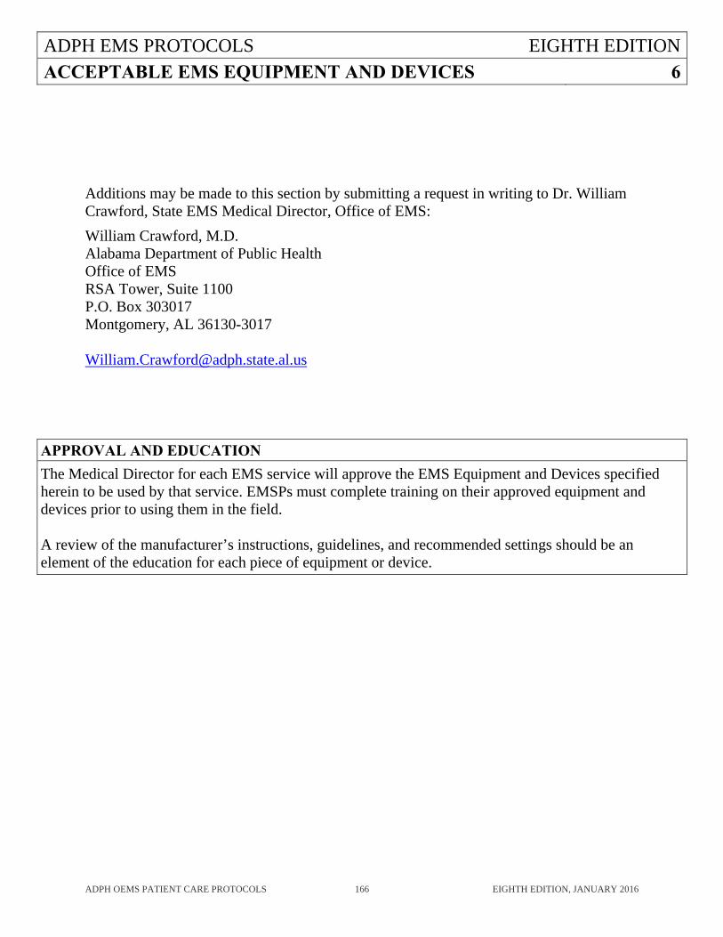
ADPH EMS PROTOCOLS EIGHTH EDITION
ACCEPTABLE EMS EQUIPMENT AND DEVICES 6
ADPH OEMS PATIENT CARE PROTOCOLS 166 EIGHTH EDITION, JANUARY 2016
Additions may be made to this section by submitting a request in writing to Dr. William Crawford, State EMS Medical Director, Office of EMS:
William Crawford, M.D. Alabama Department of Public Health Office of EMS RSA Tower, Suite 1100 P.O. Box 303017 Montgomery, AL 36130-3017 [email protected]
APPROVAL AND EDUCATION
The Medical Director for each EMS service will approve the EMS Equipment and Devices specified herein to be used by that service. EMSPs must complete training on their approved equipment and devices prior to using them in the field. A review of the manufacturer’s instructions, guidelines, and recommended settings should be an element of the education for each piece of equipment or device.
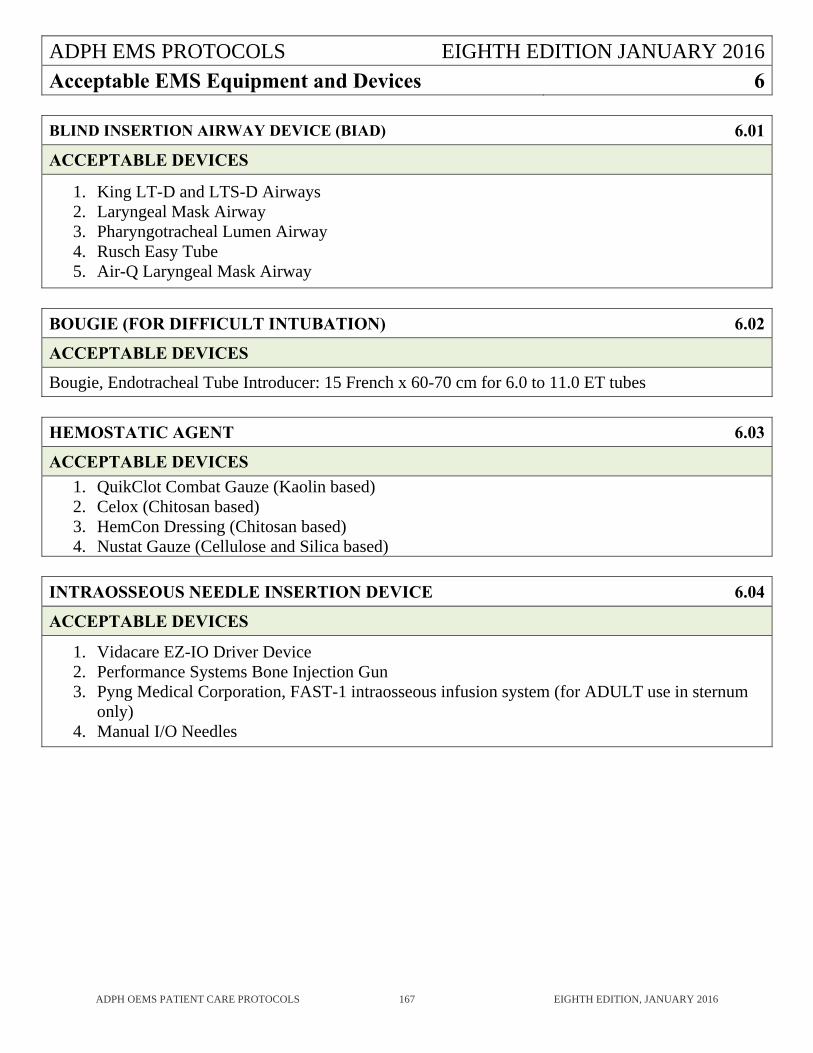
ADPH EMS PROTOCOLS EIGHTH EDITION JANUARY 2016
Acceptable EMS Equipment and Devices 6
ADPH OEMS PATIENT CARE PROTOCOLS 167 EIGHTH EDITION, JANUARY 2016
BLIND INSERTION AIRWAY DEVICE (BIAD) 6.01
ACCEPTABLE DEVICES
1. King LT-D and LTS-D Airways 2. Laryngeal Mask Airway 3. Pharyngotracheal Lumen Airway 4. Rusch Easy Tube 5. Air-Q Laryngeal Mask Airway
BOUGIE (FOR DIFFICULT INTUBATION) 6.02
ACCEPTABLE DEVICES
Bougie, Endotracheal Tube Introducer: 15 French x 60-70 cm for 6.0 to 11.0 ET tubes
HEMOSTATIC AGENT 6.03
ACCEPTABLE DEVICES
1. QuikClot Combat Gauze (Kaolin based) 2. Celox (Chitosan based) 3. HemCon Dressing (Chitosan based) 4. Nustat Gauze (Cellulose and Silica based)
INTRAOSSEOUS NEEDLE INSERTION DEVICE 6.04
ACCEPTABLE DEVICES
1. Vidacare EZ-IO Driver Device 2. Performance Systems Bone Injection Gun 3. Pyng Medical Corporation, FAST-1 intraosseous infusion system (for ADULT use in sternum
only) 4. Manual I/O Needles

ADPH EMS PROTOCOLS EIGHTH EDITION
DISASTER 7
ADPH OEMS PATIENT CARE PROTOCOLS 168 EIGHTH EDITION, JANUARY 2016
DISASTER EIGHTH EDITION JANUARY 2016Respiratory Illness/Influenza Mass Casualty Emergency
7.01
ADPH OEMS PATIENT CARE PROTOCOLS 169 EIGHTH EDITION, JANUARY 2016
DISASTER PROTOCOL IMPLEMENTATION
This protocol is to be implemented only when there is a significant respiratory disease that has impacted the health care system to the extent that hospital beds are full, few or no ventilators are available for new patients with respiratory failure, the EMS/Dispatch work force is significantly depleted due to absenteeism, and the calls for EMS support overwhelm resources to manage all calls. When the Governor proclaims a state of emergency, the ADPH OEMS will activate this protocol to provide authorization for the adjustment in the prehospital standard of care. Depending upon the Governor’s proclamation, ADPH OEMS may activate this protocol statewide or on a regional or local basis.
KEY POINTS
Treat patients using the Respiratory Illness/Influenza Protocol (3.28). Endotracheal intubation should not be performed on any patient except by direct order of the
OLMD physician. (Cat B) Because of the danger of EMS personnel becoming infected, aerosol-generating procedures
such as advanced airway procedures, use of bag-mask, and nebulizer treatments should not be performed on patients with acute febrile respiratory illness except by direct order of the OLMD physician (Cat B). CPAP when used with an expiratory filter remains Category A and does not require OLMD orders.
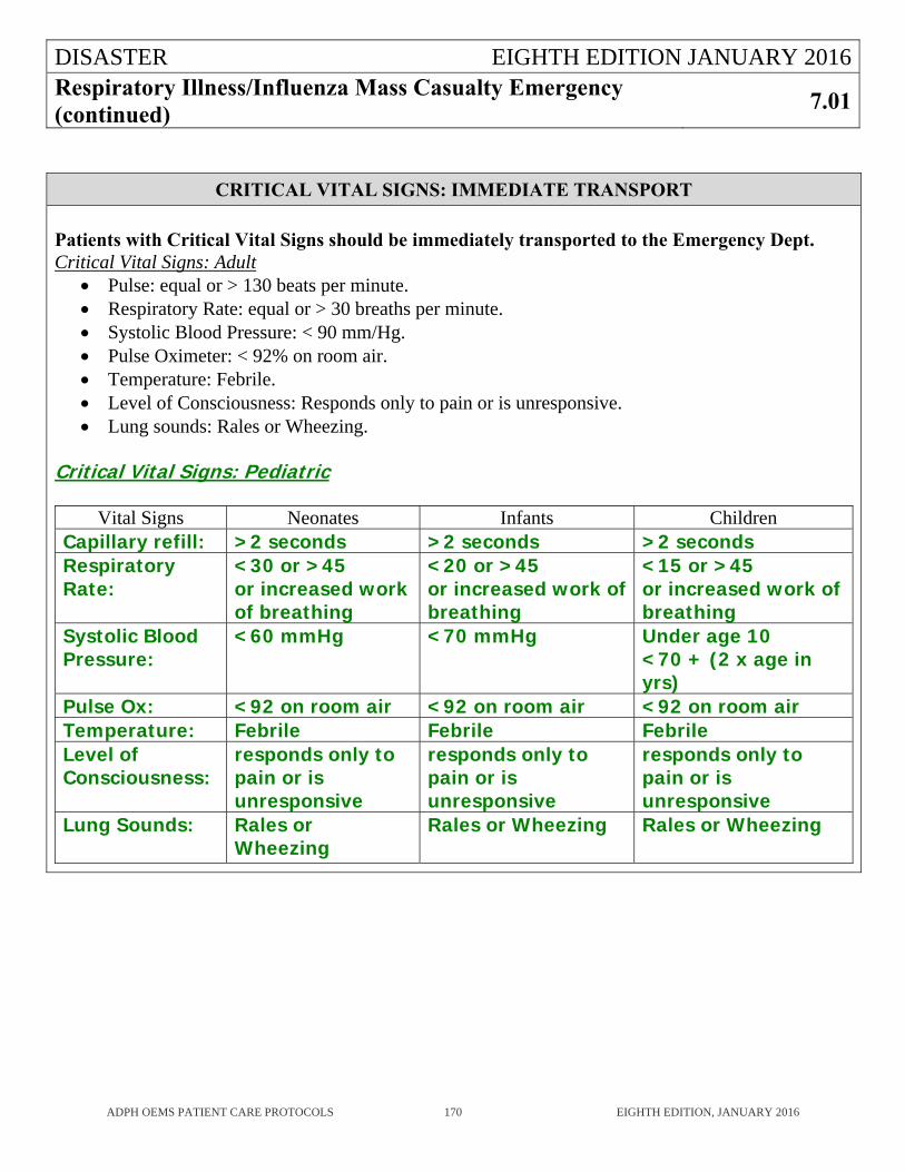
DISASTER EIGHTH EDITION JANUARY 2016Respiratory Illness/Influenza Mass Casualty Emergency (continued)
7.01
ADPH OEMS PATIENT CARE PROTOCOLS 170 EIGHTH EDITION, JANUARY 2016
CRITICAL VITAL SIGNS: IMMEDIATE TRANSPORT
Patients with Critical Vital Signs should be immediately transported to the Emergency Dept. Critical Vital Signs: Adult
Pulse: equal or > 130 beats per minute. Respiratory Rate: equal or > 30 breaths per minute. Systolic Blood Pressure: < 90 mm/Hg. Pulse Oximeter: < 92% on room air. Temperature: Febrile. Level of Consciousness: Responds only to pain or is unresponsive. Lung sounds: Rales or Wheezing.
Critical Vital Signs: Pediatric
Vital Signs Neonates Infants Children Capillary refill: >2 seconds >2 seconds >2 secondsRespiratory Rate:
<30 or >45 or increased work of breathing
<20 or >45or increased work of breathing
<15 or >45or increased work of breathing
Systolic Blood Pressure:
<60 mmHg <70 mmHg Under age 10<70 + (2 x age in yrs)
Pulse Ox: <92 on room air <92 on room air <92 on room airTemperature: Febrile Febrile Febrile Level of Consciousness:
responds only to pain or is unresponsive
responds only to pain or is unresponsive
responds only to pain or is unresponsive
Lung Sounds: Rales or Wheezing
Rales or Wheezing Rales or Wheezing
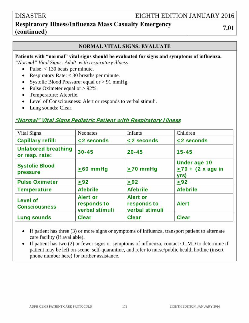
DISASTER EIGHTH EDITION JANUARY 2016Respiratory Illness/Influenza Mass Casualty Emergency (continued)
7.01
ADPH OEMS PATIENT CARE PROTOCOLS 171 EIGHTH EDITION, JANUARY 2016
NORMAL VITAL SIGNS: EVALUATE
Patients with “normal” vital signs should be evaluated for signs and symptoms of influenza. “Normal” Vital Signs: Adult with respiratory illness
Pulse: < 130 beats per minute. Respiratory Rate: < 30 breaths per minute. Systolic Blood Pressure: equal or > 91 mmHg. Pulse Oximeter equal or > 92%. Temperature: Afebrile. Level of Consciousness: Alert or responds to verbal stimuli. Lung sounds: Clear.
“Normal” Vital Signs Pediatric Patient with Respiratory Illness Vital Signs Neonates Infants Children
Capillary refill: <2 seconds <2 seconds <2 seconds Unlabored breathing or resp. rate: 30-45 20-45 15-45
Systolic Blood pressure >60 mmHg >70 mmHg
Under age 10>70 + (2 x age in yrs)
Pulse Oximeter >92 >92 >92 Temperature Afebrile Afebrile Afebrile
Level of Consciousness
Alert or responds to verbal stimuli
Alert or responds to verbal stimuli
Alert
Lung sounds Clear Clear Clear
If patient has three (3) or more signs or symptoms of influenza, transport patient to alternate care facility (if available).
If patient has two (2) or fewer signs or symptoms of influenza, contact OLMD to determine if patient may be left on-scene, self-quarantine, and refer to nurse/public health hotline (insert phone number here) for further assistance.
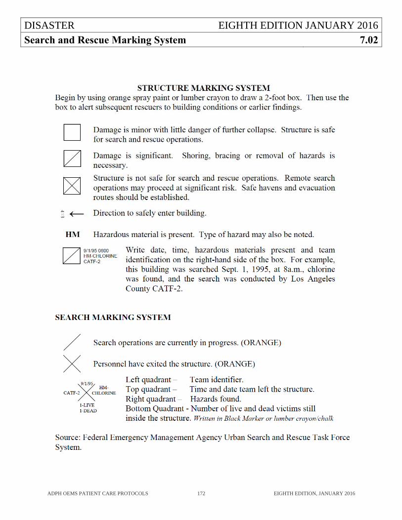
DISASTER EIGHTH EDITION JANUARY 2016
Search and Rescue Marking System 7.02
ADPH OEMS PATIENT CARE PROTOCOLS 172 EIGHTH EDITION, JANUARY 2016
DISASTER EIGHTH EDITION JANUARY 2016
Triage of Mass Casualties 7.03
ADPH OEMS PATIENT CARE PROTOCOLS 173 EIGHTH EDITION, JANUARY 2016
GUIDELINE
This protocol is to be implemented only when there is a mass casualty incident where the number of patients exceeds the capabilities of the EMS providers on the scene. As situations change and resources vary, periodic re-triage may be appropriate. This protocol is only meant to be a guideline. If desired, the EMSP may use a different mass casualty triage algorithm if it has been approved by his/her EMS agency.
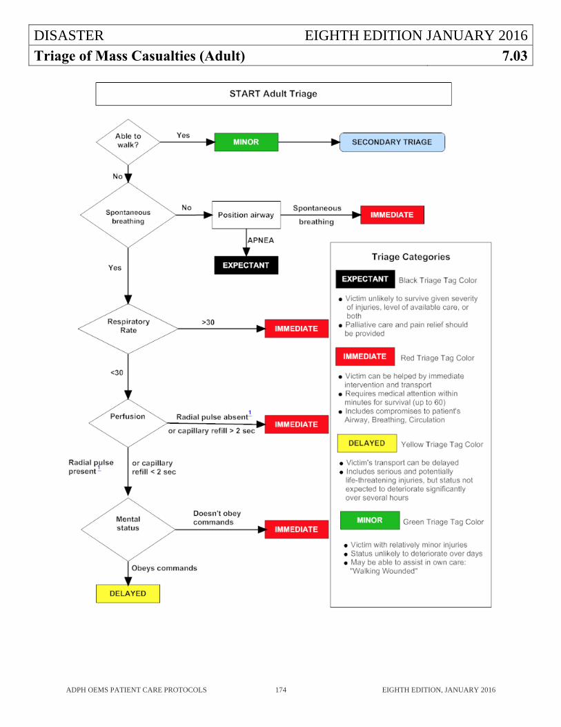
DISASTER EIGHTH EDITION JANUARY 2016
Triage of Mass Casualties (Adult) 7.03
ADPH OEMS PATIENT CARE PROTOCOLS 174 EIGHTH EDITION, JANUARY 2016
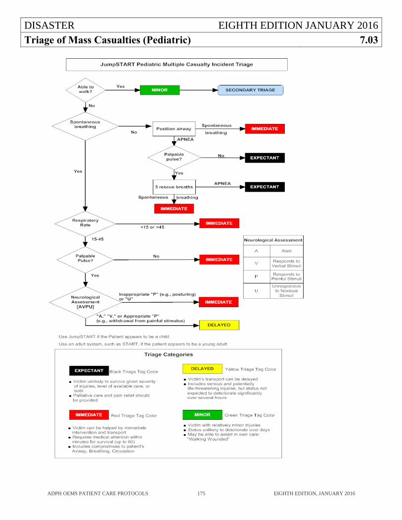
DISASTER EIGHTH EDITION JANUARY 2016
Triage of Mass Casualties (Pediatric) 7.03
ADPH OEMS PATIENT CARE PROTOCOLS 175 EIGHTH EDITION, JANUARY 2016

ADPH EMS PROTOCOLS EIGHTH EDITION
FORMS 8
ADPH OEMS PATIENT CARE PROTOCOLS 176 EIGHTH EDITION, JANUARY 2016
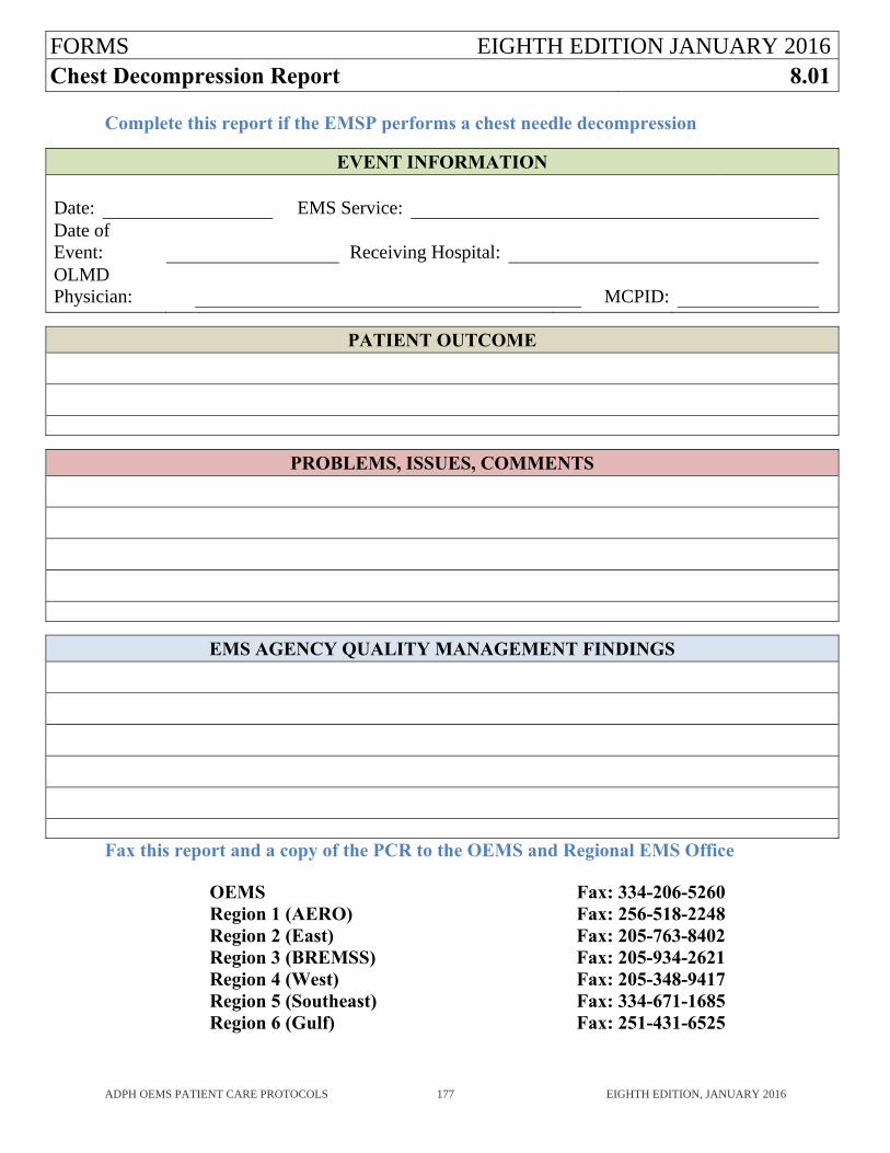
FORMS EIGHTH EDITION JANUARY 2016Chest Decompression Report 8.01
ADPH OEMS PATIENT CARE PROTOCOLS 177 EIGHTH EDITION, JANUARY 2016
Complete this report if the EMSP performs a chest needle decompression
EVENT INFORMATION
Date: EMS Service:Date of Event: Receiving Hospital:OLMD Physician: MCPID:
PATIENT OUTCOME
PROBLEMS, ISSUES, COMMENTS
EMS AGENCY QUALITY MANAGEMENT FINDINGS
Fax this report and a copy of the PCR to the OEMS and Regional EMS Office OEMS Fax: 334-206-5260 Region 1 (AERO) Fax: 256-518-2248 Region 2 (East) Fax: 205-763-8402 Region 3 (BREMSS) Fax: 205-934-2621 Region 4 (West) Fax: 205-348-9417 Region 5 (Southeast) Fax: 334-671-1685 Region 6 (Gulf) Fax: 251-431-6525
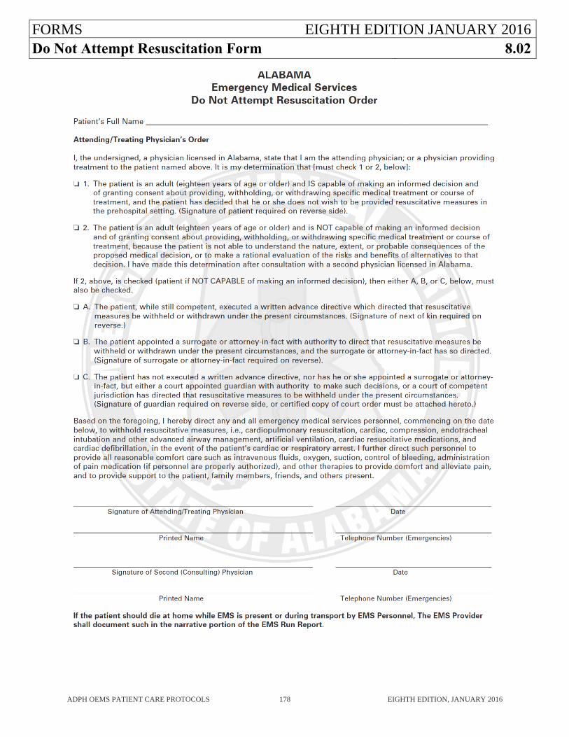
FORMS EIGHTH EDITION JANUARY 2016Do Not Attempt Resuscitation Form 8.02
ADPH OEMS PATIENT CARE PROTOCOLS 178 EIGHTH EDITION, JANUARY 2016
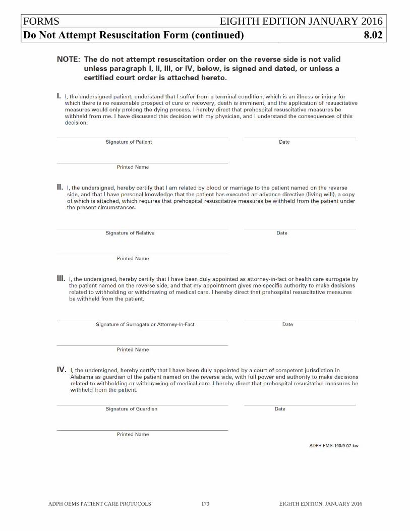
FORMS EIGHTH EDITION JANUARY 2016Do Not Attempt Resuscitation Form (continued) 8.02
ADPH OEMS PATIENT CARE PROTOCOLS 179 EIGHTH EDITION, JANUARY 2016
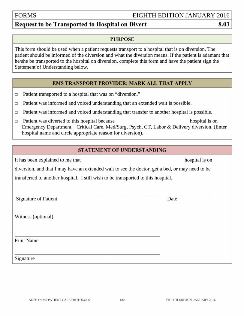
FORMS EIGHTH EDITION JANUARY 2016
Request to be Transported to Hospital on Divert 8.03
ADPH OEMS PATIENT CARE PROTOCOLS 180 EIGHTH EDITION, JANUARY 2016
PURPOSE
This form should be used when a patient requests transport to a hospital that is on diversion. The patient should be informed of the diversion and what the diversion means. If the patient is adamant that he/she be transported to the hospital on diversion, complete this form and have the patient sign the Statement of Understanding below.
EMS TRANSPORT PROVIDER: MARK ALL THAT APPLY
□ Patient transported to a hospital that was on “diversion.”
□ Patient was informed and voiced understanding that an extended wait is possible.
□ Patient was informed and voiced understanding that transfer to another hospital is possible.
□ Patient was diverted to this hospital because ____________________________ hospital is on Emergency Department, Critical Care, Med/Surg, Psych, CT, Labor & Delivery diversion. (Enter hospital name and circle appropriate reason for diversion).
STATEMENT OF UNDERSTANDING
It has been explained to me that _______________________________________ hospital is on
diversion, and that I may have an extended wait to see the doctor, get a bed, or may need to be
transferred to another hospital. I still wish to be transported to this hospital.
_______________________________________________________ ________________ Signature of Patient Date Witness (optional) ________________________________________________________ Print Name ________________________________________________________ Signature
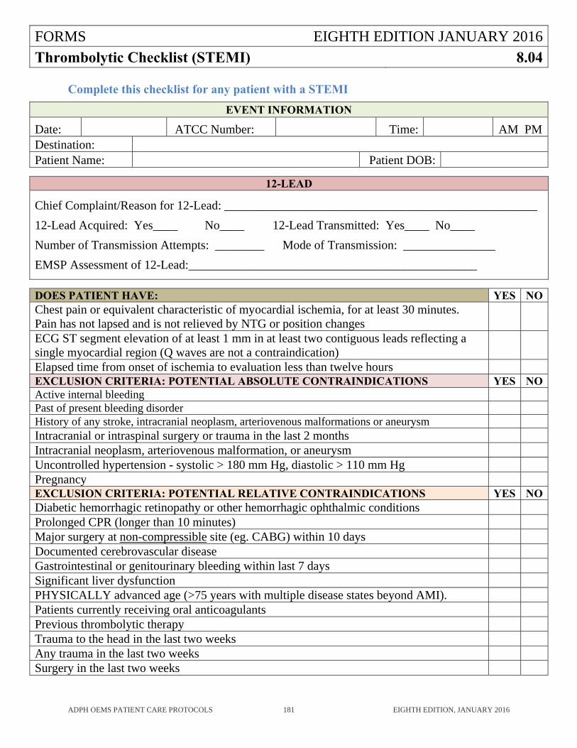
FORMS EIGHTH EDITION JANUARY 2016
Thrombolytic Checklist (STEMI) 8.04
ADPH OEMS PATIENT CARE PROTOCOLS 181 EIGHTH EDITION, JANUARY 2016
Complete this checklist for any patient with a STEMI
EVENT INFORMATION
Date: ATCC Number: Time: AM PMDestination: Patient Name: Patient DOB:
12-LEAD
Chief Complaint/Reason for 12-Lead: ___________________________________________________
12-Lead Acquired: Yes____ No____ 12-Lead Transmitted: Yes____ No____
Number of Transmission Attempts: ________ Mode of Transmission: _______________
EMSP Assessment of 12-Lead:_______________________________________________
DOES PATIENT HAVE: YES NOChest pain or equivalent characteristic of myocardial ischemia, for at least 30 minutes. Pain has not lapsed and is not relieved by NTG or position changes
ECG ST segment elevation of at least 1 mm in at least two contiguous leads reflecting a single myocardial region (Q waves are not a contraindication)
Elapsed time from onset of ischemia to evaluation less than twelve hours EXCLUSION CRITERIA: POTENTIAL ABSOLUTE CONTRAINDICATIONS YES NOActive internal bleeding Past of present bleeding disorder History of any stroke, intracranial neoplasm, arteriovenous malformations or aneurysm Intracranial or intraspinal surgery or trauma in the last 2 months Intracranial neoplasm, arteriovenous malformation, or aneurysm Uncontrolled hypertension - systolic > 180 mm Hg, diastolic > 110 mm Hg Pregnancy EXCLUSION CRITERIA: POTENTIAL RELATIVE CONTRAINDICATIONS YES NODiabetic hemorrhagic retinopathy or other hemorrhagic ophthalmic conditions Prolonged CPR (longer than 10 minutes) Major surgery at non-compressible site (eg. CABG) within 10 days Documented cerebrovascular disease Gastrointestinal or genitourinary bleeding within last 7 days Significant liver dysfunction PHYSICALLY advanced age (>75 years with multiple disease states beyond AMI). Patients currently receiving oral anticoagulants Previous thrombolytic therapy Trauma to the head in the last two weeks Any trauma in the last two weeks Surgery in the last two weeks
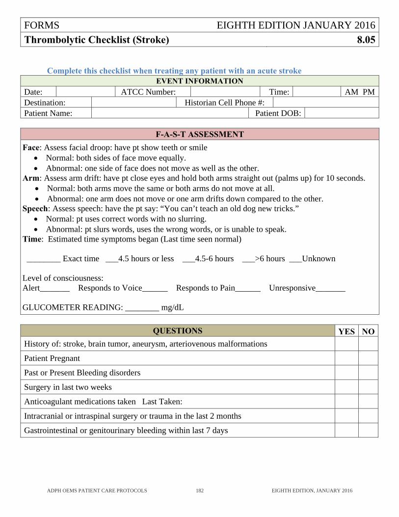
FORMS EIGHTH EDITION JANUARY 2016
Thrombolytic Checklist (Stroke) 8.05
ADPH OEMS PATIENT CARE PROTOCOLS 182 EIGHTH EDITION, JANUARY 2016
Complete this checklist when treating any patient with an acute stroke
EVENT INFORMATION
Date: ATCC Number: Time: AM PMDestination: Historian Cell Phone #: Patient Name: Patient DOB:
F-A-S-T ASSESSMENT
Face: Assess facial droop: have pt show teeth or smile Normal: both sides of face move equally. Abnormal: one side of face does not move as well as the other.
Arm: Assess arm drift: have pt close eyes and hold both arms straight out (palms up) for 10 seconds. Normal: both arms move the same or both arms do not move at all. Abnormal: one arm does not move or one arm drifts down compared to the other.
Speech: Assess speech: have the pt say: “You can’t teach an old dog new tricks.” Normal: pt uses correct words with no slurring. Abnormal: pt slurs words, uses the wrong words, or is unable to speak.
Time: Estimated time symptoms began (Last time seen normal) ________ Exact time ___4.5 hours or less ___4.5-6 hours ___>6 hours ___Unknown Level of consciousness: Alert_______ Responds to Voice______ Responds to Pain______ Unresponsive_______ GLUCOMETER READING: ________ mg/dL
QUESTIONS YES NO
History of: stroke, brain tumor, aneurysm, arteriovenous malformations
Patient Pregnant
Past or Present Bleeding disorders
Surgery in last two weeks
Anticoagulant medications taken Last Taken:
Intracranial or intraspinal surgery or trauma in the last 2 months
Gastrointestinal or genitourinary bleeding within last 7 days
ADPH EMS PROTOCOLS EIGHTH EDITION JANUARY 2016
Expanded Scope of Practice 9
ADPH OEMS PATIENT CARE PROTOCOLS 183 EIGHTH EDITION, JANUARY 2016
The Expanded Scope of Practice section of the ADPH EMS Protocols is ONLY to be utilized by those ALS Transport EMS agencies that have been specifically approved to do so by the ADPH Office of EMS. Any EMS agency that meets the specified criteria contained in these protocols may apply for these privileges. The service Medical Director for any EMS agency approved to utilize the Expanded Scope of Practice protocols must have a written plan for implementation of the Expanded Scope of Practice that must be approved by the ADPH Office of EMS. The service Medical Director for any EMS agency approved to utilize the Expanded Scope of Practice protocols must attend in person or by phone a quarterly Expanded Scope of Practice Quality Assurance conference to be coordinated by the ADPH OEMS. The patient care report for every case utilizing the Expanded Scope of Practice must be reviewed by the service Medical Director and must be sent to the ADPH OEMS for review by the State EMS Medical Director.
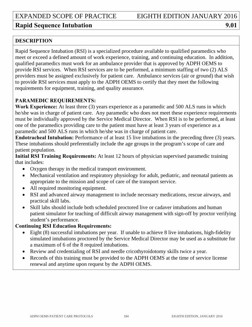
EXPANDED SCOPE OF PRACTICE EIGHTH EDITION JANUARY 2016Rapid Sequence Intubation 9.01
ADPH OEMS PATIENT CARE PROTOCOLS 184 EIGHTH EDITION, JANUARY 2016
DESCRIPTION
Rapid Sequence Intubation (RSI) is a specialized procedure available to qualified paramedics who meet or exceed a defined amount of work experience, training, and continuing education. In addition, qualified paramedics must work for an ambulance provider that is approved by ADPH OEMS to provide RSI services. When RSI services are to be performed, a minimum staffing of two (2) ALS providers must be assigned exclusively for patient care. Ambulance services (air or ground) that wish to provide RSI services must apply to the ADPH OEMS to certify that they meet the following requirements for equipment, training, and quality assurance. PARAMEDIC REQUIREMENTS: Work Experience: At least three (3) years experience as a paramedic and 500 ALS runs in which he/she was in charge of patient care. Any paramedic who does not meet these experience requirements must be individually approved by the Service Medical Director. When RSI is to be performed, at least one of the paramedics providing care to the patient must have at least 3 years of experience as a paramedic and 500 ALS runs in which he/she was in charge of patient care. Endotracheal Intubation: Performance of at least 15 live intubations in the preceding three (3) years. These intubations should preferentially include the age groups in the program’s scope of care and patient population. Initial RSI Training Requirements: At least 12 hours of physician supervised paramedic training that includes:
Oxygen therapy in the medical transport environment. Mechanical ventilation and respiratory physiology for adult, pediatric, and neonatal patients as
appropriate to the mission and scope of care of the transport service. All required monitoring equipment. RSI and advanced airway management to include necessary medications, rescue airways, and
practical skill labs. Skill labs should include both scheduled proctored live or cadaver intubations and human
patient simulator for teaching of difficult airway management with sign-off by proctor verifying student’s performance.
Continuing RSI Education Requirements: Eight (8) successful intubations per year. If unable to achieve 8 live intubations, high-fidelity
simulated intubations proctored by the Service Medical Director may be used as a substitute for a maximum of 6 of the 8 required intubations.
Review and credentialing of RSI and needle cricothyroidotomy skills twice a year. Records of this training must be provided to the ADPH OEMS at the time of service license
renewal and anytime upon request by the ADPH OEMS.
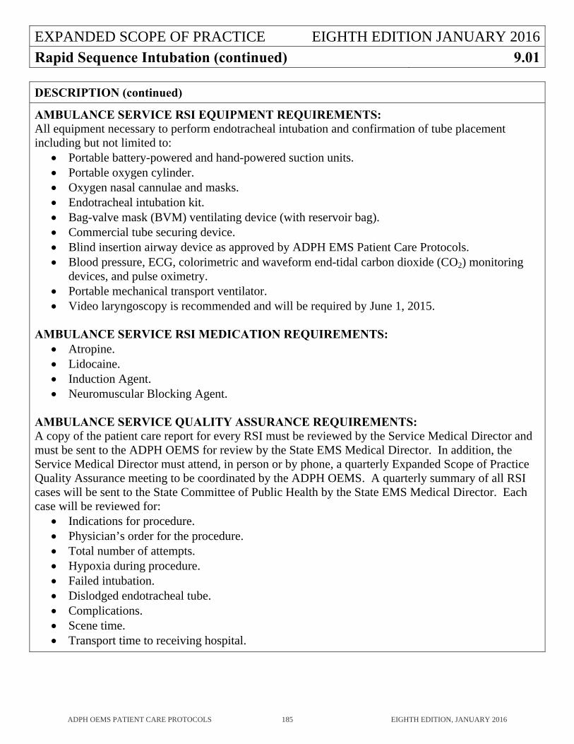
EXPANDED SCOPE OF PRACTICE EIGHTH EDITION JANUARY 2016Rapid Sequence Intubation (continued) 9.01
ADPH OEMS PATIENT CARE PROTOCOLS 185 EIGHTH EDITION, JANUARY 2016
DESCRIPTION (continued)
AMBULANCE SERVICE RSI EQUIPMENT REQUIREMENTS: All equipment necessary to perform endotracheal intubation and confirmation of tube placement including but not limited to:
Portable battery-powered and hand-powered suction units. Portable oxygen cylinder. Oxygen nasal cannulae and masks. Endotracheal intubation kit. Bag-valve mask (BVM) ventilating device (with reservoir bag). Commercial tube securing device. Blind insertion airway device as approved by ADPH EMS Patient Care Protocols. Blood pressure, ECG, colorimetric and waveform end-tidal carbon dioxide (CO2) monitoring
devices, and pulse oximetry. Portable mechanical transport ventilator. Video laryngoscopy is recommended and will be required by June 1, 2015.
AMBULANCE SERVICE RSI MEDICATION REQUIREMENTS:
Atropine. Lidocaine. Induction Agent. Neuromuscular Blocking Agent.
AMBULANCE SERVICE QUALITY ASSURANCE REQUIREMENTS: A copy of the patient care report for every RSI must be reviewed by the Service Medical Director and must be sent to the ADPH OEMS for review by the State EMS Medical Director. In addition, the Service Medical Director must attend, in person or by phone, a quarterly Expanded Scope of Practice Quality Assurance meeting to be coordinated by the ADPH OEMS. A quarterly summary of all RSI cases will be sent to the State Committee of Public Health by the State EMS Medical Director. Each case will be reviewed for:
Indications for procedure. Physician’s order for the procedure. Total number of attempts. Hypoxia during procedure. Failed intubation. Dislodged endotracheal tube. Complications. Scene time. Transport time to receiving hospital.
EXPANDED SCOPE OF PRACTICE EIGHTH EDITION JANUARY 2016Rapid Sequence Intubation (continued) 9.01
ADPH OEMS PATIENT CARE PROTOCOLS 186 EIGHTH EDITION, JANUARY 2016
INDICATIONS
A patient who requires an endotracheal tube for mechanical ventilation but in whom endotracheal intubation cannot be performed without medication assistance due to muscular rigidity or intact gag reflex. Patients should meet the following criteria for RSI:
Inability of a conscious patient to adequately ventilate or oxygenate (pulse oximetry <90%) in spite of high-flow oxygen and ventilatory assistance by BVM or CPAP.
Head injury with GCS<9 and inability to oxygenate (pulse oximetry <90%) or ventilate by high-flow oxygen.
Patients whose airway status is expected to deteriorate (airway burns, anaphylaxis, or angioedema with stridor, expanding neck hematoma, etc).
Patients with prolonged seizure activity that cannot be controlled with benzodiazepines. Combativeness or agitation that threatens the airway, spinal cord stability, and/or the transport
team’s safety.
CONTRAINDICATIONS
Ability to maintain an adequate airway with adequate oxygenation (SpO2 >90%) and ventilation with a less invasive maneuver.
Allergy to any medication that is critical to this procedure. Ability to perform intubation effectively and safely without use of these medications. Patient with contraindications to the use of succinylcholine:
o Personal or family history or malignant hyperthermia. o Burns >24 hours old. o Known hyperkalemia. o Acute crush injuries or spinal cord injuries >24 hours old. o Severe neuromuscular disorder (muscular dystrophy, amyotrophic lateral sclerosis,
multiple sclerosis, myasthenia gravis). o Penetrating eye injuries. o Acute closed-angle glaucoma.
PRECAUTIONS
Neuromuscular blocking drugs (commonly referred to as paralytics) and sedative hypnotic drugs must ONLY be administered by provider skilled in advanced airway management.
EXPANDED SCOPE OF PRACTICE EIGHTH EDITION JANUARY 2016Rapid Sequence Intubation (continued) 9.01
ADPH OEMS PATIENT CARE PROTOCOLS 187 EIGHTH EDITION, JANUARY 2016
PROCDURE
Adult (Cat B) Pediatric (Cat B) Preparation:
Identify need for RSI and obtain approval from OLMD. Administer 100% oxygen by non-rebreather mask or bag-valve-mask assisted ventilation. Determine if predictors of a difficult airway exist and prepare accordingly. Assure that all airway equipment, including backup equipment for failed airway attempts is
readily available. Complete brief neurological assessment (GCS, pupillary response, motor function). Apply monitoring devices to patient if not already done. Confirm patient IV access. Position patient, maintaining in-line cervical spine stabilization in trauma patients. Prepare appropriate medications. Prepare and test all airway equipment:
o Laryngoscope handles and blades. o Endotracheal tube, stylette, lubricant, syringe. o BVM device, oxygen delivery system. o Suction. o End-Tidal CO2 monitoring device and backup. o Commercial tube securing device. o Rescue airway device.
EXPANDED SCOPE OF PRACTICE EIGHTH EDITION JANUARY 2016Rapid Sequence Intubation (continued) 9.01
ADPH OEMS PATIENT CARE PROTOCOLS 188 EIGHTH EDITION, JANUARY 2016
PROCEDURE (continued)
Preoxygenation: Preoxygenation with 100% oxygen to achieve highest possible pulse oximetry reading prior to intubation, beginning as soon as patient care is initiated.
Spontaneous breathing patients with effective respirations: 100% oxygen by non-rebreather mask.
Patients who remain hypoxic in spite of supplemental oxygen or patients who are not spontaneously breathing: 100% oxygen by BVM positive pressure ventilation at a rate of 8-10 breaths/minute. Do not hyperventilate.
Cricoid pressure may be used in patients who are being ventilated with BVM ventilation to prevent gastric distention.
Pretreatment:
In pediatric patients, consider pretreatment with Atropine to prevent reflex bradycardia induced by direct laryngoscopy.
Consider pretreatment with Lidocaine for patients with head injury. Induction and Paralysis:
Administer one of the following induction agents: o Etomidate. o Ketamine. o Midazolam.
Administer neuromuscular blocking agent: o Succinylcholine. o Rocuronium (when Succinylcholine is contraindicated)
Protection and Positioning:
Consider application of cricoid pressure from the time the patient begins to lose consciousness until the endotracheal tube cuff is inflated.
Perform intubation when there is adequate relaxation of the jaw and airway muscles. If intubation fails, ventilate with 100% oxygen with BVM ventilation. Do not hyperventilate. After patient is reoxygenated, reattempt intubation OR move to a rescue airway (blind insertion
airway device). Typically, no more than two (2) intubation attempts per provider and three (3) attempts total by
experienced paramedics should be made before utilizing a blind insertion airway device. If a blind insertion airway device is placed successfully, leave it in place and transport the
patient. If unable to adequately ventilate via endotracheal tube or with a blind insertion airway device,
consider needle cricothyrotomy. If intubation is successful, inflate the endotracheal tube cuff with 5-10 cc of air. Secure tube with endotracheal tube holder. Ventilate the patient with 100% oxygen.
EXPANDED SCOPE OF PRACTICE EIGHTH EDITION JANUARY 2016Rapid Sequence Intubation (continued) 9.01
ADPH OEMS PATIENT CARE PROTOCOLS 189 EIGHTH EDITION, JANUARY 2016
PROCEDURE (continued)
Proof of Placement: Visualize the endotracheal tube pass through the vocal cords. Confirm tube placement using end-tidal quantitative capnography. Colorimetric Capnometry
and/or esophageal detector devices may be used as additional methods of confirmation if desired.
Auscultate the chest and epigastrium for appropriate breath sounds. Observe for symmetrical chest wall expansion with ventilation. Observe the pulse oximetry. Observe for misting in the endotracheal tube. Note compliance of chest wall. Note depth of tube in relation to teeth or lips.
Post Intubation Management:
Secure the endotracheal tube. Replace cervical collar on patient when indicated. Continue to monitor all vital signs, including waveform capnography. Consider administration of a long acting neuromuscular blocking agent. Maintain sedation as needed.
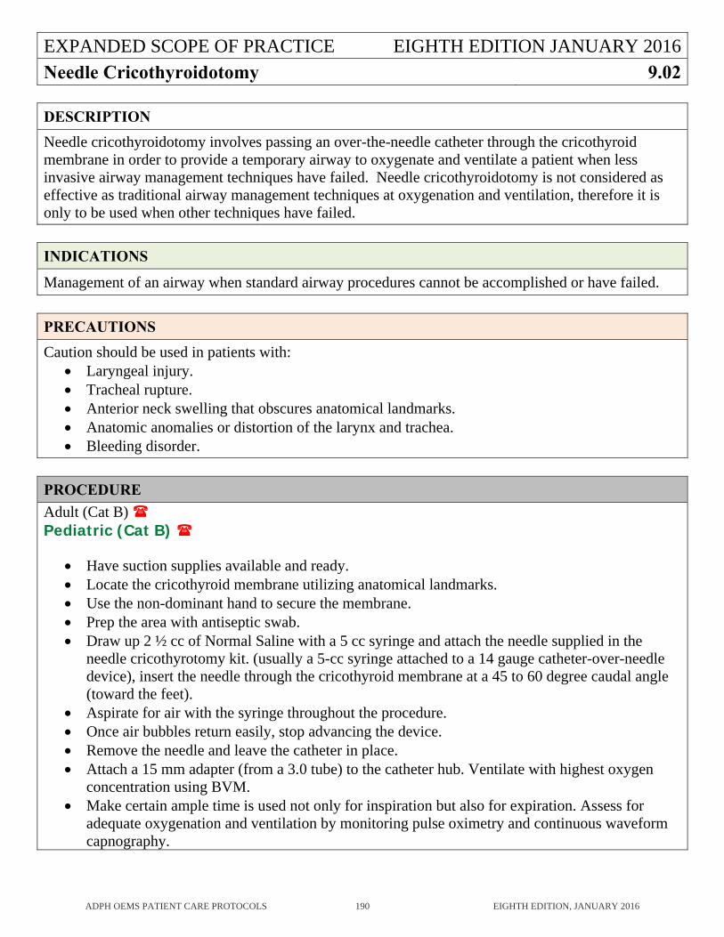
EXPANDED SCOPE OF PRACTICE EIGHTH EDITION JANUARY 2016Needle Cricothyroidotomy 9.02
ADPH OEMS PATIENT CARE PROTOCOLS 190 EIGHTH EDITION, JANUARY 2016
DESCRIPTION
Needle cricothyroidotomy involves passing an over-the-needle catheter through the cricothyroid membrane in order to provide a temporary airway to oxygenate and ventilate a patient when less invasive airway management techniques have failed. Needle cricothyroidotomy is not considered as effective as traditional airway management techniques at oxygenation and ventilation, therefore it is only to be used when other techniques have failed.
INDICATIONS
Management of an airway when standard airway procedures cannot be accomplished or have failed.
PRECAUTIONS
Caution should be used in patients with: Laryngeal injury. Tracheal rupture. Anterior neck swelling that obscures anatomical landmarks. Anatomic anomalies or distortion of the larynx and trachea. Bleeding disorder.
PROCEDURE
Adult (Cat B) Pediatric (Cat B)
Have suction supplies available and ready. Locate the cricothyroid membrane utilizing anatomical landmarks. Use the non-dominant hand to secure the membrane. Prep the area with antiseptic swab. Draw up 2 ½ cc of Normal Saline with a 5 cc syringe and attach the needle supplied in the
needle cricothyrotomy kit. (usually a 5-cc syringe attached to a 14 gauge catheter-over-needle device), insert the needle through the cricothyroid membrane at a 45 to 60 degree caudal angle (toward the feet).
Aspirate for air with the syringe throughout the procedure. Once air bubbles return easily, stop advancing the device. Remove the needle and leave the catheter in place. Attach a 15 mm adapter (from a 3.0 tube) to the catheter hub. Ventilate with highest oxygen
concentration using BVM. Make certain ample time is used not only for inspiration but also for expiration. Assess for
adequate oxygenation and ventilation by monitoring pulse oximetry and continuous waveform capnography.
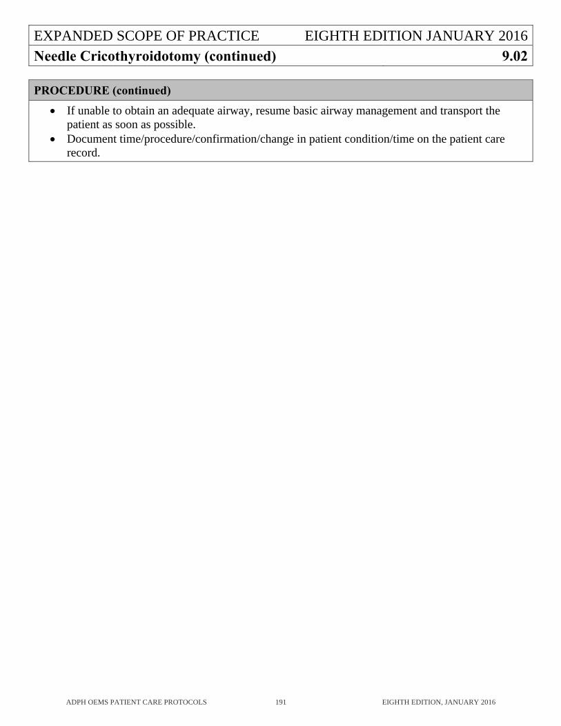
EXPANDED SCOPE OF PRACTICE EIGHTH EDITION JANUARY 2016Needle Cricothyroidotomy (continued) 9.02
ADPH OEMS PATIENT CARE PROTOCOLS 191 EIGHTH EDITION, JANUARY 2016
PROCEDURE (continued)
If unable to obtain an adequate airway, resume basic airway management and transport the patient as soon as possible.
Document time/procedure/confirmation/change in patient condition/time on the patient care record.
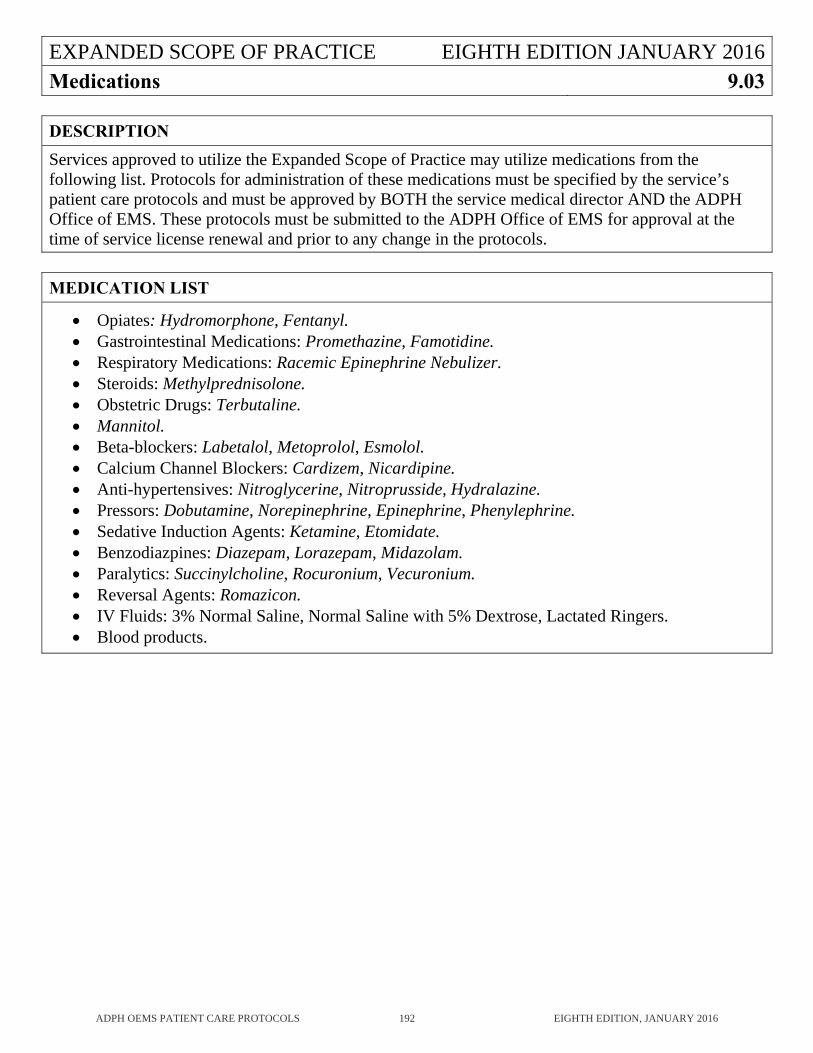
EXPANDED SCOPE OF PRACTICE EIGHTH EDITION JANUARY 2016Medications 9.03
ADPH OEMS PATIENT CARE PROTOCOLS 192 EIGHTH EDITION, JANUARY 2016
DESCRIPTION
Services approved to utilize the Expanded Scope of Practice may utilize medications from the following list. Protocols for administration of these medications must be specified by the service’s patient care protocols and must be approved by BOTH the service medical director AND the ADPH Office of EMS. These protocols must be submitted to the ADPH Office of EMS for approval at the time of service license renewal and prior to any change in the protocols.
MEDICATION LIST
Opiates: Hydromorphone, Fentanyl. Gastrointestinal Medications: Promethazine, Famotidine. Respiratory Medications: Racemic Epinephrine Nebulizer. Steroids: Methylprednisolone. Obstetric Drugs: Terbutaline. Mannitol. Beta-blockers: Labetalol, Metoprolol, Esmolol. Calcium Channel Blockers: Cardizem, Nicardipine. Anti-hypertensives: Nitroglycerine, Nitroprusside, Hydralazine. Pressors: Dobutamine, Norepinephrine, Epinephrine, Phenylephrine. Sedative Induction Agents: Ketamine, Etomidate. Benzodiazpines: Diazepam, Lorazepam, Midazolam. Paralytics: Succinylcholine, Rocuronium, Vecuronium. Reversal Agents: Romazicon. IV Fluids: 3% Normal Saline, Normal Saline with 5% Dextrose, Lactated Ringers. Blood products.