airway management of the critically ill patient...
TRANSCRIPT
Critical Care Horizons 1
Airway Management of the Critically Ill Patient:Modifications of Traditional Rapid SequenceInduction and IntubationTim Leeuwenburg1*
1Kangaroo Island Medical Centre and MedSTAR Retrieval, South Australia*Corresponding author’s email: [email protected]
Date published: 24/06/2015
Discussion about airway management is common amongst clinicians involved in critical care,regardless of background. The technique of rapid sequence induction and intubation (RSI) wasdescribed in 1970 and there are many accepted variations in modern day practice. Sadly this canlead to difficulty, particularly in the event of an airway misadventure, as clinicians may be subjectto post hoc critique from expert opinion in other disciplines, and often held to a ‘standard’ of RSIthat no longer exists. Experts may differ in opinions, and expertise in one arena may not translateto another. This paper outlines the variations in RSI practice and the rationale for deviation. Suchdiscussion is necessary, as expert opinion referring to a ‘standard’ RSI may be inappropriate for thecritically ill patient, exposing practitioners to medico-legal risk. Acknowledgement of variations inRSI practice allows the development of institutional procedures, with potential for future consensusrecommendations guided by both published studies and expert opinion.
Keywords: Airway management; Rapid sequence induction; Endotracheal intubation; Emergencyintubation; Difficult airway
Competing interests: The author declares that he has no competing interests
Released under a Creative Commons CC BY-NC-ND 4.0 license except for content otherwise specified
DOI: To be confirmed
Introduction
Rapid sequence induction and intubation (RSI) has been con-sidered the gold standard in emergency airway management.Core elements of the classical RSI include rapid induction ofanaesthesia followed by administration of a paralysing agent,techniques to minimise aspiration risk and a goal of first passplacement of a cuffed endotracheal tube in the trachea.
There is evidence for variation in how individuals, institu-tions and nations practice RSI [1]. The technique of RSI is cen-tred around reduction of risk; that of regurgitation/aspiration,and that associated with the procedure itself, including fail-ure to rapidly secure the airway, hypoxia, airway trauma, andhypotension from induction agents. Analysis of airway com-plications reveals a higher incidence of difficulty in intensivecare unit (ICU) and emergency department (ED) intubationsthan in the operating theatre (OT) (incidence of death or braindamage 38-fold higher in the ED and 58-fold higher in the ICUcompared with OT) [2–4].
RSI is a technique utilised by clinicians in anaesthesia,emergency medicine, and intensive care, both in hospital
and in the prehospital environment [5]. Variations in RSIare inevitable given the heterogeneous mix of patient pre-morbid physiology, RSI operators, teams, environment andavailable options. Indeed it is appropriate that RSI ismodified to the circumstances, particularly in the criticallyill patient [6]. Unfortunately, the existence of such appropriateheterogeneity in practice can lead to criticism, whether betweenclinical experts, between health institutions, between medicalspecialties or in the medico-legal arena [7–9].
It is not the purpose of this paper to outline a uniformstandard for RSI; rather to explore issues pertaining toexpertise, to discuss recognised variations in components ofthe RSI technique and to advocate for pragmatic modificationsfor RSI in the critically ill patient.
Individual organisations may wish to use this as a guide toformulate institutional standard operating procedures for RSIof the critically ill, as well as for training programs for thoseinvolved in emergency RSI, thus helping to mitigate recognisedcomplications of airway management.
1 Critical Care Horizons 2015; 1: 1-10
Critical Care Horizons 2
The dilemma of defining an expert
Anaesthetists are traditionally regarded as the experts in airwaymanagement, reflecting the duration of their training andpivotal role in airway management. Nevertheless, airwaymanagement is a core skill of staff in other disciplines,particularly those who are actively involved in resuscitationand emergency management, or in circumstances where aspecialist anaesthetist is not immediately available. Theappropriate degree of diligence and expertise is expected fromall providers caring for the critically ill, whether anaesthetist,intensivist, emergency physician, rural proceduralist, retrievalnurse practitioner or prehospital paramedic.
Inevitably there will be differences of opinion betweenexperts, with previously ‘indisputable truths’ in difficultairway management having been challenged in recent times[10]. This lack of scientific consensus is problematic. In the caseof an airway catastrophe, expert witnesses are usually drawnfrom those who may refer to classical RSI, reflecting theirown traditional teaching, not the current practice of modifiedRSI in the critical care arena. There are documented medico-legal critiques leading to censure, including expert opinionthat it was negligent to fail to pass a nasogastric tube pre-RSI, inappropriate use of a bolus dose of induction agent, andnegligent to omit cricoid force during RSI [11, 12]. Post hocexpert criticism can be catastrophic for individuals. It is moreappropriate to refer to the expertise of highly-trained peersregularly practicing in a similar environment. This requiresacknowledgement of variations in practice in emergencyairway management, whether in operating theatre, emergencydepartment, intensive care, prehospital or in situations limitedby available resources. Moreover there is heterogeneity inexpert opinion and Cook et al have previously described thedifficulties of contrary expert opinion in airway management,with implications for incident review, medico-legal claims andclosed claim analysis [13].
Rapid Sequence Induction and Intubation: a standard-ised process or not?
Basic airway management (maintenance of oxygenation andventilation) by use of adjuncts such as suction, oro- andnasopharyngeal airways, bag-mask ventilation and evenplacement of a laryngeal mask in the truly obtunded is wellwithin the expected competency of all clinicians working inacute care. However, RSI is expected of advanced airwaypractitioners, with indications including:
• failure to maintain airway patency by other means
• failure of airway protection
• failure of ventilation or oxygenation
• for anticipated clinical course
• to facilitate transportation
• for humanitarian reasons
The original 15-step technique of RSI was described in 1970[14], yet this form of RSI is not uniformly applied in modernpractice [15] and nor should we expect it to be. Advancesin equipment, induction drugs and paralysing agents haveallowed refinement of the technique over time, with RSImodifications made as appropriate to the clinical circumstancesof individual patients, to the skill mix of airway teams and tothe environment in which airways are managed.
Therein lies the difficulty. Despite the universal acceptanceof RSI as the ‘gold standard’ in securing the airway ina critically ill patient, the actual components of RSI areknown to differ markedly between individuals, institutionsand countries, as well as between practitioners in differentarenas (prehospital, ED, ICU or OT) [1, 16, 17]. Documentedmodifications to RSI technique include patient position, pre-oxygenation strategies, pre-RSI decompression of gastriccontents with a nasogastric tube, choice and method ofadministration of induction agent, application of cricoidpressure, choice of paralysing agent, use of manual ventilationand options for failed RSI (not least whether awakening is anoption) [18]. However the key elements of RSI remain, namely:
• pre-oxygenation or denitrogenation to prolong time tocritical desaturation
• prevention of hypoxia and hypotension during theinduction and intubation sequence
• passage of a cuffed endotracheal tube with confirmationof placement
In short, a refinement of the classical RSI technique asdefined by Stept and Safar [14] is called for, with a need fora consensus position allowing for variation in the practice ofRSI between experts, as governed by the requirements of thepatient, team and clinical circumstances. Accepted practicevariation should be understood in the context of both the needto minimise aspiration risk and to avoid complications of theRSI technique itself.
RSI of the Critically Unwell Patient
Airway Team and DynamicsRegardless of the individual expertise of the intubator, teamfactors will impact on performance of the RSI process. Teammembers should be adequately trained prior to involvement inairway management, preferably involving simulation trainingunder increasing degrees of cognitive load to allow a degree of‘stress inoculation’ and to reinforce the importance of humanfactors in performance [19].
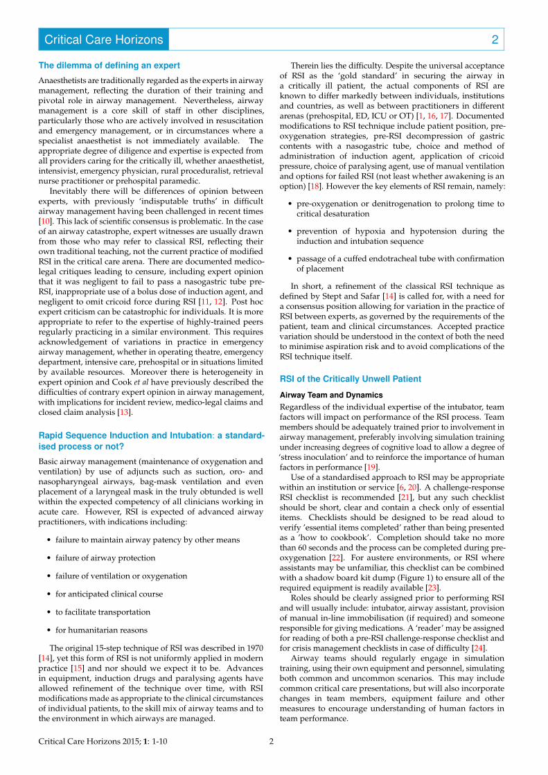
Use of a standardised approach to RSI may be appropriatewithin an institution or service [6, 20]. A challenge-responseRSI checklist is recommended [21], but any such checklistshould be short, clear and contain a check only of essentialitems. Checklists should be designed to be read aloud toverify ’essential items completed’ rather than being presentedas a ’how to cookbook’. Completion should take no morethan 60 seconds and the process can be completed during pre-oxygenation [22]. For austere environments, or RSI whereassistants may be unfamiliar, this checklist can be combinedwith a shadow board kit dump (Figure 1) to ensure all of therequired equipment is readily available [23].
Roles should be clearly assigned prior to performing RSIand will usually include: intubator, airway assistant, provisionof manual in-line immobilisation (if required) and someoneresponsible for giving medications. A ‘reader’ may be assignedfor reading of both a pre-RSI challenge-response checklist andfor crisis management checklists in case of difficulty [24].
Airway teams should regularly engage in simulationtraining, using their own equipment and personnel, simulatingboth common and uncommon scenarios. This may includecommon critical care presentations, but will also incorporatechanges in team members, equipment failure and othermeasures to encourage understanding of human factors inteam performance.
Critical Care Horizons 2015; 1: 1-10 2
Critical Care Horizons 3
RSI CHALLENGE-RESPONSE
Monitoring - BP, ECG, SpO2, ETCO2 CHECK Nasal Cannulae at 15l/min PLUS Mask O2 CHECK Pre-oxygenation for FOUR minutes CHECK Suction checked working & available CHECK Patient Positioned? RAMP OBESE CHECK
IV & DRUGS IV Cannula connected to fluid & running CHECK NIBP on contralateral arm and BP seen CHECK Spare cannula in situ CHECK INDUCTION AGENT drawn up, dose checked CHECK SUX or ROC drawn up, dose checked CHECK VASOPRESSORS drawn up, labelled CHECK POST INTUBATION drugs drawn up & labelled CHECK
INTUBATION EQUIPMENT BVM connected to oxygen CHECK Guedel & two NPO airways available CHECK Laryngoscope blade chosen, light working CHECK ET tube size chosen, cuff tested CHECK ETT preloaded on bougie, Kiwi Grip CHECK Alternate tube size chosen & cuff tested CHECK Syringe for cuff inflation CHECK Stylet & Rapi-Fit Bougie connectors available CHECK Gooseneck, filter, inline ETCO2 CHECK Tube Tie & Tape available CHECK Ventilator settings determined CHECK Difficult airway plan’s A, B, C, D discussed CHECK LMA, iLMA and Surgical Airway available CHECK
TEAM BRIEF In-line immobilisation person briefed CHECK Cricoid pressure person briefed CHECK Drug giver briefed CHECK Anticipated problems & post RSI care brief CHECK
Ventilator settings determined & switched on CHECK TIME OF INTUBATION NOTED & 30 sec DRILLS CHECK
BOUGIE with COUDE TIP (or can use FROVA OXYGENATING BOUGIE)
NASO-PHARYNGEAL & ORO-PHARYNGEAL AIRWAYS ET ADAPTOR, IN-LINE FILTER and
ETCO2 LINE or EASYCAP
CONSIDER LOADING A STRAIGHT-TO-CUFF ATRAUMATIC STYLET
LARYNGEAL MASK AIRWAY - Classic / Supreme / iLMA (FastTrach or AirQ-II)
LARYNGOSCOPE with WORKING BULB & APPROPRIATE BLADE
SELF-INFLATING BAG-VALVE-MASK CONNECTED TO HIGH FLOW OXYGEN
consider using PEEP Valve
plus
NASAL SPECS DURING INTUBATION for Apnoeic Diffusion Oxygenation
DRUGS INDUCTION AGENT
SUX or ROC VASOPRESSOR
FLUIDS RUNNING
PLAN FOR FAILED RSI ?
DIFFICULT AIRWAY TROLLEY
AVAILABLE ?
SUCTION (confirm working then
place under pillow)
KING VISION
VL
LUBE
TIESTAPE
TWO ET TUBES OF APPROPRIATE SIZE
30 degree Coude tip
RapiFit Connectors for Frova Bougie
10 or 20 ml syringe
SURG
ICAL
AIR
WAY
: CTM
mar
ked?
Pre
pare
d to
use
SCA
LPEL
-Fin
ger-S
ize 6.
0 ETT
Figure 1. Shadow Board Kit Dump with Challenge-Response Checklist
Airway Planning
Pre-RSI briefing should include planning for anticipateddifficulties. Difficult airway plans usually include directlaryngoscopy for endotracheal intubation as the primary plan,with backup which may include alternative devices suchas videolaryngoscopes or an intubating laryngeal mask tomaintain oxygenation and facilitate subsequent intubation.Rescue ventilation via bag-mask or supra-glottic devices maybe required as a bridge, but if they fail the team should beprepared to perform an emergency surgical airway.
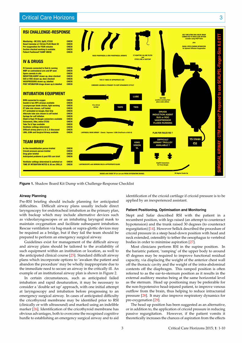
Guidelines exist for management of the difficult airwayand airway plans should be tailored to the availability ofsuch equipment within an institution or location, as well tothe anticipated clinical course [25]. Standard difficult airwayplans which incorporate options to ‘awaken the patient andabandon the procedure’ may be wholly inappropriate due tothe immediate need to secure an airway in the critically ill. Anexample of an institutional airway plan is shown in Figure 2.
In certain circumstances, such as anticipated failedintubation and rapid desaturation, it may be necessary toconsider a ’double set up’ approach, with one initial attemptat laryngoscopy and intubation before progressing to anemergency surgical airway. In cases of anticipated difficultythe cricothyroid membrane may be identified prior to RSI(clinically or with ultrasound) and marked using an indeliblemarker [26]. Identification of the cricothyroid membrane hasobvious advantages, both to overcome the recognised cognitivehurdle to establishing an emergency surgical airway and to aid
identification of the cricoid cartilage if cricoid pressure is to beapplied by an inexperienced assistant.
Patient Positioning, Optimisation and Monitoring
Stept and Safar described RSI with the patient in arecumbent position, with legs raised (an attempt to counteracthypotension) and the trunk raised 30 degrees (to counteractregurgitation) [14]. However Sellick described the procedure ofcricoid pressure in a steep head-down position with head andneck extended, ostensibly to tether the oesophagus to vertebralbodies in order to minimise aspiration [27].
Most clinicians perform RSI in the supine position. Inthe bariatric patient, ‘ramping’ of the upper body to around45 degrees may be required to improve functional residualcapacity, via displacing the weight of the anterior chest walloff the thoracic cavity and the weight of the intra-abdominalcontents off the diaphragm. This ramped position is oftenreferred to as the ear-to-sternum position as it results in theexternal auditory meatus being at the same horizontal levelas the sternum. Head up positioning may be preferable forthe non-hypotensive head-injured patient, to improve venousoutflow from the brain, thus helping to reduce intracranialpressure [28]. It may also improve respiratory dynamics forpre-oxygenation [29].
The head up position has been suggested as an alternative,or in addition to, the application of cricoid pressure in reducingpassive regurgitation. However, if the patient vomits ittheoretically increases the chances of aspiration from the effects
3 Critical Care Horizons 2015; 1: 1-10
Critical Care Horizons 4
Figure 2. Example Airway Plan
of gravity, rather than particulate matter draining from themouth.
Pregnant patients should be positioned in left lateral tiltand/or the uterus manually displaced to avoid aortocavalcompression. Regardless of whether positioned supine, headup to limit regurgitation, head down to limit aspiration or in aleft lateral position if pregnant, working suction should alwaysbe available.
For those in whom ear-to-sternum positioning is contraindi-cated (suspected spinal injury or musculoskeletal abnormalitylimiting spinal mobility), head position should minimise unnec-essary flexion-extension or lateral rotation. Spinal precautionsshould be observed for the trauma patient, which may includemanual inline stabilisation or use of an occipital pad to opti-mise laryngoscopy and minimise movement of the cervicalspine [30].
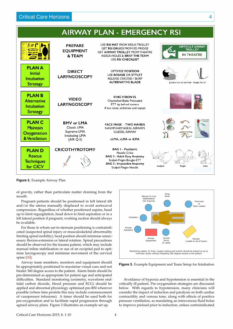
Airway team members, monitors and equipment shouldbe appropriately positioned to maximise visual cues and nothinder 360 degree access to the patient. Alarm limits should bepre-determined as appropriate for patient age and anticipateddifficulties. Standard monitoring (oximetry, waveform end-tidal carbon dioxide, blood pressure and ECG) should beapplied and abnormal physiology optimised pre-RSI whereverpossible (where time permits this may include commencementof vasopressor infusions). A timer should be used both forpre-oxygenation and to facilitate rapid progression throughagreed airway plans. Figure 3 illustrates an example set up.
Intubator
Airway Assistant
Manual In-Line Stabilisation (for trauma)
Drug Giver
Overview or
Team Leader
Cricoid Force
(if used)
Airway Equipment
Monitor (visible to all of team)
Monitoring cables, IV lines, oxygen tubing and suction should be placed so as to minimise clutter without impeding 360 degree access to the patient
Figure 3. Example Equipment and Team Setup for Intubation
Avoidance of hypoxia and hypotension is essential in thecritically ill patient. Pre-oxygenation strategies are discussedbelow. With regards to hypotension, many clinicians willconsider the impact of induction and paralysis on both cardiaccontractility and venous tone, along with effects of positivepressure ventilation, as mandating an intravenous fluid bolusto improve preload prior to induction, unless contraindicated.
Critical Care Horizons 2015; 1: 1-10 4
Critical Care Horizons 5
Measurements of blood pressure should be made regularly,with either non-invasive blood pressure set to cycle at 1 minuteintervals or placement of an arterial line if sufficient timeallows.
Pre-oxygenation
The purpose of pre-oxygenation is to denitrogenate the lungsand create a reservoir of oxygen to allow a margin of safetybefore critical desaturation during attempts to secure theairway. An excellent summary of methods to maximisepre-oxygenation and prevent desaturation during emergencyairway management is described by Weingart and Levitan[29]. Key steps include mandatory use of pre-oxygenation toextend safe apnoea time during RSI, along with appropriatepositioning, and may involve the use of positive end-expiratory pressure (PEEP). The delivery of oxygen via nasalcannulae during intubation (apnoeic diffusion oxygenation)is increasingly being adopted for airway management of thecritically ill patient [31].
The period of pre-oxygenation should adequately denitro-genate the lungs. An empiric approach applying high-flowoxygen for three minutes or eight vital capacity breaths is com-mon anaesthetic practice. However, critically ill patients mayrequire a longer period to denitrogenate and are often unableto perform eight vital capacity breaths. If available, measure-ment of expired end-tidal oxygen should be used as a guide toadequate pre-oxygenation, aiming for a value of at least 90%(FeO2 of 0.9)
Pre-oxygenation technique may be governed by availableequipment, personnel and patient requirements. Validtechniques include:
• use of a Mapleson B or C anaesthetic circuit. These lackthe separate inspiratory or expiratory ports of traditionalbag-valve-mask (BVM) devices, with exhaled gas flushedout of the circuit by high fresh gas flow via the pressure-release valve, ensuring maximal oxygen delivery [32].
• use of standard reservoir face masks on maximal oxygenflow and supplemented with nasal cannulae on maximalflow. This may be a preferred in the prehospitalenvironment, where limitations of personnel precludealternatives.
• use of standard bag-valve-mask devices commonly usedin ED, ICU or by emergency medical services. Cautionis needed as such devices may entrain room air duringspontaneous ventilation [33]. Addition of a PEEP valve tothe expiratory port of BVM assembly obviates this.
• use of existing non-invasive ventilation modes. For manycritically ill patients, RSI may represent the end result of afailure of non-invasive ventilation (NIV). NIV masks maybe left in situ and used to pre-oxygenate. CPAP/NIV maybe very useful in pre-oxygenation of the morbidly obesepatient.
On occasions, the combative patient (e.g. intoxicated,head injured, hypoxic) will thwart best attempts at bothpositioning and pre-oxygenation. Pre-treatment with smalltitrated aliquots of a sedative agent can be effective (so-called‘delayed sequence intubation’), with ketamine the preferredagent to facilitate assessment, monitoring, positioning and pre-oxygenation [34].
Choice and Timing of Induction Agent
Commonly used induction agents include thiopentone (asoriginally described by Stept and Safar), etomidate (notavailable in all countries), propofol, benzodiazepines such asmidazolam (relatively slow onset compared to other agents)and ketamine. Ketamine is gaining favour within emergencyand critical care circles due to relative cardiovascular stability[35]. It should be noted that all induction agents (includingmidazolam and ketamine) have potential for cardiovasculardepression and hypotension if too high a dose is used. Inaddition, combinations of agents may be synergistic withamplification of effect. Previous concerns of deleterious effectsof ketamine on intracranial pressure in head injury have beenchallenged and as such, use of ketamine has much to commendit for RSI in the critically ill patient [36].
Whilst the original description of RSI involved a bolusof thiopentone based on patient weight, such weight-basedcalculations may not be appropriate in critical illness due toadverse haemodynamic effects. Doses should be adjustedaccording to pre-RSI physiology, requiring dose-reductionsto as little as 10% of standard induction doses for thecritically ill patient with haemodynamic compromise [37].Both bolus dosing and titration of induction agent to loss ofconsciousness have been described. Bolus dosing from a pre-drawn syringe has the advantage of rapidity; however, there ispotential for either under- or overdosing, the former perhapscontributing to increased reports of awareness during RSI intrauma and obstetric patients, the latter risking haemodynamiccompromise. Currently, there are no data to compare thepotential aspiration risks of a longer induction time via dose-titration versus the risks of either awareness or haemodynamicinstability with a predetermined bolus technique.
Clinicians will determine the optimal choice of inductionagent for the situation, often guided by personal expertise,institutional guidelines, available agents and appropriatepatient selection. Regardless of induction agent used,delay between administration, loss of consciousness andadministration of paralysing agent may prolong the periodof aspiration risk and increased the risk of desaturation.
Adjunct Opioid Agents
Adjunct agents are not described in the traditional teaching ofRSI, yet many practitioners incorporate rapid acting opioidsto attenuate the reflex sympathetic responses to laryngoscopyand intubation. This may be especially useful in criticallyill patients with head injuries. Arguments against use ofopioids include historical concerns due to slow onset andlonger duration with older opioids, as well as concerns ofdecreased respiratory drive if intubation fails. This is less ofa concern in the critically ill patient, as options to awaken thepatient are generally not appropriate.
Lyon et al describe a modification of RSI techniqueusing adjunctive fentanyl, along with ketamine inductionand rocuronium paralysis within their prehospital service[38]. They note both superior intubating conditions anda more favourable haemodynamic response to intubation.Development of protocols for modified RSI within aninstitution, and their subsequent publication, is to beencouraged.
It should be noted that the use of opioids such as alfentaniland fentanyl may produce synergistic effects in combinationwith induction agents, and cautious dosing should be used inhaemodynamically unstable patients to minimise hypotension.
5 Critical Care Horizons 2015; 1: 1-10
Critical Care Horizons 6
Cricoid Force
Cricoid force has become an area of contention in airwaymanagement. Sellick’s original description was of a ‘firm’amount of pressure applied to the cricoid cartilage of acadaver whilst in a steep head-down position to occludethe oesophagus and prevent regurgitation of fluid into theoropharynx [27]. The procedure was repeated during inductionof 26 patients deemed at high-risk of aspiration. Noneexperienced regurgitation with application of cricoid force; 3experienced immediate reflux upon release of cricoid force aftertracheal intubation. Cricoid force was incorporated into Steptand Safar’s description of RSI and has since been consideredan essential component. Refinements describe a force of 10Napplied at the commencement of induction, increased to 30Nwith loss of consciousness [39]. Application of cricoid forceremains a recommendation during RSI from the authors of theNAP4 audit in the United Kingdom [2].
However, application of cricoid force is not consideredroutine practice in some countries or organisations. Thereare concerns that cricoid force does not effectively occlude theoesophagus and thus prevent aspiration, is variably applied byassistants (often incorrect timing, incorrect position or force)and that cricoid force can impede view at laryngoscopy thusdelaying first pass success [40–43].
Some have proposed that cricoid force is a low-riskprocedure that works in a proportion of patients but isconfounded by poor technique and relative infrequency ofregurgitation. Thus, they propose application of cricoidforce and early removal if this impedes laryngoscopy, ifthere is active vomiting, or if there is impediment of rescueventilation via laryngeal mask airway or BVM [44, 45]. Itcan be argued that in certain arenas, particularly prehospitalor with limited/untrained personnel (rural, small ED orICU) application of cricoid pressure is more likely to hinderlaryngoscopy and that the policy of ‘apply, then release’ addsadditional cognitive load to an already high-stakes tightly-coupled procedure. On this basis, some airway expertsmay opt to omit cricoid force in such circumstances, basedon limited evidence of efficacy and risk-benefit balance inregard to optimising first-pass intubation success [6, 46].Meanwhile trials are under-way to test the hypothesis that useof cricoid force during RSI in ED does not prevent aspirationand investigate the effect of such force on difficult or failedintubation [47].
A decision not to apply cricoid force may be reasonablein airway management of the critically ill patient. It isrecommended that any decision to use or omit cricoid forcebe supported by an institutional policy. Practitioners withclinical expertise in resuscitation are responsible for shapingsuch policy, mindful that this may differ from publishednational or international guidelines. Hence, despite a lackof absolute evidence of benefit, cricoid force may continueto be applied; reflecting medico-legal concerns as individualclinicians have been criticised for failing to apply cricoid forcein post-event medico-legal dissection of airway catastrophes[12]. It is essential that any expert opinion on cricoid force,as indeed any other matter in RSI, acknowledges the existingvariation in practice. At this point in time, the literature doesnot support evidence either for or against the application ofcricoid force.
Paralysis
Use of succinylcholine (a depolarising neuromuscular blocker)as the preferred agent to facilitate vocal cord relaxation andendotracheal tube passage has been the accepted norm for
RSI, with traditional teaching being that the short duration ofaction will allow return of spontaneous ventilation in the caseof a failed RSI. Whilst awakening may be an option for somepatients in the operating theatre, it is rarely the case for theunfasted, haemodynamically-compromised patient for whomRSI represents a commitment to securing the airway.
Rocuronium at a dose of 1.6 mg/kg gives the same onsetof muscle relaxation as succinylcholine and is suggestedas the preferred choice of non-depolarising neuromuscularblockers for RSI in the critically ill [48]. A commitment tofull paralysis and rapid progression to a surgical airway inthe case of failed intubation and ventilation in the criticallyill patient is congruent with pre-agreed airway plans betweenteam members, appropriate for the patient (whose pathologyrequires a cuffed tube in the trachea by whatever means) andavoids the possibility of attempting a surgical airway in acombative, coughing patient.
Manual Ventilation between Induction and Intubation
Manual ventilation has traditionally been avoided in classicalRSI, due to concerns of gastric insufflation and aspiration.However, gentle ventilation has been advocated in bothobstetric and paediatric RSI due to concerns of rapiddesaturation in these populations [49]. Anecdotal evidencefrom experienced resuscitationists includes use of gentlemanual ventilation whilst awaiting onset of paralysis as a ‘doleast harm’ approach. A decision on whether to gently ventilatewill be guided by aspiration risk - the patient with ileus, withgastroparesis or with upper gastrointestinal bleeding is clearlyat higher risk than the fasted patient. For the critically illpatient, risks of hypoxia and hypercapnia may require gentlemanual ventilation. Critically ill patients commonly havean existing metabolic acidosis with respiratory compensation,and periods of apnoea can result in significant reductions inpH which amplify haemodynamic risk. Manual ventilationattempts should be initiated with pressures less than 15 cmH2Oto minimise gastric insufflation [50]. There is potential for theuse of adjuncts such as automatic, low pressure, constant flowventilation devices to minimise ventilation pressures duringRSI [51].
Maximising First-Pass Success
It is important to appreciate that repeated attempts atlaryngoscopy may increase rates of aspiration [52]. Thus,maximising the potential for first pass success is essential inRSI of the critically ill patient.
Direct laryngoscopy using an appropriate blade and lightsource (modern day LED optics offer excellent illuminationand contrast) remains the cornerstone of intubation. Carefuland sequential visualisation of landmarks and avoidance ofrepeated attempts causing airway trauma are key skills [53].
Adjuncts such as a bougie or malleable stylet are commonlyused in cases of difficult intubation. For intubation of thecritically ill patient, such adjuncts should be used routinely.Understanding appropriate use is vital as infrequent users maynot appreciate the nuances of these devices, which are designedto facilitate navigation to the laryngeal inlet in difficult cases.
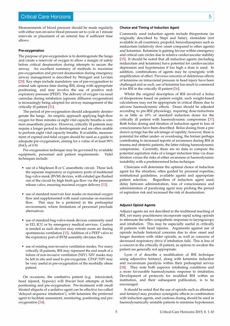
Stylets, if used, should be shaped ‘straight-to-cuff’ i.e. thestylet should remain straight as far as the proximal part ofthe endotracheal tube cuff where it should be angled to nomore than 35 degrees (angles beyond 35 degrees increasedifficulty) [54]. Traditional teaching has been to avoid pre-loading endotracheal tubes onto bougies, as the weight of thetube may impair control of the bougie tip; however, hang-upof the bougie on the endotracheal tube connector may impede
Critical Care Horizons 2015; 1: 1-10 6
Critical Care Horizons 7
Figure 4. Endotracheal Tube Pre-loaded on Bougie
smooth rail-roading of the endotracheal tube, causing delayin tube passage and risking a loss of situational awareness inthe operator. A refinement is to pre-load an endotracheal tubeonto a bougie and hold them in such a grip that control of thebougie is maintained during navigation to the laryngeal inlet(Figure 4).
It is not uncommon for the leading edge of bevel-shapedendotracheal tubes to hang up on the right arytenoid cartilage;gentle slight withdrawal and a counter-clockwise rotation ofthe endotracheal tube/bougie complex allows the free-edge toenter the glottic opening and advance.
Some advocate the use of video-laryngoscopy over directlaryngoscopy, particularly for a known or anticipated difficultairway [55]. Currently there is a plethora of available devicesavailable. Cited advantages include improved view of theglottic opening for difficult airways, allowing other membersof the team to visualise tube passage, and potential forrecording of intubation procedures for audit and training[56]. Videolaryngoscopy may afford better visualisation of theglottic opening in a difficult airway; caution is recommendedas a better view with some devices does not translate to easiertube passage unless the operator is experienced in use ofthe particular device. Additional caveats include cost, poorperformance in the presence of blood/secretions and manyrequire a different technique to traditional direct laryngoscopy.
The optimal video-laryngoscope would be low cost, havethe same technique as standard direct laryngoscopy, havesimilar blade geometry and tube passage, perform well inthe presence of both a soiled airway and in the presence ofbright sunlight. At present no such device exists. If a video-laryngoscope is used, operators must be fully aware of nuancesof the device and be trained to use in elective settings prior toan emergency [57].
Failed RSI
A difficult airway plan should be discussed and a checklistshould be completed prior to RSI such that a shared mentalmodel of actions to be undertaken exists between teammembers, both for routine and in case of difficulty [13, 58].Many such difficult airway plans exist [59, 60].
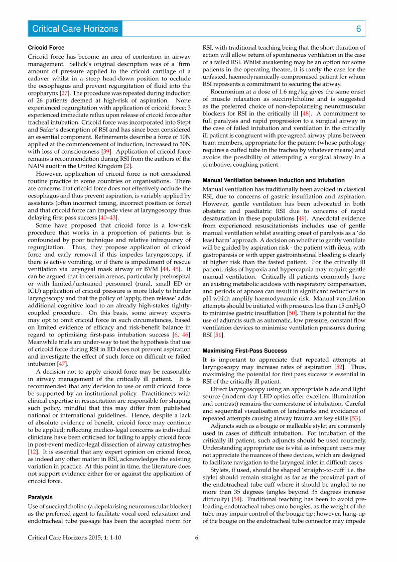
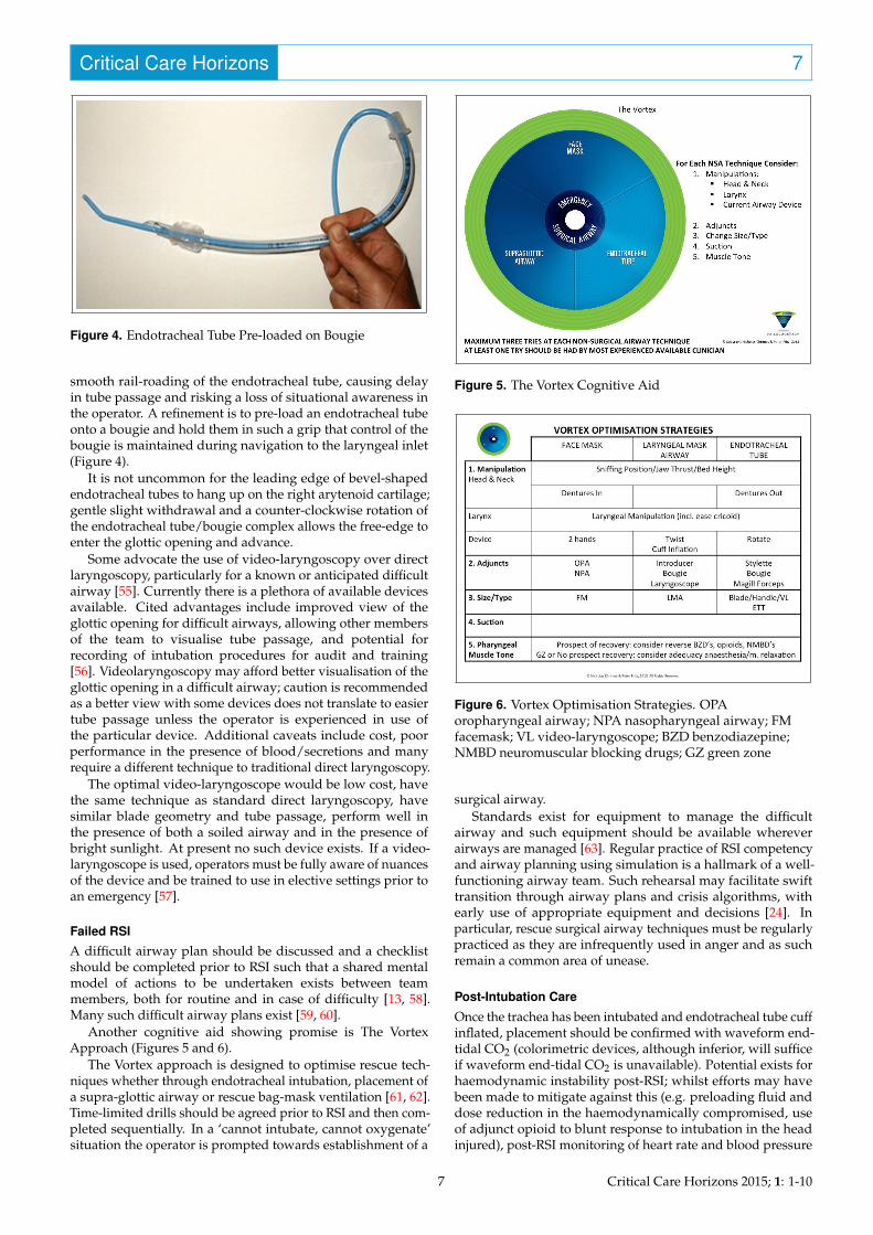
Another cognitive aid showing promise is The VortexApproach (Figures 5 and 6).
The Vortex approach is designed to optimise rescue tech-niques whether through endotracheal intubation, placement ofa supra-glottic airway or rescue bag-mask ventilation [61, 62].Time-limited drills should be agreed prior to RSI and then com-pleted sequentially. In a ‘cannot intubate, cannot oxygenate’situation the operator is prompted towards establishment of a
Figure 5. The Vortex Cognitive Aid
Figure 6. Vortex Optimisation Strategies. OPAoropharyngeal airway; NPA nasopharyngeal airway; FMfacemask; VL video-laryngoscope; BZD benzodiazepine;NMBD neuromuscular blocking drugs; GZ green zone
surgical airway.Standards exist for equipment to manage the difficult
airway and such equipment should be available whereverairways are managed [63]. Regular practice of RSI competencyand airway planning using simulation is a hallmark of a well-functioning airway team. Such rehearsal may facilitate swifttransition through airway plans and crisis algorithms, withearly use of appropriate equipment and decisions [24]. Inparticular, rescue surgical airway techniques must be regularlypracticed as they are infrequently used in anger and as suchremain a common area of unease.
Post-Intubation Care
Once the trachea has been intubated and endotracheal tube cuffinflated, placement should be confirmed with waveform end-tidal CO2 (colorimetric devices, although inferior, will sufficeif waveform end-tidal CO2 is unavailable). Potential exists forhaemodynamic instability post-RSI; whilst efforts may havebeen made to mitigate against this (e.g. preloading fluid anddose reduction in the haemodynamically compromised, useof adjunct opioid to blunt response to intubation in the headinjured), post-RSI monitoring of heart rate and blood pressure
7 Critical Care Horizons 2015; 1: 1-10
Critical Care Horizons 8
is vital. The combination of worsening acidosis from peri-intubation apnoea, the presence of hypovolaemia, the impactof induction agents on cardiac contractility and vasomotortone, and the effects of over-zealous post-intubation ventilatorsettings on right ventricular preload (reduction) and afterload(increase) is a potent trigger for haemodynamic collapse. Post-intubation ventilation and sedation plans should be previouslyagreed during airway planning, and should be enacted. RSIof the critically ill patient may pose a challenge for the mostexperienced operators and care must be taken to avoid clinicalinertia and to continue resuscitation and vigilant monitoringfor complications [34].
Summary
Rapid sequence induction and intubation has evolved sincethe original description by Stept and Safar in 1970, with manypractitioners using a modified RSI. Variations in technique existbetween individuals, specialties, institutions and countries.Whilst some components of RSI are unchanged, refinementsmay be made as appropriate to the needs of individual patients,composition of airway team and the clinical environment. Nodoubt some of the current controversies in RSI will be resolvedin time; meanwhile, the evidence-base for practice remainspredominantly based on tradition and expert opinion (Level Vevidence).
Although standardisation in procedures is to be applaudedfor the purposes of training, quality control and audit, theexistence of variation between expert practitioners should notbe a cause for inappropriate concern nor litigation. Sadly,post hoc analysis of adverse outcomes in emergency airwaymanagement may fail to acknowledge the accepted variationsin RSI practice, with expert opinion on the same case differingwidely due to individual preference, discordance in expertisebetween arenas and low quality evidence in the literature.
This paper discusses the variation in RSI practice andhighlights specific measures for consideration in the criticallyill. Acknowledgement and thorough understanding ofavailable options in airway management of the criticallyill patient should form a central component when trainingclinicians. In the absence of an agreed international standardfor RSI and with documented variation in practice, this papermay form the basis for development of agreed procedures atthe level of institutions or organisations, as well as guide futuremedico-legal opinion. Opportunity exist for development ofconsensus recommendations for airway management in thecritically ill, based on both published literature and Delphimethodology [64].
Acknowledgements
The author would like to thank the following for readingand commenting on the manuscript prior to submission:Nicholas Chrimes, Minh le Cong & Casey Parker (Australia);Daniel Kornhall (Norway); Natasha Burley, Kirsty Challen,Marietjie Slabbert & Alistair Steel (UK); Salim Rezaie & AnandSwaminathan (USA).
References
1. Dagal A, Joffe AM, Treggiari MM, Sharar SR, Tansley J,Moppett IK, et al. Rapid sequence induction practices inthe United States and the United Kingdom: a comparativesurvey study. Internet Journal of Anesthesiology.2012;30(2). Available from: https://ispub.com/IJA/30/2/13832.
2. Cook TM, Woodall N, Frerk C, Fourth National AuditProject. Major complications of airway managementin the UK: results of the Fourth National Audit Projectof the Royal College of Anaesthetists and the DifficultAirway Society. Part 1: anaesthesia. Br J Anaesth. 2011May;106(5):617–631.
3. Cook TM, Woodall N, Harper J, Benger J, Fourth NationalAudit Project. Major complications of airway managementin the UK: results of the Fourth National Audit Projectof the Royal College of Anaesthetists and the DifficultAirway Society. Part 2: intensive care and emergencydepartments. Br J Anaesth. 2011 May;106(5):632–642.
4. Cook TM, MacDougall-Davis SR. Complications andfailure of airway management. Br J Anaesth. 2012 Dec;109Suppl 1:i68–i85.
5. Kovacs G, Law JA, Ross J, Tallon J, MacQuarrie K, PetrieD, et al. Acute airway management in the emergencydepartment by non-anesthesiologists. Can J Anaesth. 2004Feb;51(2):174–180.
6. Lockey DJ, Crewdson K, Lossius HM. Pre-hospitalanaesthesia: the same but different. Br J Anaesth. 2014Aug;113(2):211–219.
7. Huitink JM. Developing expert opinion in airwaymanagement. Anaesthesia. 2011 Dec;66(12):1174; authorreply 1174–1175.
8. Stevens A. Reliability and cogency of expert witnessevidence in modern civil litigation. Anaesthesia. 2011Sep;66(9):764–768.
9. White S. Problems with expert opinion. Anaesthesia. 2011Dec;66(12):1172–1173; author reply 1173–1174.
10. Greenland KB, Irwin MG. Airway management–’spinningsilk from cocoons’ ( - Chinese idiom). Anaesthesia. 2014Apr;69(4):296–300.
11. Fitness to Practise Panel of the Medical Practitioner’sTribunal Service 17 – 26 March 2014. Medical Prac-titoner’s Tribunal Service; 2014. Available from:http://webcache.gmc-uk.org/minutesfiles/Minutes%20PUBLISHABLE%206146949%20Mar%202014.docx.
12. Hospital bosses apologise for surgery error that led towoman’s death. Boston Standard. 2013 Jul;Available from:http://goo.gl/80RrXN.
13. Cook TM, Morgan PJ, Hersch PE. Equal and opposite ex-pert opinion. Airway obstruction caused by a retrosternalthyroid mass: management and prospective internationalexpert opinion. Anaesthesia. 2011 Sep;66(9):828–836.
14. Stept WJ, Safar P. Rapid induction-intubation forprevention of gastric-content aspiration. Anesth Analg.1970 Aug;49(4):633–636.
15. Koerber JP, Roberts GEW, Whitaker R, Thorpe CM.Variation in rapid sequence induction techniques: currentpractice in Wales. Anaesthesia. 2009 Jan;64(1):54–59.
16. Morris J, Cook TM. Rapid sequence induction: a nationalsurvey of practice. Anaesthesia. 2001 Nov;56(11):1090–1097.
17. Cook T, Behringer EC, Benger J. Airway managementoutside the operating room: hazardous and incompletelystudied. Curr Opin Anaesthesiol. 2012 Aug;25(4):461–469.
18. El-Orbany M, Connolly LA. Rapid sequence inductionand intubation: current controversy. Anesth Analg. 2010May;110(5):1318–1325.
19. Kim J, Neilipovitz D, Cardinal P, Chiu M, Clinch J. A pilotstudy using high-fidelity simulation to formally evaluateperformance in the resuscitation of critically ill patients:The University of Ottawa Critical Care Medicine, High-Fidelity Simulation, and Crisis Resource Management IStudy. Crit Care Med. 2006 Aug;34(8):2167–2174.
Critical Care Horizons 2015; 1: 1-10 8
Critical Care Horizons 9
20. Standard operating procedure : Rapid sequenceintubation. UK HEMS; 2007. Available from:http://www.uk-hems.co.uk/18%20UKHEMS%20CP%20Rapid%20Sequence%20Induction%2007.pdf.
21. Booth A, Steel A, Klein J. Anaesthesia and pre-hospitalemergency medicine. Anaesthesia. 2013 Jan;68 Suppl 1:40–48.
22. Astin J, Cook TM, King EC, Bellchambers E, Bradley T.Timely safe airway management in critically ill patients.Br J Anaesth. 2013 Feb;110(2):315–316.
23. Mackenzie R, French J, Lewis S, Steel A. A pre-hospital emergency anaesthesia pre-procedure checklist.Scandinavian Journal of Trauma, Resuscitation andEmergency Medicine. 2009;17(Suppl 3):O26. Availablefrom: http://www.sjtrem.com/content/17/S3/O26.
24. Sherren PB, Tricklebank S, Glover G. Development ofa standard operating procedure and checklist for rapidsequence induction in the critically ill. Scand J TraumaResusc Emerg Med. 2014;22:41.
25. Davies L, Benger JR. Audit of advanced airwaymanagement in UK Emergency Departments followingthe Fourth National Audit Project of the Royal College ofAnaesthetists and Difficult Airway Society. Emerg Med J.2013 May;30(5):427.
26. Mallin M, Curtis K, Dawson M, Ockerse P, AhernM. Accuracy of ultrasound-guided marking of thecricothyroid membrane before simulated failed intubation.Am J Emerg Med. 2014 Jan;32(1):61–63.
27. Sellick BA. Cricoid pressure to control regurgitation ofstomach contents during induction of anaesthesia. Lancet.1961 Aug;2(7199):404–406.
28. Fessler RD, Diaz FG. The management of cerebralperfusion pressure and intracranial pressure after severehead injury. Ann Emerg Med. 1993 Jun;22(6):998–1003.
29. Weingart SD, Levitan RM. Preoxygenation and preventionof desaturation during emergency airway management.Ann Emerg Med. 2012 Mar;59(3):165–175.e1.
30. Robitaille A. Airway management in the patient withpotential cervical spine instability: continuing professionaldevelopment. Can J Anaesth. 2011 Dec;58(12):1125–1139.
31. Moran C, Karalapillai D, Darvall J, Nanuan A. Is it time forapnoeic oxygenation during endotracheal intubation incritically ill patients? Crit Care Resusc. 2014 Sep;16(3):233–235.
32. Chrimes N. Demonstration of Factors Influencing Perfor-mance of Oxygen Delivery Devices; 2013. Available from:http://monashanaesthesia.org/fio2/.
33. Kwei P, Matzelle S, Wallman D, Ong M, Weightman W. In-adequate preoxygenation during spontaneous ventilationwith single patient use self-inflating resuscitation bags.Anaesth Intensive Care. 2006 Oct;34(5):685–686.
34. Habig K, Reid C, Hanrahan B. Prehospital RSI Man-ual v2.01. Greater Sydney Area HEMS; 2012. Avail-able from: http://nswhems.files.wordpress.com/2012/12/rsimanual2-1-oct-2012.pdf.
35. Sehdev RS, Symmons DAD, Kindl K. Ketamine for rapidsequence induction in patients with head injury in theemergency department. Emerg Med Australas. 2006Feb;18(1):37–44.
36. Filanovsky Y, Miller P, Kao J. Myth: Ketamine should notbe used as an induction agent for intubation in patientswith head injury. CJEM. 2010 Mar;12(2):154–157.
37. Reich DL, Hossain S, Krol M, Baez B, Patel P, Bernstein A,et al. Predictors of hypotension after induction of generalanesthesia. Anesth Analg. 2005 Sep;101(3):622–628.
38. Lyon RM, Perkins ZB, Chatterjee D, Lockey DJ, Russell
MQ, Kent, Surrey & Sussex Air Ambulance Trust.Significant modification of traditional rapid sequenceinduction improves safety and effectiveness of pre-hospital trauma anaesthesia. Crit Care. 2015;19(1):134.
39. Stewart JC, Bhananker S, Ramaiah R. Rapid-sequenceintubation and cricoid pressure. Int J Crit Illn Inj Sci. 2014Jan;4(1):42–49.
40. Fenton PM, Reynolds F. Life-saving or ineffective? Anobservational study of the use of cricoid pressure andmaternal outcome in an African setting. Int J ObstetAnesth. 2009 Apr;18(2):106–110.
41. Harris T, Ellis D, Lockey D, Foster L. Cricoid pressure– friend or foe? Scandinavian Journal of Trauma,Resuscitation and Emergency Medicine. 2009;17(Suppl1):O5. Available from: http://www.sjtrem.com/content/17/S1/O5.
42. Maltby JR, Beriault MT. Science, pseudoscience and Sellick.Can J Anaesth. 2002 May;49(5):443–447.
43. Priebe HJ. Use of cricoid pressure during rapid sequenceinduction: Facts and fiction. Trends in Anaesthesia andCritical Care. 2012 Jun;2(3):123–127. Available from: http://linkinghub.elsevier.com/retrieve/pii/S2210844012000196.
44. Harris T, Ellis DY, Foster L, Lockey D. Cricoid pressureand laryngeal manipulation in 402 pre-hospital emergencyanaesthetics: essential safety measure or a hindrance torapid safe intubation? Resuscitation. 2010 Jul;81(7):810–816.
45. Sinha AC. Cricoid pressure: An enigma wrapped in amystery or a hand wrapped around a throat? If I can’tdisprove a lie, does it become the truth? J AnaesthesiolClin Pharmacol. 2014 Jan;30(1):1–2.
46. Ellis DY, Harris T, Zideman D. Cricoid pressurein emergency department rapid sequence trachealintubations: a risk-benefit analysis. Ann Emerg Med. 2007Dec;50(6):653–665.
47. Trethewy CE, Burrows JM, Clausen D, Doherty SR.Effectiveness of cricoid pressure in preventing gastricaspiration during rapid sequence intubation in theemergency department: study protocol for a randomisedcontrolled trial. Trials. 2012;13:17.
48. Curley GF. Rapid sequence induction with rocuronium - achallenge to the gold standard. Crit Care. 2011;15(5):190.
49. Neuhaus D, Schmitz A, Gerber A, Weiss M. Controlledrapid sequence induction and intubation - an analysis of1001 children. Paediatr Anaesth. 2013 Aug;23(8):734–740.
50. Bouvet L, Albert ML, Augris C, Boselli E, Ecochard R, Ra-billoud M, et al. Real-time detection of gastric insufflationrelated to facemask pressure-controlled ventilation usingultrasonography of the antrum and epigastric ausculta-tion in nonparalyzed patients: a prospective, randomized,double-blind study. Anesthesiology. 2014 Feb;120(2):326–334.
51. Hu X, Ramadeen A, Laurent G, So PPS, Baig E, HareGMT, et al. The effects of an automatic, low pressure andconstant flow ventilation device versus manual ventilationduring cardiovascular resuscitation in a porcine model ofcardiac arrest. Resuscitation. 2013 Aug;84(8):1150–1155.
52. Mort TC. Emergency tracheal intubation: complicationsassociated with repeated laryngoscopic attempts. AnesthAnalg. 2004 Aug;99(2):607–613.
53. Levitan RM. Airway Cam Textbook Guide to Intubationand Practical Emergency Airway Management. AirwayCam Technologies; 2004.
54. Levitan RM, Pisaturo JT, Kinkle WC, Butler K, EverettWW. Stylet bend angles and tracheal tube passage
9 Critical Care Horizons 2015; 1: 1-10
Critical Care Horizons 10
using a straight-to-cuff shape. Acad Emerg Med. 2006Dec;13(12):1255–1258.
55. Rothfield KP, Russo SG. Videolaryngoscopy: should itreplace direct laryngoscopy? a pro-con debate. J ClinAnesth. 2012 Nov;24(7):593–597.
56. De Jong A, Clavieras N, Conseil M, Coisel Y, MouryPH, Pouzeratte Y, et al. Implementation of a combovideolaryngoscope for intubation in critically ill patients: abefore-after comparative study. Intensive Care Med. 2013Dec;39(12):2144–2152.
57. Kovacs G. Airway management: "the times they are a-changin". CJEM. 2013;15(0):1–4.
58. Tobin JM, Grabinsky A, McCunn M, Pittet JF, Smith CE,Murray MJ, et al. A checklist for trauma and emergencyanesthesia. Anesth Analg. 2013 Nov;117(5):1178–1184.
59. Apfelbaum JL, Hagberg CA, Caplan RA, Blitt CD, ConnisRT, Nickinovich DG, et al. Practice guidelines formanagement of the difficult airway: an updated reportby the American Society of Anesthesiologists Task Force
on Management of the Difficult Airway. Anesthesiology.2013 Feb;118(2):251–270.
60. Henderson JJ, Popat MT, Latto IP, Pearce AC, DifficultAirway Society. Difficult Airway Society guidelines formanagement of the unanticipated difficult intubation.Anaesthesia. 2004 Jul;59(7):675–694.
61. Chrimes N. The Vortex Approach: Management of theUnanticipated Difficult Airway; 2013. Available from: http://www.vortexapproach.com/Vortex_Approach/Vortex.html.
62. Sillén A. Cognitive tool for dealing with unexpecteddifficult airway. Br J Anaesth. 2014 Apr;112(4):773–774.
63. Baker PA, Flanagan BT, Greenland KB, Morris R, OwenH, Riley RH, et al. Equipment to manage a difficultairway during anaesthesia. Anaesth Intensive Care. 2011Jan;39(1):16–34.
64. Rowe G, Wright G. The Delphi technique as a forecastingtool: issues and analysis. International Journal ofForecasting. 1999;15:353–375.
Critical Care Horizons 2015; 1: 1-10 10