aim: present in a stained blood smear and express their ...aim: to make a blood smear and to count...
TRANSCRIPT
Aim: To make a blood smear and to count the different types of leucocytes
present in a stained blood smear and express their relative
counts in percentage
Principle
- The blood contains various types of white blood cells. The number of
different types may deviate from normal acceptable range. The
percentage deviation from normal may indicate certain disease state
of the patient such as allergic and drug reactions; parasitic infection
and other types of infection. It can also identify various stages of
leukemia.
- Cell morphology and number will also differ among species.
- We will use 2 blood samples (from man and chicken).
Requirements:
• Glass slides
• Leishman stain
• Microscopes
• Disposable needles
• Vials containing anticoagulants
• Methylated-spirit
• Staining rack
Procedure
• Preparation of blood smear
• Staining of the smear
• Examination of the stained blood smear
• Identification and counting of the cells
• Method of recording and expression of the count
Note: Different staining methods are possible. (Wright’s stain, Giemsa stain, Diff Quick stain; all these are known as Romanovsky-type stains).
Preparation of the smear
• Use the blood from the finger tip prick (man) or a sample of blood
mixed with anticoagulant (collection site depends on the animal
used, refer to previous lab instruction)
• Place a drop of blood on a glass slide about 2 cm from the end of
the slide
• Hold the slide with thumb and other fingers of left hand firmly.
• Hold the spreader slide by its long sides with thumb and other
fingers of right hand and apply it over the slide by its short edge in
front of the blood drop at an angle of 45.
• Pull the spreader gently backwards till it touches the blood drop.
The blood drop spreads along the short edge of the spreader.
• Move the spreader slowly and steadily to the other end of the slide
applying uniform pressure and maintaining the angle at 45.
• Wave the slide in the air for quick drying of the smear.
• A good film - fairly uniform with a single cell thickness without
uneven streaks or gaps.
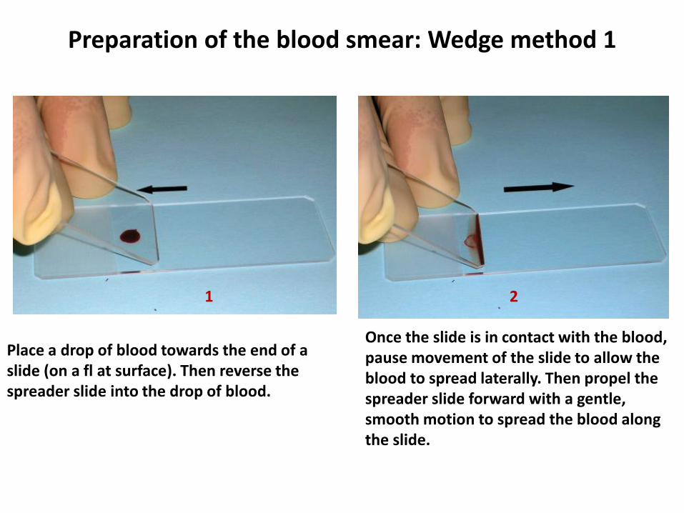
Preparation of the blood smear: Wedge method 1
Place a drop of blood towards the end of a slide (on a fl at surface). Then reverse the spreader slide into the drop of blood.
Once the slide is in contact with the blood, pause movement of the slide to allow the blood to spread laterally. Then propel the spreader slide forward with a gentle, smooth motion to spread the blood along the slide.
2 1
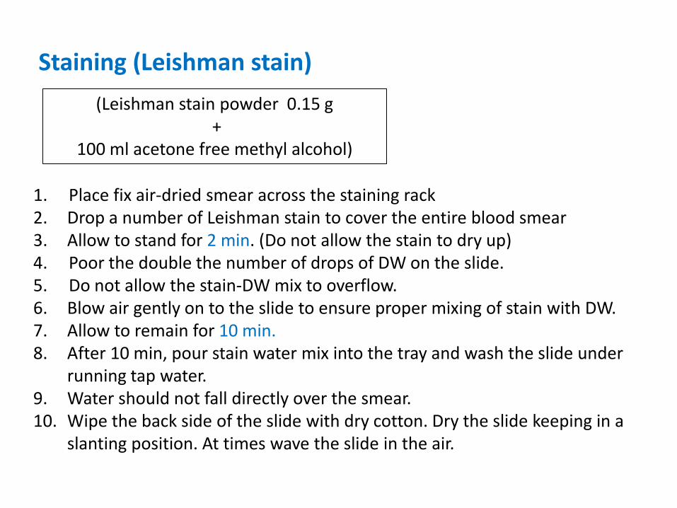
1. Place fix air-dried smear across the staining rack 2. Drop a number of Leishman stain to cover the entire blood smear 3. Allow to stand for 2 min. (Do not allow the stain to dry up) 4. Poor the double the number of drops of DW on the slide. 5. Do not allow the stain-DW mix to overflow. 6. Blow air gently on to the slide to ensure proper mixing of stain with DW. 7. Allow to remain for 10 min. 8. After 10 min, pour stain water mix into the tray and wash the slide under
running tap water. 9. Water should not fall directly over the smear. 10. Wipe the back side of the slide with dry cotton. Dry the slide keeping in a
slanting position. At times wave the slide in the air.
Staining (Leishman stain)
(Leishman stain powder 0.15 g +
100 ml acetone free methyl alcohol)
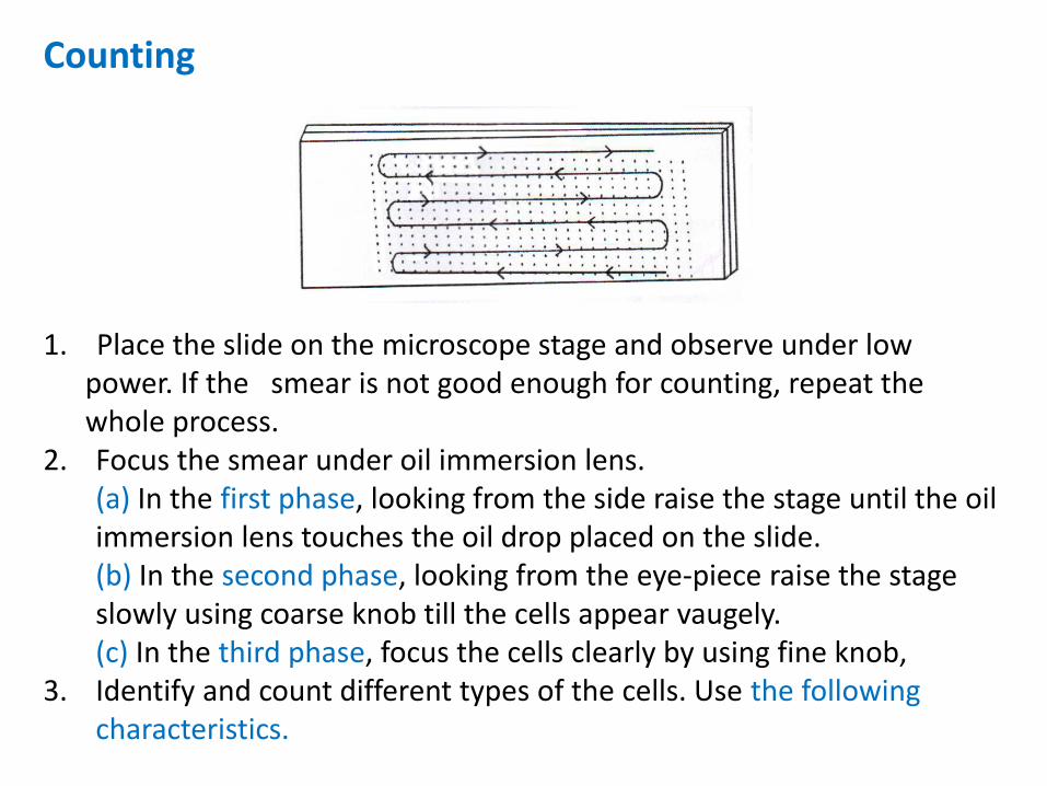
Counting
1. Place the slide on the microscope stage and observe under low power. If the smear is not good enough for counting, repeat the whole process.
2. Focus the smear under oil immersion lens. (a) In the first phase, looking from the side raise the stage until the oil
immersion lens touches the oil drop placed on the slide. (b) In the second phase, looking from the eye-piece raise the stage
slowly using coarse knob till the cells appear vaugely. (c) In the third phase, focus the cells clearly by using fine knob, 3. Identify and count different types of the cells. Use the following
characteristics.
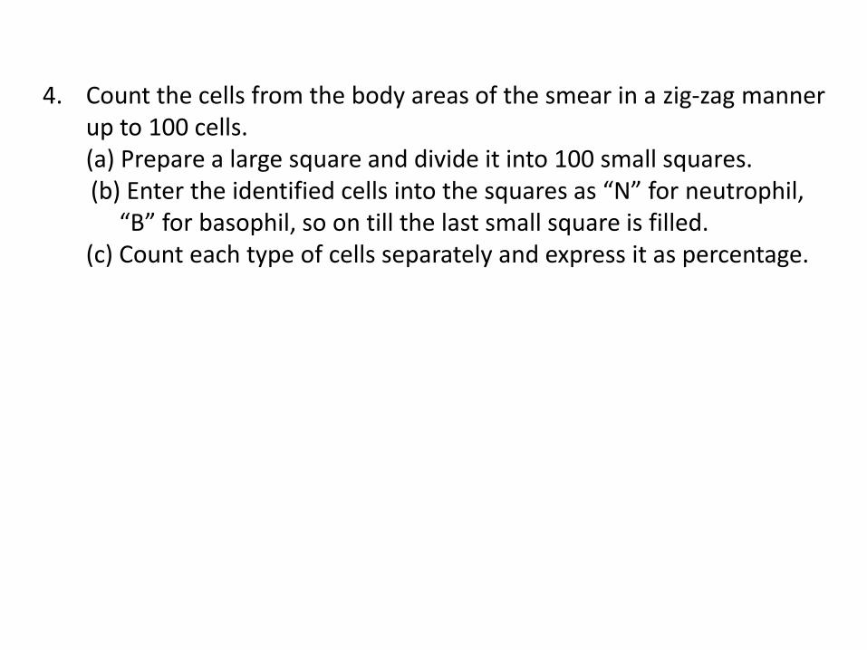
4. Count the cells from the body areas of the smear in a zig-zag manner up to 100 cells.
(a) Prepare a large square and divide it into 100 small squares. (b) Enter the identified cells into the squares as “N” for neutrophil,
“B” for basophil, so on till the last small square is filled. (c) Count each type of cells separately and express it as percentage.
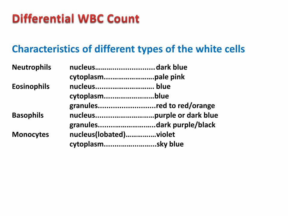
Neutrophils nucleus……….................... dark blue cytoplasm....………………….pale pink Eosinophils nucleus........…………………. blue cytoplasm.....…………………blue granules...........................red to red/orange Basophils nucleus.........…………………purple or dark blue granules........…………….….. dark purple/black Monocytes nucleus(lobated)………….…violet cytoplasm........……...……...sky blue
Characteristics of different types of the white cells
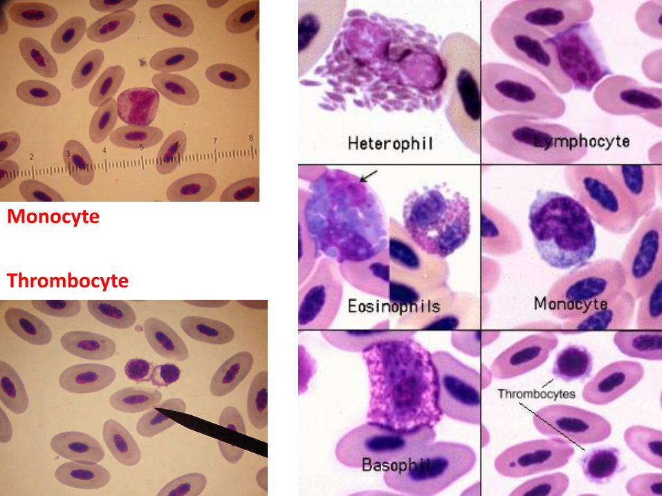
Monocyte
Thrombocyte
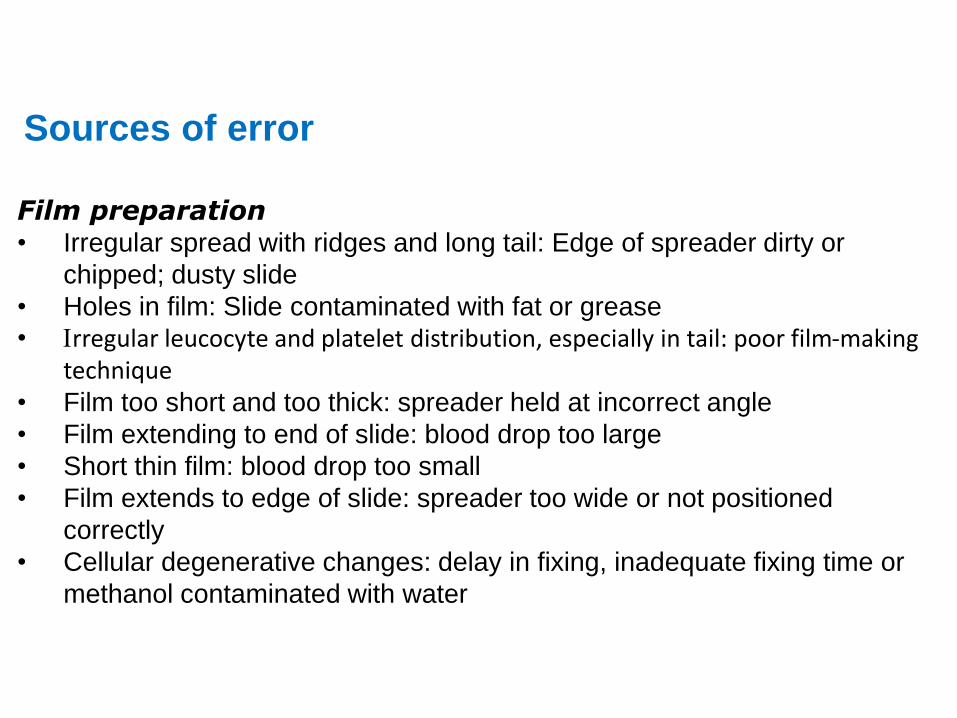
Sources of error
Film preparation
• Irregular spread with ridges and long tail: Edge of spreader dirty or
chipped; dusty slide
• Holes in film: Slide contaminated with fat or grease
• Irregular leucocyte and platelet distribution, especially in tail: poor film-making technique
• Film too short and too thick: spreader held at incorrect angle
• Film extending to end of slide: blood drop too large
• Short thin film: blood drop too small
• Film extends to edge of slide: spreader too wide or not positioned
correctly
• Cellular degenerative changes: delay in fixing, inadequate fixing time or
methanol contaminated with water
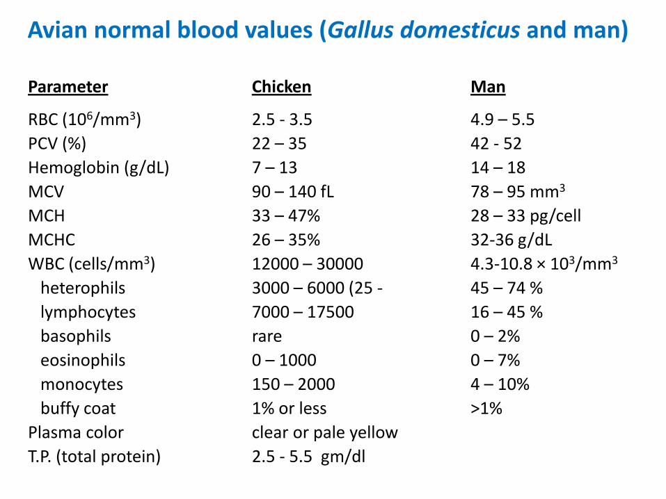
Parameter Chicken Man
RBC (106/mm3) 2.5 - 3.5 4.9 – 5.5
PCV (%) 22 – 35 42 - 52
Hemoglobin (g/dL) 7 – 13 14 – 18
MCV 90 – 140 fL 78 – 95 mm3
MCH 33 – 47% 28 – 33 pg/cell
MCHC 26 – 35% 32-36 g/dL
WBC (cells/mm3) 12000 – 30000 4.3-10.8 × 10³/mm3
heterophils 3000 – 6000 (25 - 45 – 74 %
lymphocytes 7000 – 17500 16 – 45 %
basophils rare 0 – 2%
eosinophils 0 – 1000 0 – 7%
monocytes 150 – 2000 4 – 10%
buffy coat 1% or less >1%
Plasma color clear or pale yellow
T.P. (total protein) 2.5 - 5.5 gm/dl
Avian normal blood values (Gallus domesticus and man)
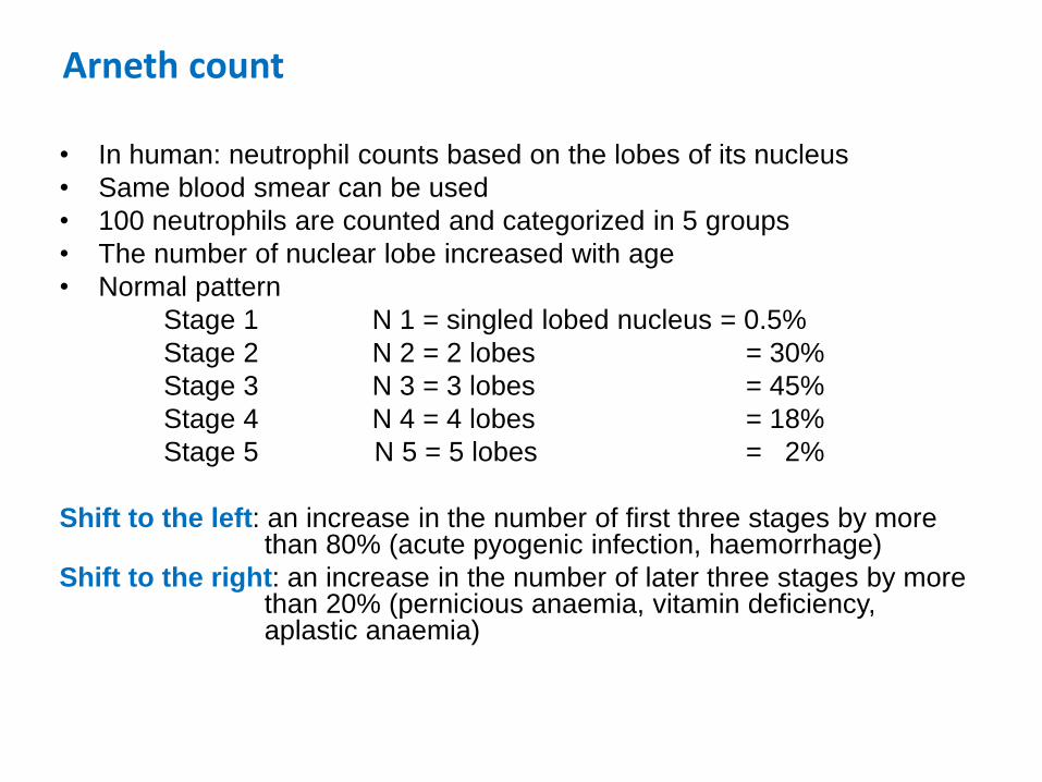
Arneth count
• In human: neutrophil counts based on the lobes of its nucleus
• Same blood smear can be used
• 100 neutrophils are counted and categorized in 5 groups
• The number of nuclear lobe increased with age
• Normal pattern
Stage 1 N 1 = singled lobed nucleus = 0.5%
Stage 2 N 2 = 2 lobes = 30%
Stage 3 N 3 = 3 lobes = 45%
Stage 4 N 4 = 4 lobes = 18%
Stage 5 N 5 = 5 lobes = 2%
Shift to the left: an increase in the number of first three stages by more than 80% (acute pyogenic infection, haemorrhage)
Shift to the right: an increase in the number of later three stages by more than 20% (pernicious anaemia, vitamin deficiency, aplastic anaemia)
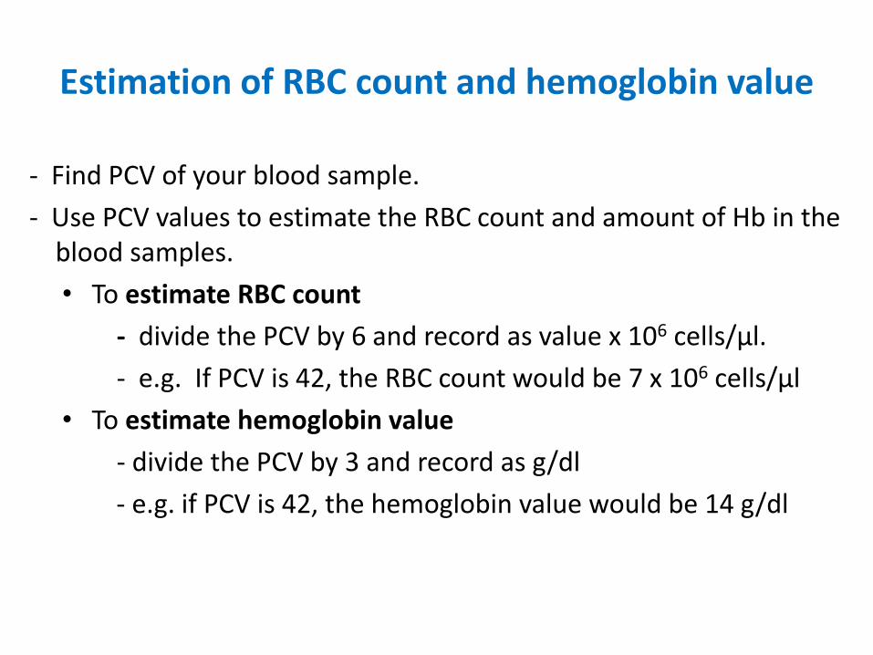
Estimation of RBC count and hemoglobin value
- Find PCV of your blood sample.
- Use PCV values to estimate the RBC count and amount of Hb in the blood samples.
• To estimate RBC count
- divide the PCV by 6 and record as value x 106 cells/µl.
- e.g. If PCV is 42, the RBC count would be 7 x 10⁶ cells/µl
• To estimate hemoglobin value
- divide the PCV by 3 and record as g/dl
- e.g. if PCV is 42, the hemoglobin value would be 14 g/dl
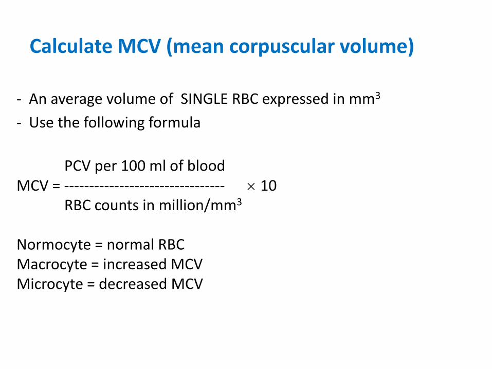
Calculate MCV (mean corpuscular volume)
- An average volume of SINGLE RBC expressed in mm3
- Use the following formula
PCV per 100 ml of blood MCV = -------------------------------- 10 RBC counts in million/mm3 Normocyte = normal RBC Macrocyte = increased MCV Microcyte = decreased MCV
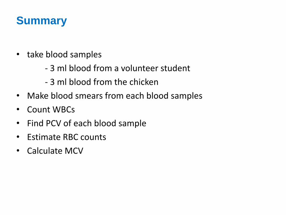
Summary
• take blood samples
- 3 ml blood from a volunteer student
- 3 ml blood from the chicken
• Make blood smears from each blood samples
• Count WBCs
• Find PCV of each blood sample
• Estimate RBC counts
• Calculate MCV
Format of the report
DVT 1073 Fisiologi Haiwan II
Lab work no. ____
Name: ____________ No. Matrik: ______________ Date: ________
• Title of the laboratory work.
• Aim
• Requirements
• Procedures
• Observation and findings
• Discussion/Comments/conclusion
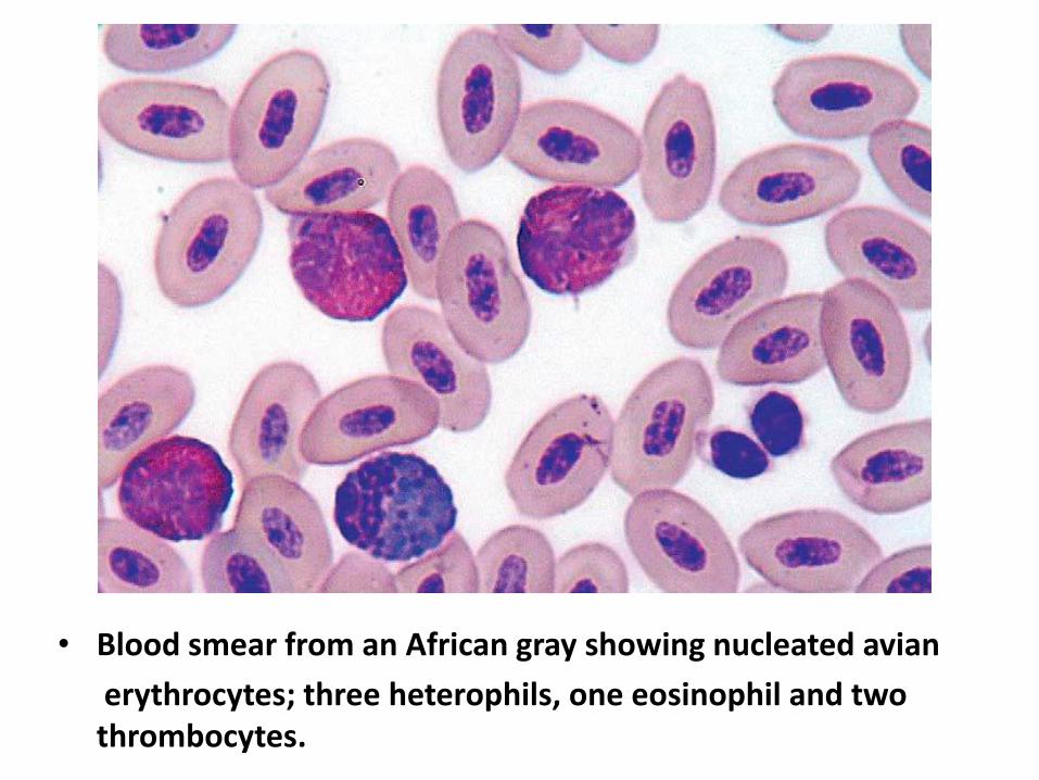
• Blood smear from an African gray showing nucleated avian
erythrocytes; three heterophils, one eosinophil and two thrombocytes.