advances in animal and veterinary sciences research ...€¦ · hence fodder constraint has already...
TRANSCRIPT
NE USAcademic Publishers
Advances in Animal and Veterinary Sciences
February 2016 | Volume 4 | Issue 2 | Page 78
INTRODUCTION
Plants form the major source of nourishment for the livestock. Animals often feed on the highly palatable
and highly nutritious fodder, however during times of scarcity, will be forced to feed on the less palatable, noxious or toxic weeds (Prasad et al., 2005). Kerala, the tiny south-ern Indian state has severe scarcity of cultivable land and hence fodder constraint has already broken the backbone of livestock industry. This will affect the production perfor-mance of the crossbred cattle of the state. Chromolaena od-orata (L.) King and Robins formerly known as Eupatorium odoratum L. (family Asteraceae) is a fast growing perennial weed found mostly in farm lands (Aro et al., 2009). It is
commonly called as ‘Communist Pacha’ in Malayalam and has an even distribution throughout Kerala. Cattle occa-sionally graze on this plant due to the fodder scarcity.
The medicinal uses of the weed were largely exploited by traditional herbalists and were found useful in the man-agement of burns, soft tissue wounds, skin infections (Phan et al., 2000; Phan et al., 2001; Panda and Ghosh, 2010), inflammation (Habtemariam, 2001; Owoyele et al., 2005; Ayyanar and Ignacimuthu, 2009; Hanh et al., 2011), malaria (Pisutthanan et al., 2005), diarrhea and diabetes (Odugbemi, 2006; Akinmoladun and Akinloye, 2007). In traditional medicine, decoction of the leaf was used as a cough remedy and as an ingredient with lemon grass and
Research Article
Abstract | Toxicological potential of Chromolaena odorata, a fast growing perennial weed was assessed in mice. Meth-anolic extract of C. odorata did not cause any mortality of mice. However, the oral feeding of extract resulted in seda-tion, loss of appetite, enteritis and decreased weight gain in treated mice. Significant elevation (P<0.05) of the serum glutamic oxalo-acetic transaminase (SGOT) and serum glutamic pyruvic transaminase (SGPT) was observed along with decrease in the serum superoxide dismutase (SOD) and catalase. No significant variation in the haematological parameters was observed in the treated mice except for the increase in counts of monocytes, lymphocytes as well as clotting and bleeding times. Gross pathological examination of extract treated mice revealed subcutaneous hemor-rhages in different parts of the body, pale and friable liver, soft and congested lungs and kidneys. Histopathology of the liver revealed centrilobular necrosis with varying degrees of degenerative changes ranging from cloudy swelling to vacuolar changes of the periportal hepatic cells, congestion of the portal vein and proliferation of endothelial cells. Hence, the methanolic extract of invasive form of C. odorata found in the Wayanad region of Western Ghats of Indian subcontinent possesses severe toxic potential when given orally in mice.
Keywords | C. odorata, Enteritis, Centrilobular necrosis, Liver, Haemorrhages, Kidney, Lungs
Praseena Paulose1, sanis Juliet1, samraJ suJith1, mechery sini1, thirumangalath meethal Divya1, suresh n nair1, leena chanDrasekhar2, mamPillikalam PraDeeP3, aJith Jacob george3, reghu ravinDran4*
Evaluation of Toxicological Potential of Methanolic Extract of Chromolaena odorata found in the Western Ghats of Indian Subcontinent Orally in Mice
Editor | Kuldeep Dhama, Indian Veterinary Research Institute, Uttar Pradesh, India.Received | November 27, 2015; Revised | December 12, 2015; Accepted | December 14, 2015; Published | January 23, 2016 *Correspondence | reghu ravinDran, Kerala Veterinary and Animal Sciences University, Pookode, Wayanad, Kerala, India; Email: [email protected] | Paulose P, Juliet S, Sujith S, Sini M, Divya TM, Nair SN, Chandrasekhar L, Pradeep M, George AJ, Ravindran R (2016). Evaluation of toxicological potential of methanolic extract of Chromolaena odorata found in the Western Ghats of Indian subcontinent orally in mice. Adv. Anim. Vet. Sci. 4(2): 78-84. DOI | http://dx.doi.org/10.14737/journal.aavs/2016/4.2.78.84ISSN (Online) | 2307-8316; ISSN (Print) | 2309-3331
Copyright © 2016 Paulose et al. This is an open access article distributed under the Creative Commons Attribution License, which permits unrestricted use, distribution, and reproduction in any medium, provided the original work is properly cited.
1Department of Veterinary Pharmacology and Toxicology; 2Department of Veterinary Anatomy and Histology; 3Vet-erinary Pathology; 4Veterinary Parasitology, College of Veterinary and Animal Sciences, Kerala Veterinary and Ani-mal Sciences University, Pookode, Wayanad, Kerala- 673576, India.
NE USAcademic Publishers
Advances in Animal and Veterinary Sciences
February 2016 | Volume 4 | Issue 2 | Page 79
guava leaves for the treatment of malaria and decoction of flowers was used as tonic, antipyretic and heart tonic (Vital and Rivera, 2009). In addition, anthelmintic and antimicro-bial activities of the leaves of the plant were widely studied (Debashisha et al., 2010; Naidoo et al., 2011; Natheer et al., 2012). The fresh leaf was ground into a paste and applied topically on affected places to heal wounds (Kilani, 2006). The plant also possessed antibacterial, astringent, anti-oxidant, antipyretic and diuretic activities (Caceres et al., 1995; Gopinath et al., 2009). Besides, the hydroalcoholic extract of C. odorata was reported to possess hypoglycaemic activity and thereby reducing the cardiovascular risks (Og-bonnia et al., 2010). Additionally, the aqueous extract of leaves of C. odorata also protected against the development of atherosclerosis and coronary heart disease, as well as the dyslipidemic conditions that characterized obesity, hyper-tension and diabetes mellitus ( Jude and Catherine, 2011). Furthermore, the immunopotentiating activities of the C. odorata leaf extract on the innate immunity of Balb/C mice were also reported (Leonora and Elena, 2012). The use of C. odorata in the formulation of diet for layers, monogastric animals and West African dwarf goats (Aro et al., 2009) were previously reported.
C. odorata extract owes its biological activities to the presence of a mixture of secondary plant products com-prising of polyphenols with flavonoids as the major con-stituent followed by alkaloids. At least ten different fla-vonoids, including 3, 5, 4’-trihydroxy-7-methoxyflavone, 5, 7, 3’- trihydroxy-5 -methoxyflavone and 3, 5, 7- trihy-droxy-4’-methoxyflavone (Ling et al., 2007) were isolated and characterized.
Previous studies conducted in our own laboratory, con-firmed the wound healing, anti-inflammatory as well as antimicrobial potentials of the plant leaf extracts (Unpub-lished). The dermal toxicity studies also confirmed that there was no cutaneous toxicity (Unpublished). However, the presence of sesquiterpene lactones and pyrrolizidine al-kaloids in the plant was speculated as the cause of the acute toxicity resulting in death of cattle and goats in Karnataka (Prasad et al., 2005).
The increasing use of traditional medicines necessitates more scientific evidences of their harmlessness; hence there is an urgent need for systematic approach in evalu-ating their efficacy and safety profile. The secondary plant metabolites like other xenobiotics, are usually detoxified in the liver. In this process, they may damage the liver or form an active metabolite that will cause toxicity to other organs (Flanagan et al., 2007). Thus, the present study was un-dertaken to evaluate the toxic potential of the methanolic extract of C. odorata found in the Western Ghats of the Indian subcontinent orally in mice.
MATERIALS AND METHODS
Plant materialThe fresh aerial parts of Chromolaena odorata were collect-ed from the campus of College of Veterinary and Animal Sciences, Pookode, Wayanad district, Kerala. The plant was identified and authenticated by a botanist and a vouch-er specimen deposited at Calicut University Herbarium (CALI, Accession number: 6629), Calicut, Kerala for fur-ther reference.
chemicalsAll the chemicals, kits and solvents used in this study were obtained from M/s Merck India, Ltd (Bombay, India).
PreParation of Plant extractThe collected aerial parts of C. odorata were dried under shade and pulverized using a mechanical grinder. The pow-dered plant material (~2 kg) was extracted in methanol us-ing a soxhlet extraction apparatus attached with rotary vac-uum evaporator. The solvent was removed under reduced pressure (74 mbar) at 31°C using a rotary vacuum evap-orator (Rotavac, Buchi, Switzerland) followed by drying at room temperature. The resulting dry methanolic extract was weighed. The per cent yield was calculated as 12.5 per cent (w/w). The extract was then stored in a refrigerator at 4°C. The appropriate dose of the dried extract was dis-solved in distilled water and administered orally to animals limiting the final volume to 1mL.
animalsTwenty four male or female Balb/c mice weighing 20-25 g were obtained from the Small Animal Breeding Station, Pookode. All the animals were housed in an animal room under standard laboratory conditions of 21-22°C, light (12 h light/dark cycle) and humidity (50 + 10 per cent). The animals were provided standard laboratory food and water ad libitum. They were acclimatized for 7 days prior to ex-periments. The experiments were performed according to the guiding principles in the use of animals in toxicology and approved by the Institutional Animal Ethics Commit-tee.
Phytochemical evaluation of the cruDe extractPhytochemical analysis of the extract was performed on a qualitative basis and on TLC plate (Harborne, 1991).
exPerimental DesignThe study was conducted for a period of 14 days. On the day of dosing, all animals were observed for mortality and clinical signs for the first 10 min, 30 min, 1, 2, 4 and 6 h after dosing. Thereafter, the animals were observed twice daily for mortality and clinical signs, for 14 days. The tests
NE USAcademic Publishers
Advances in Animal and Veterinary Sciences
February 2016 | Volume 4 | Issue 2 | Page 80
were performed between 9 AM and 1 PM. Initial and final body weights of all animals were also recorded.
The mice were randomly divided into four groups each comprising of six animals. Group I animals served as con-trol and received sterile distilled water orally for 14 days. Group II animals received carbon tetrachloride (CCl4) at 0.5 mL/kg body weight in olive oil in 1:1 ratio as intra-peritoneal injection (single day). Group III and IV ani-mals received methanolic extract of C. odorata at the rate of 250 mg/Kg and 500 mg/Kg orally for 14 days. The body weights of these animals prior to and after the experiments were also noted.
Prior to sacrifice of treated animals, blood was withdrawn from the carotid artery with or without anticoagulant into sterile vials. Blood samples without anticoagulant were al-lowed to coagulate at 37οC for 30 minutes, refrigerated for 3hr followed by centrifugation at 5000 rpm for separation of serum. Serum samples were analyzed for SGOT and SGPT. Blood samples collected in vials containing diso-dium salt of ethylenediaminetetraacetic acid (EDTA sodi-um) at the rate of 1 mg/mL were used for the estimation of haematological parameters like total leukocyte count (TLC), total erythrocyte count (TEC), haemoglobin con-centration (Hb), erythrocyte sedimentation rate (ESR), bleeding and clotting time and differential leucocyte count (DLC) (Schlam, 1975).
The animals of each group were sacrificed by anesthetic over dosage using ether. The liver and kidney were rapid-ly dissected, placed in ice cold normal saline for the es-timation of specific enzymes like superoxide dismutase (Mimami and Yoshikawa, 1979) (SOD) and catalase (Co-hen et al., 1970). The fresh tissues were analyzed immedi-ately after collection. The tissues (1g) were homogenized using glass homogenizer with 9 mL of ice cold normal saline and were used for the enzyme estimation. The liver, kidney and lungs were promptly excised and fixed in 10 per cent formal saline for histopathological examination. The specimens were processed by standard procedure after embedding in paraffin wax. The blocks were sectioned at 5 µm thickness, stained using haematoxylin and eosin meth-od (Smit and Burton, 1977) and examined microscopically.
statistical analysis of DataData were expressed as the mean ± S.E.M. (Standard Er-ror of Mean). Statistical differences between control and treated groups were tested by one way ANOVA, followed by post-hoc analysis using standard SPSS software. For post hoc analysis Duncan’s test was used. A value of P < 0.05 was considered to indicate statistical significance.
RESULTS AND DISCUSSION
In the present study, the phytochemical analysis of the crude methanolic extract of C. odorata revealed the presence of alkaloids, steroids, glycosides, flavonoids, diterpenes, tri-terpenes and phenolic compounds. In general, secondary metabolites played protective roles (e.g. anti-oxidation, free radical scavenging, UV light-absorption, and anti-prolifer-ation) and defended the plant against microorganisms such as bacteria, fungi and viruses. Many of these metabolites were bitter and / or toxic to potential herbivores, some-times affecting even the herbivore’s central and peripheral nervous systems (Rattan, 2010). The secondary metabolites were further subdivided into a number of distinct groups on the basis of their chemical structure and synthetic path-ways. The most prevalent phytochemical groups being al-kaloids, terpenes and phenolic compounds. Plant-derived alkaloids, by function and chemical nature, were generally toxic to mammals. Terpenes, on the other hand exhibited a wide range of effects within the CNS by their interactions with the noradrenergic system, cholinesterase inhibition and multiple direct interactions with the GABA system, including blockade of GABA-gated chloride channels and agonistic and antagonistic, direct and allosteric binding to GABAA receptors (Rattan, 2010).
On gross evaluation of toxicity, no mortality was recorded until fourteen days in any of the groups. The animals of ex-tract treated groups (group III and IV) exhibited sedation, 45 minutes post-treatment which persisted for 1.5 hours. An initial increase in appetite was observed in these ani-mals which progressively decreased at the end of observa-tion period. After the fifth day, the animals showed signs of enteritis and the whole perennial region of the extract treated animals were stained with faeces. By 14th day of the experiment all the animals were emaciated, totally exhaust-ed and anorectic. The animals showed ‘tucked up’ abdomen and difficulty in breathing. The skin textures were lost and the hairs were curled in. Therefore, the toxicities observed in the present study were attributed to the presence of al-kaloids and terpenes. Studies have further revealed that morbidity and mortality associated with exposure to terpe-nes were largely related to the degree of CNS depression. Terpenes being local irritants were also capable of causing GI signs and symptoms.
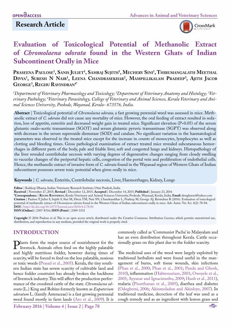
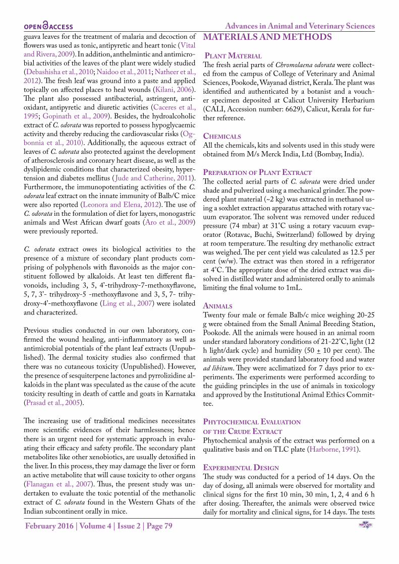
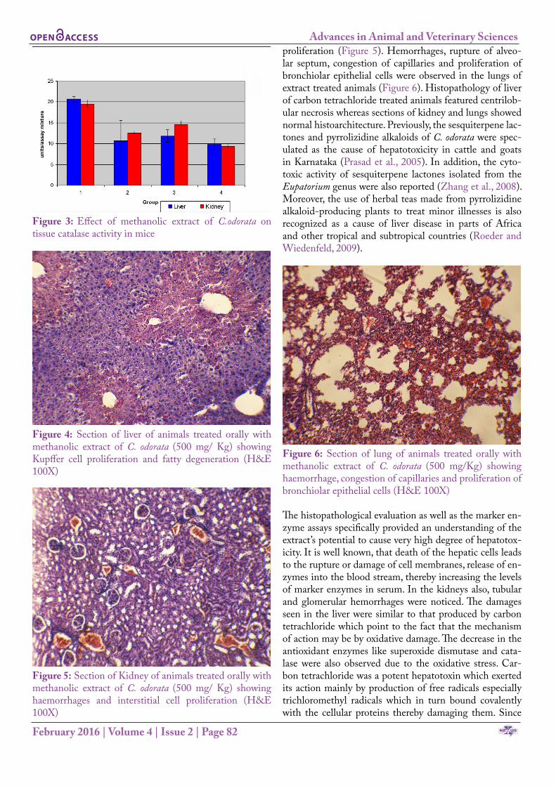
Assay of the biomarker enzymes revealed that the SGOT (AST) and SGPT (ALT) were significantly (P<0.05) ele-vated in all experimental groups compared to the control group (Figure 1). Whereas, there was a significant (P<0.05) decrease in the levels of antioxidant enzymes (SOD and catalase) (Figure 2 and 3) in the treated groups compared to control groups. However, no significant changes in the total erythrocyte and leucocyte counts as well as hemo-globin were observed in the treated animals compared to
NE USAcademic Publishers
Advances in Animal and Veterinary Sciences
February 2016 | Volume 4 | Issue 2 | Page 81
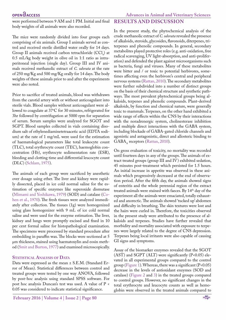
Table 1: Effect of methanolic extract of C. odorata on total erythrocyte count, total leucocyte count and haemoglobulin in mice Group TEC (million/mm3) TLC (thousands /mm3) Hb (g/dl)I- Control 9.79 + 1.09a 10400 + 110.12a 11.8+ 1.34a
II- CCl4 @ 0.5 mL/kg 13.12+ 0.93b 8900 +100.12b 12.4 +1.94a
III – Methanolic extract @ 250 mg/ kg 13.79+ 0.87b 14600 + 400.64c 13.4+3.38a
IV- Methanolic extract @500 mg /kg 13.55+ 0.77b 8900 + 110.11b 13.6+3.42a
TEC = Total erythrocyte count; TLC = total leucocyte count; Hb = haemoglobulin; CCl4 = Carbon tetra chloride; n = 6; Values are Mean ± SEM, means bearing different superscripts a, b or c (P<0.05).
Table 2: Effect of methanolic extract of C. odorata on the differential leucocyte count in mice Group Lymphocytes (%) Neutrophils (%) Monocytes (%) Eosinophils (%)I- Control 89.50 + 1.05a 10.50 + 1.04a 0 0
II- CCl4 @ 0.5 mL/kg 84.33 + 2.59a 15.67 + 2.58b 0 0
III – Methanolic extract @ 250 mg/ kg 81.50 + 0.55ab 16.0 + 0.89b 2.50 + 0.84a 0
IV- Methanolic extract @500 mg /kg 62.17 + 1.6b 31.33 + 1.37c 6.50 + 1.05b 0CCL4=Carbon tetra chloride; n = 6; Values are Mean ± SEM, means bearing different superscripts a, b or c (P<0.05).
the control (Table 1). The clotting time as well as bleed-ing time were long in treated group compared to control. Methanolic extract of C. odorata at concentration of 500 mg/Kg significantly (P<0.05) altered the counts of lym-phocytes, neutrophils and monocytes in all treated groups compared to control (Table 2). The increase in the total leukocyte count was significant (P<0.05) in carbon tetra-chloride treated and group IV as well compared to control.
Figure 1: Effect of methanolic extract of C.odorata on serum AST and ALT in mice
Gross pathological examination of animals of extract treat-ed group III and IV revealed subcutaneous hemorrhage in different parts of the body. The severity of changes were noted more in animals that received higher quantity of the extract. The liver was pale and friable with white spots on the surface. The lungs and the kidneys were congested and soft in consistency. The capsule was adhered to the kidney and was not peeling off easily. The intestinal loops were
bloated with severe catarrhal enteritis. Foci of hemorrhage were observed in the intestinal epithelium. However, no subcutaneous hemorrhage was observed in carbon tetra-chloride treated group. The ballooning of the intestinal loops and catarrhal enteritis revealed the gastrointestinal toxic potential of the extract.
Figure 2: Effect of methanolic extract of C.odorata on tissue superoxide dismutase activity in mice
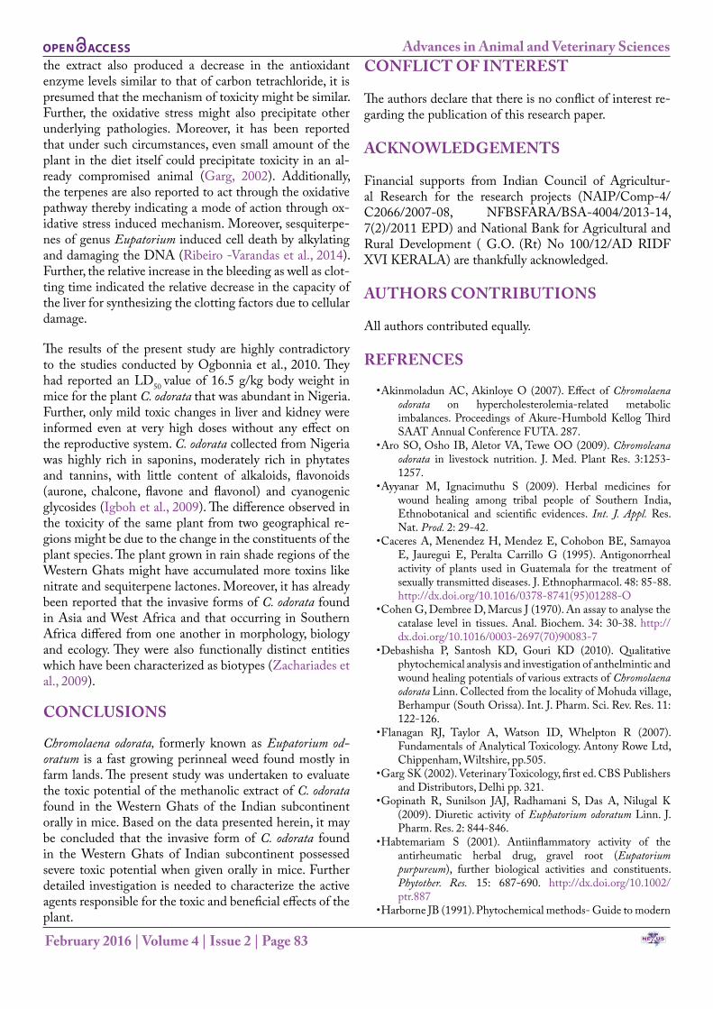
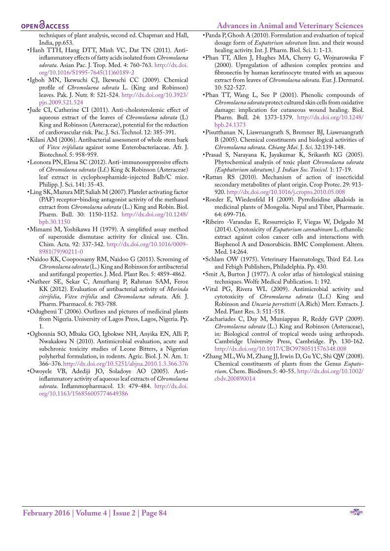
Histopathological examination of the liver sections fur-ther revealed centrilobular necrosis with varying degrees of degenerative changes ranging from cloudy swelling to vacuolar changes of the periportal hepatic cells, congestion of the portal vein and proliferation of endothelial cells in experimental groups. Varying degrees of necrosis were ob-served in hepatocytes. The degree of necrosis was severe in liver of group IV animals to an extent that the cellu-lar architecture were completely lost (Figure 4). Section of kidney of methanolic extract treated animals revealed glomerular and tubular hemorrhages and interstitial cell
NE USAcademic Publishers
Advances in Animal and Veterinary Sciences
February 2016 | Volume 4 | Issue 2 | Page 82
Figure 3: Effect of methanolic extract of C.odorata on tissue catalase activity in mice
Figure 4: Section of liver of animals treated orally with methanolic extract of C. odorata (500 mg/ Kg) showing Kupffer cell proliferation and fatty degeneration (H&E 100X)
Figure 5: Section of Kidney of animals treated orally with methanolic extract of C. odorata (500 mg/ Kg) showing haemorrhages and interstitial cell proliferation (H&E 100X)
proliferation (Figure 5). Hemorrhages, rupture of alveo-lar septum, congestion of capillaries and proliferation of bronchiolar epithelial cells were observed in the lungs of extract treated animals (Figure 6). Histopathology of liver of carbon tetrachloride treated animals featured centrilob-ular necrosis whereas sections of kidney and lungs showed normal histoarchitecture. Previously, the sesquiterpene lac-tones and pyrrolizidine alkaloids of C. odorata were spec-ulated as the cause of hepatotoxicity in cattle and goats in Karnataka (Prasad et al., 2005). In addition, the cyto-toxic activity of sesquiterpene lactones isolated from the Eupatorium genus were also reported (Zhang et al., 2008). Moreover, the use of herbal teas made from pyrrolizidine alkaloid-producing plants to treat minor illnesses is also recognized as a cause of liver disease in parts of Africa and other tropical and subtropical countries (Roeder and Wiedenfeld, 2009).
Figure 6: Section of lung of animals treated orally with methanolic extract of C. odorata (500 mg/Kg) showing haemorrhage, congestion of capillaries and proliferation of bronchiolar epithelial cells (H&E 100X)
The histopathological evaluation as well as the marker en-zyme assays specifically provided an understanding of the extract’s potential to cause very high degree of hepatotox-icity. It is well known, that death of the hepatic cells leads to the rupture or damage of cell membranes, release of en-zymes into the blood stream, thereby increasing the levels of marker enzymes in serum. In the kidneys also, tubular and glomerular hemorrhages were noticed. The damages seen in the liver were similar to that produced by carbon tetrachloride which point to the fact that the mechanism of action may be by oxidative damage. The decrease in the antioxidant enzymes like superoxide dismutase and cata-lase were also observed due to the oxidative stress. Car-bon tetrachloride was a potent hepatotoxin which exerted its action mainly by production of free radicals especially trichloromethyl radicals which in turn bound covalently with the cellular proteins thereby damaging them. Since
NE USAcademic Publishers
Advances in Animal and Veterinary Sciences
February 2016 | Volume 4 | Issue 2 | Page 83
the extract also produced a decrease in the antioxidant enzyme levels similar to that of carbon tetrachloride, it is presumed that the mechanism of toxicity might be similar. Further, the oxidative stress might also precipitate other underlying pathologies. Moreover, it has been reported that under such circumstances, even small amount of the plant in the diet itself could precipitate toxicity in an al-ready compromised animal (Garg, 2002). Additionally, the terpenes are also reported to act through the oxidative pathway thereby indicating a mode of action through ox-idative stress induced mechanism. Moreover, sesquiterpe-nes of genus Eupatorium induced cell death by alkylating and damaging the DNA (Ribeiro -Varandas et al., 2014). Further, the relative increase in the bleeding as well as clot-ting time indicated the relative decrease in the capacity of the liver for synthesizing the clotting factors due to cellular damage.
The results of the present study are highly contradictory to the studies conducted by Ogbonnia et al., 2010. They had reported an LD50 value of 16.5 g/kg body weight in mice for the plant C. odorata that was abundant in Nigeria. Further, only mild toxic changes in liver and kidney were informed even at very high doses without any effect on the reproductive system. C. odorata collected from Nigeria was highly rich in saponins, moderately rich in phytates and tannins, with little content of alkaloids, flavonoids (aurone, chalcone, flavone and flavonol) and cyanogenic glycosides (Igboh et al., 2009). The difference observed in the toxicity of the same plant from two geographical re-gions might be due to the change in the constituents of the plant species. The plant grown in rain shade regions of the Western Ghats might have accumulated more toxins like nitrate and sequiterpene lactones. Moreover, it has already been reported that the invasive forms of C. odorata found in Asia and West Africa and that occurring in Southern Africa differed from one another in morphology, biology and ecology. They were also functionally distinct entities which have been characterized as biotypes (Zachariades et al., 2009).
CONCLUSIONS
Chromolaena odorata, formerly known as Eupatorium od-oratum is a fast growing perinneal weed found mostly in farm lands. The present study was undertaken to evaluate the toxic potential of the methanolic extract of C. odorata found in the Western Ghats of the Indian subcontinent orally in mice. Based on the data presented herein, it may be concluded that the invasive form of C. odorata found in the Western Ghats of Indian subcontinent possessed severe toxic potential when given orally in mice. Further detailed investigation is needed to characterize the active agents responsible for the toxic and beneficial effects of the plant.
CONFLICT OF INTEREST
The authors declare that there is no conflict of interest re-garding the publication of this research paper.
ACKNOWLEDGEMENTS
Financial supports from Indian Council of Agricultur-al Research for the research projects (NAIP/Comp-4/C2066/2007-08, NFBSFARA/BSA-4004/2013-14, 7(2)/2011 EPD) and National Bank for Agricultural and Rural Development ( G.O. (Rt) No 100/12/AD RIDF XVI KERALA) are thankfully acknowledged.
AUTHORS CONTRIBUTIONS
All authors contributed equally.
REFRENCES
• Akinmoladun AC, Akinloye O (2007). Effect of Chromolaena odorata on hypercholesterolemia-related metabolic imbalances. Proceedings of Akure-Humbold Kellog Third SAAT Annual Conference FUTA. 287.
• Aro SO, Osho IB, Aletor VA, Tewe OO (2009). Chromoleana odorata in livestock nutrition. J. Med. Plant Res. 3:1253-1257.
• Ayyanar M, Ignacimuthu S (2009). Herbal medicines for wound healing among tribal people of Southern India, Ethnobotanical and scientific evidences. Int. J. Appl. Res. Nat. Prod. 2: 29-42.
• Caceres A, Menendez H, Mendez E, Cohobon BE, Samayoa E, Jauregui E, Peralta Carrillo G (1995). Antigonorrheal activity of plants used in Guatemala for the treatment of sexually transmitted diseases. J. Ethnopharmacol. 48: 85-88. http://dx.doi.org/10.1016/0378-8741(95)01288-O
• Cohen G, Dembree D, Marcus J (1970). An assay to analyse the catalase level in tissues. Anal. Biochem. 34: 30-38. http://dx.doi.org/10.1016/0003-2697(70)90083-7
• Debashisha P, Santosh KD, Gouri KD (2010). Qualitative phytochemical analysis and investigation of anthelmintic and wound healing potentials of various extracts of Chromolaena odorata Linn. Collected from the locality of Mohuda village, Berhampur (South Orissa). Int. J. Pharm. Sci. Rev. Res. 11: 122-126.
• Flanagan RJ, Taylor A, Watson ID, Whelpton R (2007). Fundamentals of Analytical Toxicology. Antony Rowe Ltd, Chippenham, Wiltshire, pp.505.
• Garg SK (2002). Veterinary Toxicology, first ed. CBS Publishers and Distributors, Delhi pp. 321.
• Gopinath R, Sunilson JAJ, Radhamani S, Das A, Nilugal K (2009). Diuretic activity of Euphatorium odoratum Linn. J. Pharm. Res. 2: 844-846.
• Habtemariam S (2001). Antiinflammatory activity of the antirheumatic herbal drug, gravel root (Eupatorium purpureum), further biological activities and constituents. Phytother. Res. 15: 687-690. http://dx.doi.org/10.1002/ptr.887
• Harborne JB (1991). Phytochemical methods- Guide to modern
NE USAcademic Publishers
Advances in Animal and Veterinary Sciences
February 2016 | Volume 4 | Issue 2 | Page 84
techniques of plant analysis, second ed. Chapman and Hall, India, pp.653.
• Hanh TTH, Hang DTT, Minh VC, Dat TN (2011). Anti-inflammatory effects of fatty acids isolated from Chromolaena odorata. Asian Pac. J. Trop. Med. 4: 760-763. http://dx.doi.org/10.1016/S1995-7645(11)60189-2
• Igboh MN, Ikewuchi CJ, Ikewuchi CC (2009). Chemical profile of Chromolaena odorata L. (King and Robinson) leaves. Pak. J. Nutr. 8: 521-524. http://dx.doi.org/10.3923/pjn.2009.521.524
• Jude CI, Catherine CI (2011). Anti-cholesterolemic effect of aqueous extract of the leaves of Chromolaena odorata (L) King and Robinson (Asteraceae), potential for the reduction of cardiovascular risk. Pac. J. Sci. Technol. 12: 385-391.
• Kilani AM (2006). Antibacterial assessment of whole stem bark of Vitex trifoliata against some Enterobacteriaceae. Afr. J. Biotechnol. 5: 958-959.
• Leonora PN, Elena SC (2012). Anti-immunosuppressive effects of Chromolaena odorata (Lf.) King & Robinson (Asteraceae) leaf extract in cyclophosphamide-injected Balb/C mice. Philipp. J. Sci. 141: 35-43.
• Ling SK, Mazura MP, Saliah M (2007). Platelet activating factor (PAF) receptor–binding antagonist activity of the methanol extract from Chromolaena odorata (L.) King and Robin. Biol. Pharm. Bull. 30: 1150-1152. http://dx.doi.org/10.1248/bpb.30.1150
• Mimami M, Yoshikawa H (1979). A simplified assay method of superoxide dismutase activity for clinical use. Clin. Chim. Acta. 92: 337-342. http://dx.doi.org/10.1016/0009-8981(79)90211-0
• Naidoo KK, Coopoosamy RM, Naidoo G (2011). Screening of Chromolaena odorata (L.) King and Robinson for antibacterial and antifungal properties. J. Med. Plant Res. 5: 4859-4862.
• Natheer SE, Sekar C, Amutharaj P, Rahman SAM, Feroz KK (2012). Evaluation of antibacterial activity of Morinda citrifolia, Vitex trifolia and Chromolaena odorata. Afr. J. Pharm. Pharmacol. 6: 783-788.
• Odugbemi T (2006). Outlines and pictures of medicinal plants from Nigeria. University of Lagos Press, Lagos, Nigeria. Pp. 1.
• Ogbonnia SO, Mbaka GO, Igbokwe NH, Anyika EN, Alli P, Nwakakwa N (2010). Antimicrobial evaluation, acute and subchronic toxicity studies of Leone Bitters, a Nigerian polyherbal formulation, in rodents. Agric. Biol. J. N. Am. 1: 366–376. http://dx.doi.org/10.5251/abjna.2010.1.3.366.376
• Owoyele VB, Adediji JO, Soladoye AO (2005). Anti-inflammatory activity of aqueous leaf extracts of Chromolaena odorata. Inflammopharmacol. 13: 479-484. http://dx.doi.org/10.1163/156856005774649386
• Panda P, Ghosh A (2010). Formulation and evaluation of topical dosage form of Eupatorium odoratum linn. and their wound healing activity. Int. J. Pharm. Biol. Sci. 1: 1-13.
• Phan TT, Allen J, Hughes MA, Cherry G, Wojnarowska F (2000). Upregulation of adhesion complex proteins and fibronectin by human keratinocyte treated with an aqueous extract from leaves of Chromolaena odorata. Eur. J. Dermatol. 10: 522-527.
• Phan TT, Wang L, See P (2001). Phenolic compounds of Chromolaena odorata protect cultured skin cells from oxidative damage: implication for cutaneous wound healing. Biol. Pharm. Bull. 24: 1373-1379. http://dx.doi.org/10.1248/bpb.24.1373
• Pisutthanan N, Liawruangrath S, Bremner BJ, Liawruangrath B (2005). Chemical constituents and biological activities of Chromolaena odorata. Chiang Mai. J. Sci. 32:139-148.
• Prasad S, Narayana K, Jayakumar K, Srikanth KG (2005). Phytochemical analysis of toxic plant Chromolaena odorata (Euphatorium odoratum). J. Indian Soc. Toxicol. 1: 17-19.
• Rattan RS (2010). Mechanism of action of insecticidal secondary metabolites of plant origin. Crop Protec. 29: 913-920. http://dx.doi.org/10.1016/j.cropro.2010.05.008
• Roeder E, Wiedenfeld H (2009). Pyrrolizidine alkaloids in medicinal plants of Mongolia. Nepal and Tibet, Pharmazie. 64: 699-716.
• Ribeiro -Varandas E, Ressurreição F, Viegas W, Delgado M (2014). Cytotoxicity of Eupatorium cannabinum L. ethanolic extract against colon cancer cells and interactions with Bisphenol A and Doxorubicin. BMC Complement. Altern. Med. 14:264.
• Schlam OW (1975). Veterinary Haematology, Third Ed. Lea and Febigh Publishers, Philadelphia. Pp. 430.
• Smit A, Burton J (1977). A color atlas of histological staining techniques. Wolfe Medical Publication. 1: 192.
• Vital PG, Rivera WL (2009). Antimicrobial activity and cytotoxicity of Chromolaena odorata (L.f.) King and Robinson and Uncaria perrottetti (A.Rich) Merr. Extracts. J. Med. Plant Res. 3: 511-518.
• Zachariades C, Day M, Muniappan R, Reddy GVP (2009). Chromolaena odorata (L.) King and Robinson (Asteraceae), in: Biological control of tropical weeds using arthropods. Cambridge University Press, Cambridge. Pp. 130-162. http://dx.doi.org/10.1017/CBO9780511576348.008
• Zhang ML, Wu M, Zhang JJ, Irwin D, Gu YC, Shi QW (2008). Chemical constituents of plants from the Genus Eupato-rium. Chem. Biodivers.5: 40-55. http://dx.doi.org/10.1002/cbdv.200890014