adrenal gland scintigraphy -...
TRANSCRIPT
A
A
AcecmsrIttctmmwaum
*
†A
2
drenal Gland Scintigraphy
nca M. Avram, MD,* Lorraine M. Fig, MBChB, MPH,*,† and Milton D. Gross, MD*,†
There is no question that high-resolution imaging techniques have revolutionized theapproach to diagnostic imaging. Computed tomography (CT) and magnetic resonanceimaging provide exquisite images of the adrenal glands and offer the best initial imagingapproach in the evaluation of patients with suspected adrenal disease. However, anassessment of anatomy is only a portion of the diagnostic effort, which begins with abiochemical evaluation to establish the presence of adrenal gland dysfunction. With aconfirmed biochemical diagnosis in hand, a logical and stepwise diagnostic approach canbe tailored to a particular patient. Where scintigraphy fits in the evaluation of diseases ofthe adrenal cortex and medulla in the context of high-resolution imaging and whichradiopharmaceuticals should be deployed has changed substantially during the last 2decades. Adrenal functional imaging has evolved from classic planar scintigraphy tosingle-photon emission computed tomography (SPECT) and positron emission tomography(PET) using tracers that, by targeting specific metabolic or synthetic processes within thegland, have depicted adrenal pathophysiology. New PET/CT and SPECT/CT technologiesintegrate anatomic and functional information and redefine the radiotracer principle in thelarger context of high resolution anatomic imaging.Semin Nucl Med 36:212-227 © 2006 Elsevier Inc. All rights reserved.
rbessisehomPtitapdptarwo
drenal gland scintigraphy uses radiopharmaceuticalswith specific imaging characteristics for the adrenal
ortex and adrenal medulla. These radiopharmaceuticalsnter adrenal hormone synthetic pathways and act as pre-ursor or precursor-like compounds, mimic native hor-ones, or demonstrate affinity for specific endocrine tis-
ue receptors and, in doing so, provide informationegarding target tissue endocrine function(s) (Table 1).1,2
ntegrating information obtained from anatomic and func-ional imaging has always been essential for characteriza-ion of adrenal disease. Computed tomography (CT) isurrently the primary method of imaging the adrenals, andhe widespread use of CT technology in clinical practiceakes it an ideal instrument for evaluating adrenal glandorphology (Table 2).3 In addition to imaging patientsith abnormal adrenal function, the use of CT for general
bdominal imaging has led to the incidental discovery ofnsuspected adrenal masses (incidentalomas) that, inany patients, require further characterization.
Department of Radiology, Division of Nuclear Medicine, University ofMichigan, Ann Arbor, MI.
Department of Veterans Affairs Health Systems, Ann Arbor, MI.ddress reprint requests to Anca M. Avram, MD, Division of Nuclear Med-
icine, Department of Radiology, B1G 505G University Hospital, 1500 E.Medical Center Drive, Ann Arbor, Michigan 48109-0028. E-mail:
12 0001-2998/06/$-see front matter © 2006 Elsevier Inc. All rights reserved.doi:10.1053/j.semnuclmed.2006.03.004
Magnetic resonance imaging (MRI) has value in high-esolution imaging and in tissue characterization and cane used alone or in conjunction with other modalities tovaluate adrenal dysfunction (Table 2).3 Other modalities,uch as adrenal vein hormone sampling, have value inelected cases in which neither anatomic nor functionalmaging can discern the site(s) of adrenal hormonal hyper-ecretion. Despite the sharp anatomic detail of CT or MRI,valuation with adrenal scintigraphy in conjunction withormonal analysis is used not only in defining the functionf adrenal lesions but also in the diagnosis and staging ofalignant neoplasms of adrenal origin (Table 3).1,4
ositron emission tomography (PET)/CT and single-pho-on emission computed tomography (SPECT)/CT are newmaging techniques that permit the simultaneous acquisi-ion of anatomic and functional information, resulting inccurate localization and metabolic characterization ofathophysiological processes. The introduction of new ra-iopharmaceuticals and reintroduction of older radio-harmaceuticals in the context of fused anatomic-func-ional imaging have considerably expanded the field ofdrenal gland scintigraphy. In this review, we discuss ad-enal gland imaging and recent developments in the field,ith the goal of demonstrating the importance and valuef an approach that integrates the functional and anatomic
valuation of adrenal disease.
PFTsacwhacwacaa
ll1l
tttlcu(abaN
nb(trftr
T
1
1
1
7
1
1
1
9
8
1
9
1
9
1
1
1
1
1
1
1
1
1
1
1
1
1
1
RL*†
Adrenal gland scintigraphy 213
hysiological Basis ofunctional Adrenal Imaging
he adrenal glands are paired retroperitoneal endocrinetructures localized superior and medial to the kidneys. Thedrenal glands consist of 2 embryologically, morphologi-ally, and functionally distinct units: the adrenal cortex,hich is of mesodermal origin and secrets steroid-derivedormones, and the medulla, which is of neural crest originnd secretes norepinephine and epinephrine. The adrenalortex contains 2 regions: the subcapsular zona glomerulosa,hich secretes mineralocorticoids (principally aldosterone)
nd the deeper zona fascicularis and reticularis, which se-retes glucocorticosteroids (principally cortisol) and weakndrogens (dehydroepiandrosterone [DHEA], dehydroepi-ndrosterone sulfate [DHEA-S], and androstenedione).
Steroid hormone biosynthesis starts with cholesterol de-ivered to the adrenal cortical cells by circulating low-densityipoproteins. Cholesterol analog, like 131I-6�-iodomethyl-9-norcholesterol (NP-59), are incorporated in low-density
able 1 Radiopharmaceuticals for Adrenal Gland Imaging
Radiopharmaceutical Me31I-19-iodocholesterol Uptake me31I-6-iodocholesterol31I-6�-iodomethylnorcholesterol5Se-selenomethylnorcholesterol31I, 123I, 111In, 99mTc-low density lipoproteins23I-tyr3-octreotide Somatosta
11In-/111In- DOTA-tyr3-octreotide*†0Y-DOTA-tyr3-octreotide*†6Y-DOTA-tyr3-octreotide*†14mIn-octreotide*0Y-DOTA-lanreotide*†11In-/111In-DOTA-lanreotide†9mTc-HYNIC-tyr3-octreotide†23I-vasoactive intestinal peptide (123I-VIP) Hormone23I-metaiodobenzylguanidine (123I-MIBG) Neuronal b
31I-metaiodobenzylguanidine (131I-MIBG)†25I-metaiodobenzylguanidine (125I-MIBG)†31I-aminoiodobenzylguanidine (131I-AIBG)†1C-epinephrine Catechola1C-hydroxyephedrine (11C-HED) Catechola1C-phenylephrine Catechola8F-dopamine Catechola8F-fluorodeoxyglucose (18F-FDG) Glucose a
1C-acetate TCA interm1C-etiomidate Enzyme in1C-metiomate31I-metyrapone
eprinted with permission from Gross et al.4
DL, low-density lipoprotein.Therapeutic or potential therapeutic radiopharmaceutical.Tricarboxcylic acid cycle intermediate.
ipoproteins and accumulated by adrenocortical cells u
hrough a receptor-mediated process.5 Once located withinhe adrenal cortex, the 19-norcholesterol derivatives are es-erified, like native cholesterol, but are not further metabo-ized. In the syndromes of excess production of glucocorti-oids, mineralocorticoids, and adrenal androgens, NP-59ptake is facilitated by adrenocorticotrophic hormoneACTH) in the inner zones of the cortex and the rennin–ngiotensin system in the outer cortex and has been shown toe quantitatively related to steroid hormone output.5,6 Drugsnd conditions that interfere with adrenocortical uptake ofP-59 are listed in Table 4.The adrenal medulla synthesizes catecholamines (norepi-
ephrine and epinephrine) that are stored in membrane-ound storage vesicles in association with soluble proteinschromogranins) and nucleotides forming the neurosecre-ory granules. Under neural or humoral (eg, angiotensin, se-otonin, histamine) stimulation, catecholamines are releasedrom the vesicles by exocytosis and diffuse into the circula-ion. Within the medulla, catecholamines are recycled andestored in secretory vesicles by an energy-dependent type I
c ActivityTarget(s)/Mechanism(s) of
Uptake
by LDL receptor Adrenal cortex via the LDL-receptor+ ++ ++ ++ +alog Neuroendocrine via somatostatin-
receptor+ ++ ++ ++ ++ ++ ++ +
Neuroendocrine via VIP-receptorr Neuroendocrine via active transport
into neurosecretory granules+ ++ ++ +
+nalog +nalog +
+Adrenal cortex as a metabolic
intermediatee* +
Adrenal cortical enzyme inhibitor+ ++ +
taboli
diated
tin an
locke
minemine amine aminenalog
ediathibitor
ptake mechanism specific to the adrenal medulla and sym-
paav
lnamyM(hbchnp1
ponaol1
lst1
lc
twc
T
U
A
C
M
T
A
A
RN
214 A.M. Avram, L.M. Fig, and M.D. Gross
athetic nervous system. Circulating catecholamines are in-ctivated by a type II uptake mechanism in the extra-adrenalnd extraneuronal tissues, followed by rapid metabolic con-ersion to metanephrines.
Metaiodobenzylguanidine (MIBG) is a guanethidine ana-og that structurally is similar to norepinephine. It is recog-ized by type I uptake mechanism and stored in the catechol-mine storage vesicles and has been used to image the adrenaledulla and sympathomimetic tissues for more than 20
ears.7-9 There are certain medications that interfere withIBG uptake and should be avoided before MIBG imaging
Table 5). With the exception of labetalol, which has an in-ibitory action on the type I uptake mechanism, adrenergiclockade does not interfere with the uptake of MIBG, andlinical studies can be safely performed during treatment forypercatecholaminemia.10-12 New positron-emitting radio-uclides that target the catecholamine synthesis or reuptakeathway include 11C-epinephrine, 11C-hydroxyepinephrine,
8F-fluorodopamine (18F-FDA), and 18F-fluorodihydroxy-henylalanine (18F-DOPA). These tracers are catecholaminesr catecholamine analogs that are actively transported in theeurosecretory granules via the type I uptake mechanismnd, by exploiting the high spatial resolution of PET technol-gy, have demonstrated high sensitivity and specificity forocalization of pheochromocytomas.13 Among these agents,8F-FDA has been shown to be superior to 131I-MIBG forocalization of metastatic pheochromocytoma.14 However,ome malignant pheochromocytomas have failed to concen-rate both MIBG and 18F-FDA and have been imaged with8F-fluorodeoxyglucose (FDG) or 111In-pentetreotide, whichikely are related to tumor dedifferentiation and lack of cate-holamine uptake mechanism expression.15,16
FDG is a nonspecific tumor imaging agent that has becomehe primary positron-emitting radionuclide and is usedidely in the diagnosis and staging of a variety of malignan-
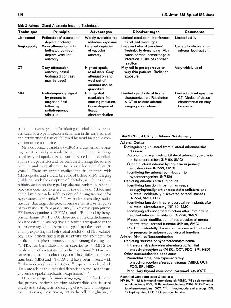
able 2 Adrenal Gland Anatomic Imaging Techniques
Technique Principle Advantages
ltrasound Reflection of ultrasound,depicts anatomy
Widely available,radiation expos
ngiography X-ray attenuation withiodinated contrast,depicts vascularanatomy
Detailed depictioof vascularanatomy
T X-ray attenuation,anatomy based(iodinated contrastmay be used)
Highest spatialresolution. X-raattenuation andwashout ofcontrast can bequantified
RI Radiofrequency signalby protons inmagnetic fieldfollowingradiofrequencystimulus
High spatialresolution. Noionizing radiatioSome degree otissuecharacterization
Disadvantages Comments
noure
Limited resolution. Interferenceby fat and bowel gas
Limited utility
n Invasive (arterial puncture).Technically demanding. Maycause adrenal hemorrhage orinfarction. Risks of contrastreaction
Generally obsolete foradrenal localization
yMay fail in postoperative or
very thin patients. Radiationexposure.
Very widely used
n.f
Limited specificity of tissuecharacterization. Resolution< CT in routine adrenalimaging applications.
Limited advantages overCT. Modes of tissuecharacterization maybe useful.
ies. FDG is a glucose analog, enters the cells like glucose, is
able 3 Clinical Utility of Adrenal Scintigraphy
drenal CortexDistinguishing unilateral from bilateral adrenocortical
diseaseAutonomous asymmetric, bilateral adrenal hyperplasia
in hypercortisolism (NP-59, SMC)Subtle bilateral adrenal hyperplasia in primary
aldosteronism (NP-59, SMC)Identifying the adrenal contribution to
hyperandrogenism (NP-59)Depicting adrenal cortical function
Identifying function in benign vs spaceoccupying/malignant or metastatic unilateral andbilateral incidentally discovered adrenal masses(NP-59, SMC, FDG)
Identifying function in adrenocortical re-implants afterbilateral adrenalectomy (NP-59, SMC)
Identifying adrenocortical function after intra-arterialalcohol infusion for ablation (NP-59, SMC)
Preoperative identification of suppression of normalcontralateral adrenal function (NP-59, SMC)
Predict incidentally discovered masses with potentialto progress to autonomous adrenal function
drenal Medulla/NeuroendocrineDepicting sources of hypercatecholaminemia
Intra-adrenal/extra-adrenal/metastatic/familialpheochromocytomas (MIBG, OCT, FDG, EPI, HED)
Other neuroendocrine neoplasmsNeuroblastoma, non-hypersecretory
pheochromocytoma/paragangliomas (MIBG, OCT,FDG, EPI, HED)
Medullary thyroid carcinoma, carcinoid, etc (OCT)
eprinted with permission Gross et al.4
P-59, 131I-6�-iodomethyl-norcholesterol; †SMC, 75Se-selenomethyl-norcholesterol; FDG, 18F-fluorodeoxyglucose; MIBG, 131I(123I)-meta-iodobenzylguanidine; OCT, 123I, 11In-octreotide and analogs; EPI,
11C-epinephrine; HED, 11C-hydroxyephedrine.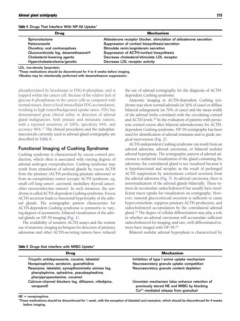
ptgnrdgwamd
FCdarffsodAnAin
ua
td
dboatdug
aaeaiAtnmfiehrgirm
T
L*† ression
T
N*
Adrenal gland scintigraphy 215
hosphorylated by hexokinase to FDG-6-phosphate, and israpped within the cancer cell. Because of the relative lack oflucose-6-phosphatase in the cancer cells as compared withormal tissues, there is focal intracellular FDG accumulation,esulting in high tumor/background uptake ratios. FDG hasemonstrated great clinical utility in detection of adrenalland malignancies, both primary and metastatic tumors,ith a reported sensitivity of 100%, specificity 94%, and
ccuracy 96%.17 The clinical procedures and the radiophar-aceuticals currently used in adrenal gland scintigraphy areescribed in Table 6.
unctional Imaging of Cushing Syndromeushing syndrome is characterized by excess cortisol pro-uction, which often is associated with varying degrees ofdrenal androgen overproduction. Cushing syndrome mayesult from stimulation of adrenal glands by excess ACTHrom the pituitary (ACTH-producing pituitary adenoma) orrom an extrapituitary tumor (ectopic ACTH syndrome, eg,mall cell lung cancer, carcinoid, medullary thyroid cancer,ther neuroendocrine tumors). In such instances, the syn-rome is called ACTH-dependent Cushing syndrome. ExcessCTH secretion leads to functional hypertrophy of the adre-al glands. The scintigraphic pattern characteristic forCTH-dependent Cushing syndrome is symmetric to vary-
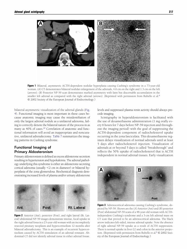
ng degrees of asymmetric, bilateral visualization of the adre-al glands on NP-59 imaging (Fig. 1).The availability of sensitive ACTH assays and the routine
se of anatomic imaging techniques for detection of pituitarydenomas and other ACTH-secreting tumors have reduced
able 4 Drugs That Interfere With NP-59 Uptake*
Drug
Spironolactone AldosteKetoconazole SuppresDiuretics; oral contraceptives StimulaGlucocorticoids (eg, dexamethasone)† SuppresCholesterol-lowering agents DecreasHypercholesterolemia/genetic Decreas
DL, low-density lipoprotein.These medications should be discontinued for 4 to 6 weeks beforeStudies may be intentionally performed with dexamethasone supp
able 5 Drugs that interfere with MIBG Uptake*
Drug
Tricyclic antidepressants, cocaine, labetalolNorepinephrine, serotonin, guanethidineReserpine, labetalol, symapthomimetic amines (eg,
phenylephrine, ephedrine, pseudoephedrine,phenylpropanolamine, cocaine)
Calcium-channel blockers (eg, diltiazem, nifedipine,verapamil)
E � norepinephine.These medications should be discontinued for 1 week, with the exce
before imaging.
he use of adrenal scintigraphy for the diagnosis of ACTH-ependent Cushing syndrome.Anatomic imaging in ACTH-dependent Cushing syn-
rome may show normal adrenals (in 30% of cases) or diffuseilateral enlargement (in 70% of cases) and the mean widthf the adrenal limbs correlated with the circulating cortisolnd ACTH levels.18 In the evaluation of patients with persis-ent cortisol excess after bilateral adrenalectomy for ACTH-ependent Cushing syndrome, NP-59 scintigraphy has beensed for identification of adrenal remnants and to guide sur-ical intervention (Fig. 2).
ACTH-independent Cushing syndrome can result from andrenal adenoma, adrenal carcinoma, or bilateral nodulardrenal hyperplasia. The scintigraphic pattern of adrenal ad-noma is unilateral visualization of the gland containing thedenoma; the contralateral gland is not visualized because its hypofunctional and atrophic as the result of prolongedCTH suppression by autonomous cortisol secretion from
he adrenal adenoma (Fig. 3). In adrenal carcinoma, there isonvisualization of the adrenal glands bilaterally. These tu-ors do accumulate radiocholesterol but usually have insuf-cient tracer uptake for visualization on scintigraphy. How-ver, tumoral glucocorticoid secretion is sufficient to causeypercortisolism, suppress pituitary ACTH production, andadiocholesterol accumulation by the contralateral adrenalland.19 The degree of cellular differentiation may play a rolen whether an adrenal carcinoma will accumulate sufficientadiocholesterol for imaging, and rare, well-differentiated tu-ors have imaged with NP-59.20
Bilateral nodular adrenal hyperplasia is characterized by
Mechanism
eceptor blocker, stimulation of aldosterone secretionf cortisol biosynthesis/secretion
in/angiotensin secretionf ACTH/cortisol biosynthesislesterol/stimulate LDL receptor
receptor activity
ng..
Mechanism
Inhibition of type I amine uptake mechanismNeurosecretory granule uptake competitionNeurosecretory granule content depletion
Uncertain mechanism (also enhance retention ofpreviously stored NE and MIBG by blockingCa2�-mediated release from granules)
f labetalol and reserpine, which should be discontinued for 4 weeks
rone rsion o
te rension oe choe LDL
imagi
ption o
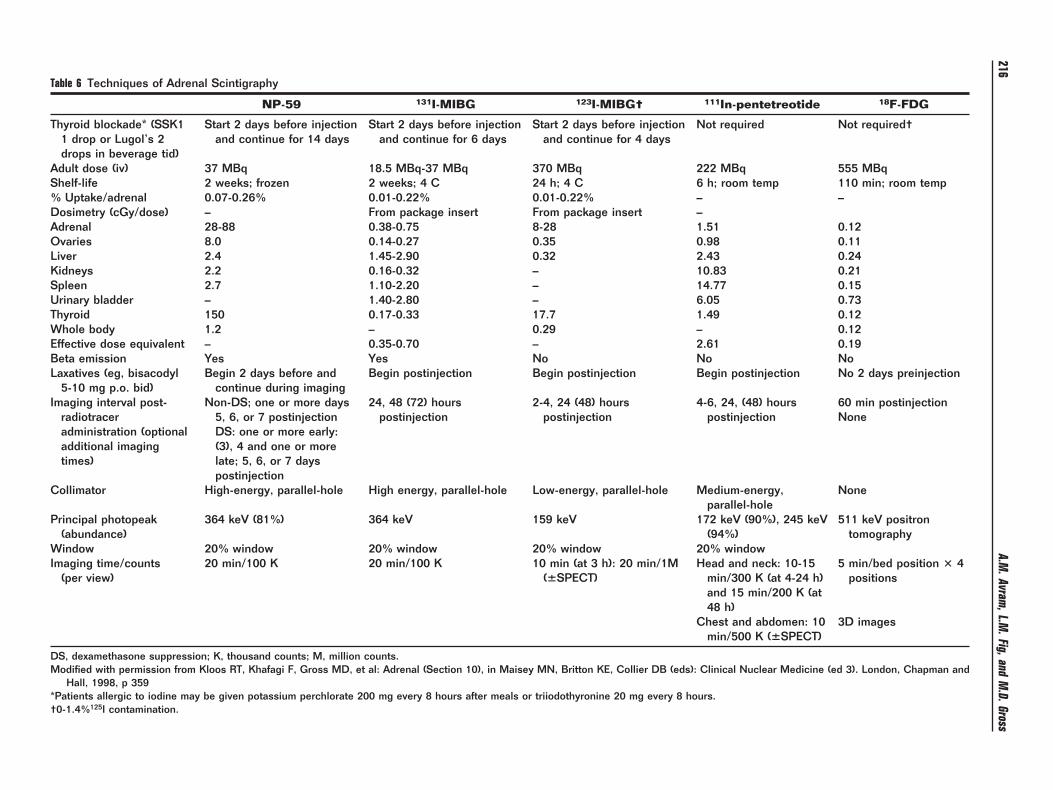
Table 6 Techniques of Adrenal Scintigraphy
NP-59 131I-MIBG 123I-MIBG† 111In-pentetreotide 18F-FDG
Thyroid blockade* (SSK11 drop or Lugol’s 2drops in beverage tid)
Start 2 days before injectionand continue for 14 days
Start 2 days before injectionand continue for 6 days
Start 2 days before injectionand continue for 4 days
Not required Not required†
Adult dose (iv) 37 MBq 18.5 MBq-37 MBq 370 MBq 222 MBq 555 MBqShelf-life 2 weeks; frozen 2 weeks; 4 C 24 h; 4 C 6 h; room temp 110 min; room temp% Uptake/adrenal 0.07-0.26% 0.01-0.22% 0.01-0.22% – –Dosimetry (cGy/dose) – From package insert From package insert –Adrenal 28-88 0.38-0.75 8-28 1.51 0.12Ovaries 8.0 0.14-0.27 0.35 0.98 0.11Liver 2.4 1.45-2.90 0.32 2.43 0.24Kidneys 2.2 0.16-0.32 – 10.83 0.21Spleen 2.7 1.10-2.20 – 14.77 0.15Urinary bladder – 1.40-2.80 – 6.05 0.73Thyroid 150 0.17-0.33 17.7 1.49 0.12Whole body 1.2 – 0.29 – 0.12Effective dose equivalent – 0.35-0.70 – 2.61 0.19Beta emission Yes Yes No No NoLaxatives (eg, bisacodyl
5-10 mg p.o. bid)Begin 2 days before and
continue during imagingBegin postinjection Begin postinjection Begin postinjection No 2 days preinjection
Imaging interval post-radiotraceradministration (optionaladditional imagingtimes)
Non-DS; one or more days5, 6, or 7 postinjectionDS: one or more early:(3), 4 and one or morelate; 5, 6, or 7 dayspostinjection
24, 48 (72) hourspostinjection
2-4, 24 (48) hourspostinjection
4-6, 24, (48) hourspostinjection
60 min postinjectionNone
Collimator High-energy, parallel-hole High energy, parallel-hole Low-energy, parallel-hole Medium-energy,parallel-hole
None
Principal photopeak(abundance)
364 keV (81%) 364 keV 159 keV 172 keV (90%), 245 keV(94%)
511 keV positrontomography
Window 20% window 20% window 20% window 20% windowImaging time/counts
(per view)20 min/100 K 20 min/100 K 10 min (at 3 h): 20 min/1M
(�SPECT)Head and neck: 10-15
min/300 K (at 4-24 h)and 15 min/200 K (at48 h)
5 min/bed position � 4positions
Chest and abdomen: 10min/500 K (�SPECT)
3D images
DS, dexamethasone suppression; K, thousand counts; M, million counts.Modified with permission from Kloos RT, Khafagi F, Gross MD, et al: Adrenal (Section 10), in Maisey MN, Britton KE, Collier DB (eds): Clinical Nuclear Medicine (ed 3). London, Chapman and
Hall, 1998, p 359*Patients allergic to iodine may be given potassium perchlorate 200 mg every 8 hours after meals or triiodothyronine 20 mg every 8 hours.†0-1.4%125I contamination.
216A.M
.Avram,L.M
.Fig,andM
.D.Gross
b4coimtti
FPProcpo
lc
teoAoi5ari
gy.)
Fetrbtd
Fp(iCaoTt
Adrenal gland scintigraphy 217
ilateral asymmetric visualization of the adrenal glands (Fig.). Functional imaging is most important in these cases be-ause anatomic imaging may cause the misidentifiation ofnly the largest adrenal nodule as a unilateral adenoma, fail-ng to correctly denote the bilateral nature of the process in as
any as 40% of cases.20 Correlation of anatomic and func-ional information will avoid an inappropriate and noncura-ive, unilateral adrenalectomy. Table 7 summarizes the imag-ng patterns in Cushing syndrome.
unctional Imaging ofrimary Aldosteronismrimary aldosteronism is defined as excess aldosterone secretionesulting in hypertension and hypokalemia. The adrenal pathol-gy underlying this syndrome is either an aldosterone-secretingortical adenoma (usually �2 cm in diameter) or bilateral hy-erplasia of the zona glomerulosa. Biochemical diagnosis dem-nstrating increased levels of plasma and/or urinary aldosterone
Figure 1 Bilateral, asymmetric ACTH-dependent nodulwoman. (A) CT demonstrates bilateral nodular enlargem(arrows). (B) Posterior NP-59 scan demonstrates markesmaller left adrenal as compared with the right adrena© 2002 Society of the European Journal of Endocrinolo
igure 2 Anterior (Ant), posterior (Post), and right lateral (Rt. Lat-ral) abdominal NP-59 images demonstrate intense, focal uptake inhe right adrenal fossa in a 23-year-old woman with an incompletelyesected pituitary neoplasm and hypercortisolism 12 months afterilateral adrenalectomy. This is an example of recurrent hypercor-isolism caused by ACTH stimulation of an adrenal remnant. Ab-
ominal CT did not identify adrenal tissue in either adrenal fossae. eevels and suppressed plasma renin activity should always pre-ede imaging.
Scintigraphy in hyperaldosteronism is facilitated withhe use of dexamethasone administration (1 mg orally ev-ry 6 hours for 7 days before NP-59 injection and through-ut the imaging period) with the goal of suppressing theCTH-dependent component of radiocholesterol uptakeccurring in the zona fasciculata. This dexamethasone reg-men delays visualization of normal adrenals until at least
days after radiocholesterol injection. Visualization ofdrenals at or beyond 5 days is called “breakthrough” andeflects the 50% uptake of radiocholesterol that is ACTHndependent in normal adrenal tissues. Early visualization
erplasia causing Cushing’s syndrome in a 73-year-oldthe adrenals, 4.6 cm on the right and 1.3 cm on the leftmetry with faint but discernable accumulation in the
ws). (Reprinted with permission from Rubello et al.8
igure 3 Adrenocortical adenoma causing Cushing’s syndrome, de-icted by NP-59. Shown are the (A) Anterior (Ant) and (B) posteriorPost) abdominal NP-59 scans of a 48-year-old woman with ACTH-ndependent Cushing’s syndrome and a 3-cm left adrenal mass onT scan that proved to be an adrenocortical adenoma. The blackrrow indicates left-sided, intense adrenal uptake. Note the absencef right adrenal NP-59 uptake as a result of ACTH suppression.here is normal uptake in liver (L) and colon in the anterior projec-
ion. (Reprinted with permission from Rubello et al.8 © 2002 Soci-
ar hypent ofd asyml (arro
ty of the European Journal of Endocrinology.)
((pmruddm
FH
Esorep
T
C*†‡§¶
218 A.M. Avram, L.M. Fig, and M.D. Gross
before the fifth day after injection) in a unilateral patternaldosteronoma; Fig. 5) or bilateral pattern (adrenal hy-erplasia; Fig. 6) before day 5 after injection is the hall-ark of primary aldosteronism. For high diagnostic accu-
acy, medications that may increase radiocholesterolptake in normal adrenal tissues, such as spironolactone,iuretics, and oral contraceptives (Table 3), should beiscontinued 4 to 6 weeks before imaging. Table 8 sum-arizes the imaging patterns in primary aldosteronism.
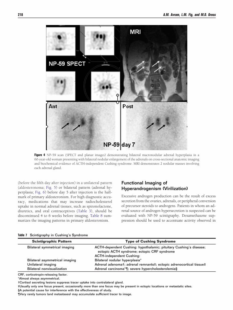
Figure 4 NP-59 scan (SPECT and planar images) dem60-year-old woman presenting with bilateral nodular enand biochemical evidence of ACTH-independent Cushieach adrenal gland.
able 7 Scintigraphy in Cushing’s Syndrome
Scintigraphic Pattern
Bilateral symmetrical imaging ACTH-depeectopic A
ACTH-indepBilateral asymmetrical imaging Bilateral noUnilateral imaging Adrenal adeBilateral nonvisualization Adrenal car
RF, corticotropin-releasing factor.Almost always asymmetrical.Cortisol secreting lesions suppress tracer uptake into contralateraUsually only one focus present, occasionally more than one focusA potential cause for interference with the effectiveness of study.
Very rarely tumors (and metastases) may accumulate sufficient tracer tounctional Imaging ofyperandrogenism (Virilization)
xcessive androgen production can be the result of excessecretion from the ovaries, adrenals, or peripheral conversionf precursor steroids to androgens. Patients in whom an ad-enal source of androgen hypersecretion is suspected can bevaluated with NP-59 scintigraphy. Dexamethasone sup-ression should be used to accentuate activity observed in
ting bilateral macronodular adrenal hyperplasia in aent of the adrenals on cross-sectional anatomic imagingdrome. MRI demonstrates 2 nodular masses involving
Type of Cushing Syndrome
Cushing: hypothalamic; pituitary Cushing’s disease;yndrome; ectopic CRF syndrome
nt Cushing:hyperplasia*†; adrenal remnants‡; ectopic adrenocortical tissue‡a*¶; severe hypercholesterolemia§
.e present in ectopic locations or metastatic sites.
onstralargemng syn
ndentCTH sende
dularnomacinom
l glandmay b
image.

aa
mwlatlevfibsio
tszstnm(rc
arasans
FtbraapccmSiaw
TAA
et al.8 ©
Adrenal gland scintigraphy 219
utonomous adrenal tissues while suppressing physiologicctivity in the ACTH-dependent zona fascicularis.
Three patterns of adrenocortical visualization (on dexa-ethasone suppression) were demonstrated in a study of 37omen with varying degrees of hyperandrogenism: Early bi-
ateral visualization (�5 days) was found in 15 patients, anddrenal vein testosterone gradients were found in 6 of 6 pa-ients where these measurements were made, confirming bi-ateral zona reticularis adrenal hyperplasia. In 5 patients,arly (�5 days) unilateral or markedly asymmetric adrenalisualization was seen, and adrenal adenomas were con-rmed in 4 of the 5 patients. In the remaining 17 patients,ilateral late visualization was observed (�5 days), and thesecans were considered normal.21 Occasionally, NP-59 imag-ng has been successfully used in the identification of bothvarian (arrhenoblastoma) and testicular neoplasms.22-24
A new approach to functional imaging of the adrenal cor-ex used PET with 11C-metomidate (MTO).25-27 MTO bindspecifically to 11�-hydroxylase (CYP 11B1, P45011�), an en-yme that is essential in the biosynthesis of cortisol and aldo-terone and regulated by ACTH. Because of its highly specificargeting, MTO has been used to distinguish lesions of adre-ocortical origin. In a study of 16 patients with adrenalasses 11C-MTO clearly separated 13 adrenocortical lesions
including both nonsecreting and hypersecretory lesions rep-esented by cortical adenomas, cortical carcinoma, and ma-
Figure 5 Left adrenal aldosteronoma depicted with dexamebiochemical evidence of hyperaldosteronism and a left admass (black arrow). Shown are the anterior (B, Ant) and pinjection and anterior (D) and posterior (E) abdominal NPuptake (black arrows) occurs early, before day 5 afteringallbladder (GB). (Reprinted with permission from Rubello
ronodular hyperplasia) from 3 noncortical lesions (benign e
nd malignant pheochromocytoma and metastasis to the ad-enal). However, 11C-MTO PET could not distinguish benigndrenocortical tumors and adrenocortical cancer.26 In thistudy, FDG-PET separated all malignant lesions from benigndrenal masses, showing a specificity, sensitivity, and diag-ostic accuracy of 100% for characterization (malignant ver-us benign) of adrenal masses.26
Another study comparing the results of 11C-MTO PET andDG-PET for evaluation of adrenal lesions studied 21 pa-ients with incidentally discovered adrenal masses, includingoth hormonally active and inactive adrenal adenomas, ad-enal cancer, pheochromocytoma, cyst, lipoma, lymphoma,nd adrenal metastases.27 11C-MTO identified all lesions ofdrenocortical origin, with the highest tracer uptake (ex-ressed as standardized uptake value [SUV]) in adrenocorti-al carcinoma (SUVmedian 28), followed by hypersecretoryortical adenomas (SUVmedian 12.7) and nonsecretory adeno-as (SUVmedian 12.2); the noncortical tumors demonstrated
UVmedian 5.7. 18F-FDG-PET showed increased tracer activityn pheochromocytomas and adrenocortical carcinoma, butll inactive adenomas and most hypersecreting adenomasere difficult to detect against background activity.27
he Incidentally Discovereddrenal Mass (Adrenal Incidentaloma)drenal adenomas are relatively common (2-9%) in the gen-
e suppression NP-59 imaging. A 57-year-old woman withass. Abdominal CT (A) demonstrates a 2-cm left adrenalr (C, Post) abdominal NP-59 scans on the third day afterans on the fifth day after injection. Abnormal left adrenal(B and C). Normal uptake in liver (L), bowel (B), and2002 Society of the European Journal of Endocrinology.)
thasonrenal mosterio-59 sc
jection
ral population, and the routine use of high-resolution imag-

iaptilomncwn4c
wc
sfaopnnD
cdmf�hltltetmngbmriam1eieas
aicowpmd
ai
Freeratce©
T
SU
S
*
220 A.M. Avram, L.M. Fig, and M.D. Gross
ng techniques (ultrasound, CT, and MRI) for evaluation ofbdominal symptoms leads to identification of many unsus-ected adrenal lesions.17 These lesions require further inves-igation for detecting hormonal hypersecretion and for mak-ng the critical distinction between benign and malignantesions. In the absence of a known malignancy, 70% to 94%f adrenal incidentalomas are nonsecreting benign adeno-as.28 In the clinical setting of a known extra-adrenal malig-
ancy, 50% are adrenal metastases, mostly derived from car-inomas of the lung, breast, stomach, ovary, and kidneys, asell as leukemia, lymphomas, and melanomas. Other adre-al masses seen in decreasing prevalence are adrenal cysts in% to 22%, myelolipoma in 7% to 15%, and pheochromo-ytoma in 0% to 11%.28
Diagnostic evaluation of adrenal incidentalomas beginsith biochemical evaluation to assess for hormone hyperse-
retion because hormonally active adrenal masses require
igure 6 Bilateral adrenal hyperplasia causing primary hyperaldoste-onism in a 35-year-old woman with hypertension, biochemicalvidence of hyperaldosteronism, and an outside CT report of bilat-ral adrenal thickening. Shown are the anterior (A, Ant) and poste-ior (B, Post) abdominal NP-59 scan on the fourth day after injectionnd the anterior (C) and posterior (D) abdominal NP-59 scans onhe fifth day after injection. Bilateral adrenal visualization on day 4onfirmed on day 5 after injection (black arrows) documents bilat-ral hyperfunction. (Reprinted with permission from Rubello et al.8
2002 Society of the European Journal of Endocrinology.)
able 8 Imaging in Hyperaldosteronism
Scintigraphic Pattern
ymmetrical bilateral early imaging (before day 5)nilateral early imaging (before day 5)
ymmetrical late imaging (on or after day 5);nondiagnostic pattern
Should be excluded by measurement of renin and aldosterone levels and
urgical excision. Laboratory screening should consider aunctional analysis of all zones of the adrenal cortex and thedrenal medulla and include serum cortisol, plasma ACTH,vernight dexamethasone suppression test, fractionatedlasma catecholamines, 24-hour urine for metanephrine andormetanephrine, serum sodium, and potassium, plasma re-in, serum aldosterone and adrenal androgens (serumHEA-S).CT and MRI provide important anatomic information in
haracterization of incidentalomas. CT densitometry, based onetecting the presence and amount of lipid within an adrenalass, is useful for initial differentiation of adrenal adenomas
rom metastases. On unenhanced CT a density measurement10 Hounsfield units (HU) defines a lipid-rich adenoma and
as 71% sensitivity and 98% specificity for diagnosing a benignesion.29 If the mass is inhomogenous or has a density �10 HU,he diagnosis is uncertain (lipid-poor adenoma or malignantesion) and contrast-enhanced CT scan is performed to assesshe washout of initial enhancement in the lesion. Adenomasnhance significantly after the intravenous contrast administra-ion and show more rapid washout of contrast than adrenaletastases. Standard contrast-enhanced CT images of the adre-al glands are obtained approximately 60 seconds after the be-inning of bolus intravenous injection of contrast material, thiseing the only time when the attenuation value of adenomas andetastases are nearly identical. Adenomas have a much more
apid loss of enhancement, as early as 5 minutes after contrastnjection, and attenuation values at 15 minutes after contrastdministration can be used to differentiate adenomas from otherasses: masses with an attenuation value less than 30 to 40 H on
5-minute delayed contrast-enhanced CT are almost always ad-nomas.30 In addition to the delayed CT attenuation value itself,t is also possible to calculate the percentage washout of initialnhancement: the optimal threshold for enhancement washoutt 15 minutes is 60%, resulting in a sensitivity of 88% and apecificity of 96% for the diagnosis of adenoma.30
Chemical shift MRI also is used to differentiate adrenaldenomas from metastases, and it is the preferred anatomicmaging method in children and pregnant women. Any lipid-ontaining tissue shows a signal loss caused by cancellationf the signal from fat and water on opposed-phase comparedith in-phase images. Visual analysis of in-phase and op-osed-phase imaging for the detection of lipid within adrenalasses showed a sensitivity of 78% and specificity of 87% foriagnosing adrenal adenoma.31
Despite the excellent anatomic and structural detail the CTnd MR techniques provide, functional adrenal imaging us-ng targeted radionuclides, such as NP-59, MIBG, and FDG,
Type of Aldosteronism
lateral autonomous hyperplasia; secondary aldosteronism*ilateral adenoma (Conn’s tumor) unilateral malignant
aldosterone secreting tumor (rare)ormal adrenals; dexamethasone suppressiblealdosteronism (rare)
BiUn
N
should not require imaging.
oa
gdtatgaood(msasa
teFbd
r5ndv
twttttcpinsMcFPf
ctp
T
CD
N
C uptake
Adrenal gland scintigraphy 221
ffers the best diagnostic sensitivity and specificity for char-cterization of incidental adrenal lesions.
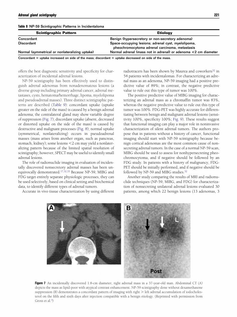
NP-59 scintigraphy has been effectively used to distin-uish adrenal adenomas from nonadenomatous lesions (aiverse group including primary adrenal cancer, adrenal me-astases, cysts, hematoma/hemorrhage, lipoma, myelolipomand pseudoadrenal masses). Three distinct scintigraphic pat-erns are described (Table 9): concordant uptake (uptakereater on the side of the mass) is caused by a benign adrenaldenoma; the contralateral gland may show variable degreef suppression (Fig. 7); discordant uptake (absent, decreasedr distorted uptake on the side of the mass) is caused byestructive and malignant processes (Fig. 8); normal uptakesymmetrical, nonlateralizing) occurs in pseudoadrenalasses (mass arises from another organ, such as pancreas,
tomach, kidney); some lesions �2 cm may yield a nonlater-lizing pattern because of the limited spatial resolution ofcintigraphy; however, SPECT may be useful to identify smalldrenal lesions.
The role of radionuclide imaging in evaluation of inciden-ally discovered nonsecretory adrenal masses has been un-quivocally demonstrated.17,32,33 Because NP-59, MIBG andDG target entirely separate physiologic processes, they cane used selectively, based on clinical setting and biochemicalata, to identify different types of adrenal tumors.Accurate in vivo tissue characterization by using different
able 9 NP-59 Scintigraphic Patterns in Incidentaloma
Scintigraphic Pattern
oncordant Beiscordant Sp
pormal (symmetrical or nonlateralizing uptake) No
oncordant � uptake increased on side of the mass; discordant �
Figure 7 An incidentally discovered 1.8-cm diameter, rdepicts the mass as lipid-poor with atypical contrast enhsuppression (B) demonstrates a concordant pattern of imterol on the fifth and sixth days after injection compati
Gross et al.3)adiotracers has been shown by Maurea and coworkers32 in4 patients with incidentalomas. For characterizing an adre-al mass as an adenoma, NP-59 imaging had a positive pre-ictive value of 89%; in contrast, the negative predictivealue to rule out this type of tumor was 100%.
The positive predictive value of MIBG imaging for charac-erizing an adrenal mass as a chromaffin tumor was 83%,hereas the negative predictive value to rule out this type of
umor was 100%. FDG-PET was highly accurate for differen-iating between benign and malignant adrenal lesions (sensi-ivity 100%, specificity 100%; Fig. 9). These results suggesthat functional imaging can play a major role in noninvasiveharacterization of silent adrenal tumors. The authors pro-ose that in patients without a history of cancer, functional
maging should start with NP-59 scintigraphy because be-ign cortical adenomas are the most common cause of non-ecreting adrenal tumors. In the case of a normal NP-59 scan,IBG should be used to assess for nonhypersecreting pheo-
hromocytoma, and if negative should be followed by anDG study. In patients with a history of malignancy, FDG-ET should be initially performed, and if negative should beollowed by NP-59 and MIBG studies.32
Another study comparing the results of MRI and radionu-lide techniques (NP-59, MIBG, and FDG) for characteriza-ion of nonsecreting unilateral adrenal lesions evaluated 30atients, among which 22 benign lesions (13 adenomas, 3
Etiology
hypersecretory or non-secretory adenoma)cupying lesions; adrenal cyst, myelolipoma,romocytoma adrenal carcinoma, metastasisdrenal (mass not in adrenal) or adenoma <2 cm diameter
decreased on side of the mass.
renal mass in a 37-year-old man. Abdominal CT (A)ent. NP-59 scintigraphy done without dexamethasonewith right � left adrenal accumulation of iodocholes-
th a benign etiology. (Reprinted with permission from
nign (ace-ocheochrmal a
ight adancemaging
ble wi
cmatQinn
6cMo
fda
222 A.M. Avram, L.M. Fig, and M.D. Gross
ysts, 2 myelolipomas, and 4 pheochromocytomas) and 8alignant lesions (4 adrenocortical carcinomas, 1 sarcoma,
nd 3 metastases) were finally diagnosed (based on histopa-hology, biopsy or 2-year clinical-imaging follow-up).33
ualitative MR evaluation showed T2 signal hyperintensityn 46% of adenomas and 100% of pheochromocytomas; sig-al intensity loss on chemical-shift imaging in 100% of ade-omas; gadolinium enhancement in 100% of pheos and in
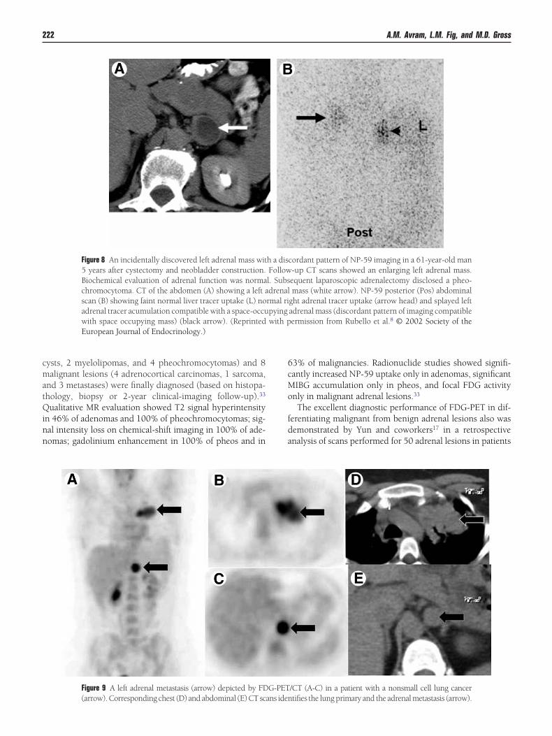
Figure 8 An incidentally discovered left adrenal mass wit5 years after cystectomy and neobladder construction.Biochemical evaluation of adrenal function was normachromocytoma. CT of the abdomen (A) showing a left ascan (B) showing faint normal liver tracer uptake (L) noadrenal tracer acumulation compatible with a space-occuwith space occupying mass) (black arrow). (ReprintedEuropean Journal of Endocrinology.)
Figure 9 A left adrenal metastasis (arrow) depicted by FD
(arrow). Corresponding chest (D) and abdominal (E) CT scans iden3% of malignancies. Radionuclide studies showed signifi-antly increased NP-59 uptake only in adenomas, significantIBG accumulation only in pheos, and focal FDG activity
nly in malignant adrenal lesions.33
The excellent diagnostic performance of FDG-PET in dif-erentiating malignant from benign adrenal lesions also wasemonstrated by Yun and coworkers17 in a retrospectivenalysis of scans performed for 50 adrenal lesions in patients
cordant pattern of NP-59 imaging in a 61-year-old man-up CT scans showed an enlarging left adrenal mass.equent laparoscopic adrenalectomy disclosed a pheo-mass (white arrow). NP-59 posterior (Pos) abdominal
ght adrenal tracer uptake (arrow head) and splayed leftadrenal mass (discordant pattern of imaging compatibleermission from Rubello et al.8 © 2002 Society of the
/CT (A-C) in a patient with a nonsmall cell lung cancer
h a disFollowl. Subsdrenalrmal ripyingwith p
G-PET
tifies the lung primary and the adrenal metastasis (arrow).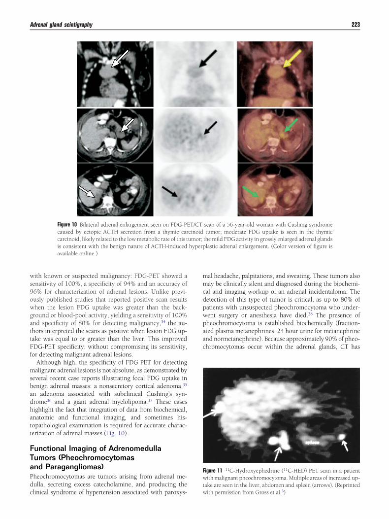
ws9owgattFf
msbadhatt
FTaPdc
mmcdpwpaac
Fwt
Adrenal gland scintigraphy 223
ith known or suspected malignancy: FDG-PET showed aensitivity of 100%, a specificity of 94% and an accuracy of6% for characterization of adrenal lesions. Unlike previ-usly published studies that reported positive scan resultshen the lesion FDG uptake was greater than the back-round or blood-pool activity, yielding a sensitivity of 100%nd specificity of 80% for detecting malignancy,34 the au-hors interpreted the scans as positive when lesion FDG up-ake was equal to or greater than the liver. This improvedDG-PET specificity, without compromising its sensitivity,or detecting malignant adrenal lesions.
Although high, the specificity of FDG-PET for detectingalignant adrenal lesions is not absolute, as demonstrated by
everal recent case reports illustrating focal FDG uptake inenign adrenal masses: a nonsecretory cortical adenoma,35
n adenoma associated with subclinical Cushing’s syn-rome36 and a giant adrenal myelolipoma.37 These casesighlight the fact that integration of data from biochemical,natomic and functional imaging, and sometimes his-opathological examination is required for accurate charac-erization of adrenal masses (Fig. 10).
unctional Imaging of Adrenomedullaumors (Pheochromocytomasnd Paragangliomas)heochromocytomas are tumors arising from adrenal me-ulla, secreting excess catecholamine, and producing the
Figure 10 Bilateral adrenal enlargement seen on FDG-PEcaused by ectopic ACTH secretion from a thymic carcarcinoid, likely related to the low metabolic rate of this tis consistent with the benign nature of ACTH-inducedavailable online.)
linical syndrome of hypertension associated with paroxys- w
al headache, palpitations, and sweating. These tumors alsoay be clinically silent and diagnosed during the biochemi-
al and imaging workup of an adrenal incidentaloma. Theetection of this type of tumor is critical, as up to 80% ofatients with unsuspected pheochromocytoma who under-ent surgery or anesthesia have died.28 The presence ofheochromocytoma is established biochemically (fraction-ted plasma metanephrines, 24 hour urine for metanephrinend normetanephrine). Because approximately 90% of pheo-hromocytomas occur within the adrenal glands, CT has
igure 11 11C-Hydroxyephedrine (11C-HED) PET scan in a patientith malignant pheochromocytoma. Multiple areas of increased up-
ake are seen in the liver, abdomen and spleen (arrows). (Reprinted
scan of a 56-year-old woman with Cushing syndrometumor; moderate FDG uptake is seen in the thymic
the mild FDG activity in grossly enlarged adrenal glandslastic adrenal enlargement. (Color version of figure is
T/CTcinoidumor;hyperp
ith permission from Gross et al.3)

bcmshtpfcapphm
ppimhpg
aaocp1
aic
Ftyra
Fstd
224 A.M. Avram, L.M. Fig, and M.D. Gross
een used as the primary imaging study. Most pheochromo-ytomas are readily detected on CT because they typicallyeasure 2 to 5 cm. in diameter and display regions of necro-
is or hemorrhage. Most pheochromocytomas have an unen-anced attenuation greater than 10 HU. Although adminis-ration of intravenous contrast was traditionally avoided inatients with known or suspected pheochromocytoma forear of inducing a hypertensive crisis, newer nonionic IVontrast agents showed no significant increases in catechol-mine levels in either control subjects or patients withheochromocytomas.38 Contrast-enhanced CT of adrenalheochromocytoma commonly shows inhomogeneous en-ancement of a solid mass, similar to the finding in adrenaletastasis or adrenal cortical carcinoma.MRI is considered the anatomic imaging of choice for
heochromocytoma due to its excellent anatomic detail, theotential for better tissue characterization and the ability of
maging in multiple planes. On MRI, most pheochromocyto-as are hypointense on T1-weighted images and markedlyyperintense on T2-weighted images. Although initial re-orts suggested that pheochromocytomas could be distin-uished by their marked hyperintensity on T2-weighted im-
igure 12 MIBG scan demonstrating intensely focal tracer activity inhe left adrenal gland consistent with pheochromocytoma in a 56-ear-old woman with hypertension, elevated plasma norepineph-ine levels, and a 3-cm left adrenal mass seen on cross-sectionalnatomic imaging.
Figure 13 Malignant, metastatic pheochromocytoma dembilateral adrenalectomy and persistent hypertension cosuperior vena cava obstruction. (A) Anterior chest and amultiple, abnormal foci of 123I-MIBG in metastatic pheoregions. (B) Chest CT identifies the superior mediastin
printed with permission from Rubello et al.8 © 2002 Society oges, considerable overlap with other neoplasms, includingdrenal cortical carcinomas, was demonstrated in up to 33%f cases.39 Functional imaging of pheochromocytoma in-ludes MIBG scintigraphy (using 131I or 123I) and newerositron emitters 18F-FDA, 18F-DOPA, 11C-epinephrine, and
1C-hydroxyephedrine (Fig. 11).13
Sporadic intra-adrenal pheochromocytomas are depicteds intensely focal MIBG uptake (Fig. 12). MIBG scintigraphys of particular value in imaging metastatic disease (the mostommon sites are the skeleton, lymph nodes, lung, and peri-
ted by 123I-MIBG and CT in a 31-year-old woman afterted by renal insufficiency and recent development ofn scan. L � normal liver uptake whereas arrows depictocytoma deposits in the mediastinum and para-aortic
s responsible for SVC obstruction (white arrow). (Re-
igure 14 FDG-PET/CT scan demonstrating vertebral and bilateralacral osseous metastases (arrows) in a 63-year-old man with nega-ive MIBG scan and elevated plasma catecholamine levels who un-erwent resection of a pelvic paraganglioma 3 years previously.
onstramplicabdomechromal mas
f the European Journal of Endocrinology.)
tmfdapf
aSwmmpaw
ipawn
moc
a4tcmtlnioooan1
tctr
CDts
spleen
Adrenal gland scintigraphy 225
oneum) and in the localization of extra-adrenal pheochro-ocytomas (Fig. 13). These lesions occur at many locations
rom the base of the skull to the pelvis and are frequently notetected by computed tomography due to their small sizend close relationship to other structures.40 MIBG scintigra-hy has demonstrated 87% sensitivity and 99% specificityor detection of pheochromocytomas.
Most pheochromocytomas, whether benign or malignant,re metabolically active, and can be imaged with FDG-PET.hulkin and coworkers41 identified pheochromocytomasith FDG in 22 of 29 patients (76%), FDG uptake beingore common in malignant than benign pheochromocyto-as (Fig 14). Interestingly, some pheochromocytomas thatoorly concentrated MIBG were well visualized with FDG,nd all pheochromocytomas that failed to accumulate FDGere detected with MIBG.Discordant results between FDG imaging and MIBG imag-
ng, as well as 18F-FDA imaging, were demonstrated in 5atients with metastatic pheochromocytoma. Both 123I MIBGnd 18F-FDA grossly underestimated the extent of diseasehen compared with CT and MRI. FDG-PET showed lesionsot detected on either 123I-MIBG or 18F-FDA scans.15
When choosing the optimal imaging modality for pheochro-ocytoma, it is important to consider that approximately 25%
f patients with apparently sporadic pheochromocytoma are
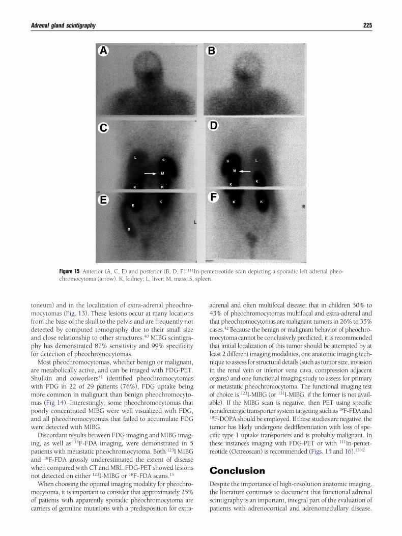
Figure 15 Anterior (A, C, E) and posterior (B, D, F) 111
chromocytoma (arrow). K, kidney; L, liver; M, mass; S,
arriers of germline mutations with a predisposition for extra- p
drenal and often multifocal disease; that in children 30% to3% of pheochromocytomas multifocal and extra-adrenal andhat pheochromocytomas are malignant tumors in 26% to 35%ases.42 Because the benign or malignant behavior of pheochro-ocytoma cannot be conclusively predicted, it is recommended
hat initial localization of this tumor should be attempted by ateast 2 different imaging modalities, one anatomic imaging tech-ique to assess for structural details (such as tumor size, invasion
n the renal vein or inferior vena cava, compression adjacentrgans) and one functional imaging study to assess for primaryr metastatic pheochromocytoma. The functional imaging testf choice is 123I-MIBG (or 131I-MIBG, if the former is not avail-ble). If the MIBG scan is negative, then PET using specificoradrenergic transporter system targeting such as 18F-FDA and
8F-DOPA should be employed. If these studies are negative, theumor has likely undergone dedifferentiation with loss of spe-ific type 1 uptake transporters and is probably malignant. Inhese instances imaging with FDG-PET or with 111In-pentet-eotide (Octreoscan) is recommended (Figs. 15 and 16).13,42
onclusionespite the importance of high-resolution anatomic imaging.
he literature continues to document that functional adrenalcintigraphy is an important, integral part of the evaluation of
tetreotide scan depicting a sporadic left adrenal pheo-.
In-pen
atients with adrenocortical and adrenomedullary disease.
Fibsdapa
R
1
1
1
1
1
1
1
1
1
1
2
2
2
2
2
2
2
2
tastases
226 A.M. Avram, L.M. Fig, and M.D. Gross
unctional scintigraphy complements anatomy-based imag-ng and facilitates diagnostic localization. The creation of hy-rid imaging devices, either PET or SPECT with CT will allowimultaneous functional and anatomic evaluation of adrenalysfunction and will serve as the basis for the re-inventionnd exploitation of older radiopharmaceuticals and the im-etus for the development of novel radiopharmaceuticals fordrenocortical and adrenomedulla imaging.
eferences1. Gross MD, Shapiro B, Bui C, et al: Adrenal scintigraphy and metaiodo-
benzylguanidine therapy of neuroendocrine tumors, in Sandler RE,Coleman JA, Patton FJ, et al (eds): Diagnostic Nuclear Medicine (ed 4).Philadelphia, Lippincott Williams and Wilkins, 2003, pp 715-734
2. Gross MD, Bui C, Shapiro B: Adrenocortical scintigraphy, in Ell PJ,Gambhir SS (eds): Nuclear Medicine in Clinical Diagnosis and Treat-ment (ed 3). Edinburgh, Churchill and Livingstone, 2004, pp 45-52
3. Gross MD, Korobkin M, Hussain H, et al: Adrenal gland imaging, inJameson JL, DeGroot LJ (eds): Endocrinology (ed 5). Philadelphia,W.B. Saunders, 2005, pp 2425-2453
4. Gross MD, Rubello D, Shapiro B: Is there a future for adrenal scintig-raphy? Nucl Med Commun 23:197-202, 2002
5. Gross MD, Valk TW, Thrall JH, et al: The role of pharmacologic ma-nipulation in adrenal cortical scintigraphy. Semin Nucl Med 11:128-148, 1981
6. Gross MD, Shapiro B: Scintigraphic studies in adrenal hypertension.Semin Nucl Med 19:122-143, 1990
7. Shapiro B, Gross MD, Shulkin BL: Radioisotope diagnosis and therapyof malignant pheochromocytomas. Trends Endocrinol Metab 12:469-475, 2001
8. Rubello D, Bui C, Casara D, et al: Functional scintigraphy of the adrenalgland. Eur J Endocrinol 147:13-28, 2002
9. Gross MD, Shapiro B, Shreve P: Radionuclide imaging of the adrenalcortex. Q J Nucl Med 43:224-232, 1999
0. Sisson JC, Frager MS, Valk TW, et al. Scintigraphic localization ofpheochromocytoma. N Engl J Med 305:12, 1981
1. Shapiro B, Wieland DM, Brown LE, et al: 131I-meta-iodobenzylgua-indine (MIBG) adrenal medullary scintigraphy: interventional studies,in Spencer RP (ed): Interventional Nuclear Medicine. New York, Grune& Stratton, 1983, pp 451-481
2. Khafagi FA, Shapiro B, Fig LM, et al: Labetalol reduces iodine-131MIBG uptake by pheochromocytoma and normal tissues. J Nucl Med30:481-489, 1989
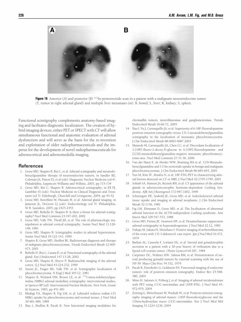
Figure 16 Anterior (A) and posterior (B) 111In-pentetreot(T, tumor in right adrenal gland) and multiple liver me
3. Ilias I, Shulkin B, Pacak K: New functional imaging modalities for
chromaffin tumors, neuroblastomas and ganglioneuromas. TrendsEndocrinol Metab 16:66-72, 2005
4. Ilias I, Yu J, Carrasquillo JA, et al: Superiority of 6-18F-fluorodopaminepositron emission tomography versus 131-I metaiodobenzylguanidinescintigraphy in the localization of metastatic pheochromocytoma.J Clin Endocrinol Metab 88:4083-4087.2003
5. Mamede M, Carrasquillo JA, Chen CC, et al: Discordant localization of2-[18F]-fluoro-2-deoxy-D-glucose in 6-[18F]-fluorodopamine- and[123I]-metaiodobenzylguanidine-negative metastatic pheochromocy-toma sites. Nucl Med Commun 27:31-36, 2006
6. Van der Harst E, de Herder WW, Bruining HA et al: 123I-Metaiodo-benzylguanidine and 111In-octreotide uptake in benign and malignantpheochromocytomas. J Clin Endocrinol Metab 86:685-693, 2001
7. Yun M, Kim W, Alnafisi N, et al: 18F-FDG PET in characterizing adre-nal lesions detected on CT or MRI. J Nucl Med 42:1795-1799, 2001
8. Sohaib SA, Hanson JA, Reznek RH, et al: CT appearance of the adrenalglands in adrenocorticotrophic hormone-dependent Cushing Syn-drome. AJR Am J Roentgenol 172:997-1002, 1999
9. Schteingart DE, Seabold JE, Gross MD, et al. Iodocholesterol adrenaltissue uptake and imaging in adrenal neoplasms. J Clin EndocrinolMetab 52:1156, 1981
0. Fig LM, Ehrmann D, Gross MD, et al: The localization of abnormaladrenal function in the ACTH-independent Cushing syndrome. AnnIntern Med 109:547-553, 1988
1. Gross MD, Freitas JE, Swanson DP, et al: Dexamethasone suppressionadrenal scintigraphy in hyperandrogenism. J Nucl Med 22:12, 1981
2. Nakajo M, Sakata H, Shinohara S: Positive imaging of arrhenoblastomaof the ovary with 131-I-aldosterol: case report. Jpn J Nucl Med 16:472,1979
3. Barkan AL, Cassorla F, Loriaux DL, et al: Steroid and gonadotrophinsecretion in a patient with a 30-year history of virilization due to alipoid-cell ovarian tumor. Obstet Gynecol 64:287, 1984
4. Carpenter DC, Wahner HW, Salassa RM, et al: Demonstration of ste-roid producing gonadal tumors by external scanning with the use ofNP-59. Mayo Clin Proc 54:332, 1979
5. Pacak K, Eisenhofer G, Goldstein DS: Functional imaging of endocrinetumors: role of positron emission tomography. Endocr Rev 25:568-580, 2004
6. Minn H, Salonen A, Friberg J, et al: Imaging of adrenal incidentalomaswith PET using (11)C-metomidate and (18)F-FDG. J Nucl Med 45:972-979, 2004
7. Zettinig G, Mitterhauser M, Wadsak W, et al: Positron emission tomog-raphy imaging of adrenal masses: (18)F-fluorodeoxyglucose and the11beta-hydroxylase tracer (11)C-metomidate. Eur J Nucl Med Mol
n in a patient with a malignant neuroendocrine tumor(m). B, bowel; L, liver; K, kidney; S, spleen.
ide sca
Imaging 31:1224-1230, 2004
2
2
3
3
3
3
3
3
3
3
3
3
4
4
4
Adrenal gland scintigraphy 227
8. Kloos RT, Gross MD, Francis IR, et al: Incidentally discovered adrenalmasses. Endocr Rev 16:460-484, 1995
9. Boland GW, Lee MJ, Gazelle GS, et al. Characterization of adrenalmasses using unenhanced CT: an analysis of the CT literature. AJR Am JRoentgenol 171:201-204, 1998
0. Korobkin M, Brodeur FJ, Francis IR, et al. CT time-attenuation washoutcurves of adrenal adenomas and nonadenomas. AJR Am J Roentgenol170:747-752, 1998
1. Outwater EK, Siegelman ES, Radecki PD, et al: Distinction betweenbenign and malignant adrenal masses: value of T1-weighted chemical-shift MR imaging. AJR Am J Roentgenol 165:579-583, 1995
2. Maurea S, Klain M, Mainolfi C, et al: The diagnostic role of radionuclideimaging in evaluation of patients with nonhypersecreting adrenalmasses. J Nucl Med 42:884-892, 2001
3. Maurea S, Caraco C, Klain M, et al: Imaging characterization of non-hypersecreting adrenal masses. Comparison between MR and radionu-clide techniques. Q J Nucl Med Mol Imaging 48:188-197, 2004
4. Erasmus JJ, Patz EF Jr., McAdams HP et al: Evaluation of adrenal massesin patients with bronchogenic carcinoma using 18F-FDG PET. AJRAm J Roentgenol 168:1357-1360, 1997
5. Rao SK, Caride VJ, Ponn R, et al: F-18 fluorodeoxyglucose positron
emission tomography-positive benign adrenal cortical adenoma:imaging features and pathologic correlation. Clin Nucl Med 29:300-302, 2004
6. Shimizu A, Oriuchi N, Tsushima Y, et al: High [18F] 2-fluoro-2-deoxy-D-glucose (FDG) uptake of adrenocortical adenoma showing subclin-ical Cushing’s syndrome. Ann Nucl Med 17:403-406, 2003
7. Ludwig V, Rice MH, Martin WH, et al: 2-Deoxy-2-[18F]fluoro-D-glu-cose positron emission tomography uptake in a giant adrenal myeloli-poma. Mol Imaging Biol 4:355-358, 2002
8. Mukherjee JJ, Peppercorn PD, Reznek RH, et al: Pheochromocytoma:Effect of nonionic contrast medium in CT on circulating catecholaminelevels. Radiology 202:227-231, 1997
9. Francis IR, Korobkin M: Pheochromocytoma. Radiol Clin N Am 34:1101-1112, 1996
0. Shapiro B, Sisson JC, Kalff V, et al: The location of middle mediastinalpheochromocytomas. J Thorac Cardiovasc Surg 87:814, 1984
1. Shulkin BL, Thompson NW, Shapiro B, et al: Pheochromocytomas:imaging with 2-[fluorine-18]fluoro-2-deoxy-D-glucose PET. Radiology212:35-41, 1999
2. Ilias I, Pacak K: Current approaches and recommended algorithm forthe diagnostic localization of pheochromocytoma. J Clin Endocrinol
Metab 89:479-491, 2004