adenosine triphosphatases of thermophilic archaeal double...
TRANSCRIPT

LUND UNIVERSITY
PO Box 117221 00 Lund+46 46-222 00 00
Adenosine triphosphatases of thermophilic archaeal double-stranded DNA viruses.
Happonen, Lotta; Erdmann, Susanne; Garrett, Roger A; Butcher, Sarah J
Published in:Cell & bioscience
DOI:10.1186/2045-3701-4-37
2014
Link to publication
Citation for published version (APA):Happonen, L., Erdmann, S., Garrett, R. A., & Butcher, S. J. (2014). Adenosine triphosphatases of thermophilicarchaeal double-stranded DNA viruses. Cell & bioscience, 4(Jul 23), [37]. https://doi.org/10.1186/2045-3701-4-37
General rightsCopyright and moral rights for the publications made accessible in the public portal are retained by the authorsand/or other copyright owners and it is a condition of accessing publications that users recognise and abide by thelegal requirements associated with these rights.
• Users may download and print one copy of any publication from the public portal for the purpose of private studyor research. • You may not further distribute the material or use it for any profit-making activity or commercial gain • You may freely distribute the URL identifying the publication in the public portalTake down policyIf you believe that this document breaches copyright please contact us providing details, and we will removeaccess to the work immediately and investigate your claim.
Download date: 12. Jan. 2020

Cell & BioscienceHapponen et al. Cell & Bioscience 2014, 4:37http://www.cellandbioscience.com/content/4/1/37
REVIEW Open Access
Adenosine triphosphatases of thermophilicarchaeal double-stranded DNA virusesLotta J Happonen1, Susanne Erdmann2, Roger A Garrett2 and Sarah J Butcher3*
Abstract
Adenosine triphosphatases (ATPases) of double-stranded (ds) DNA archaeal viruses are structurally related to theAAA+ hexameric helicases and translocases. These ATPases have been implicated in viral life cycle functions such asDNA entry into the host, and viral genome packaging into preformed procapsids. We summarize bioinformaticalanalyses of a wide range of archaeal ATPases, and review the biochemical and structural properties of thosearchaeal ATPases that have measurable ATPase activity. We discuss their potential roles in genome delivery into thehost, virus assembly and genome packaging in comparison to hexameric helicases and packaging motors frombacteriophages.
Keywords: ATPase, Archaeal, Virus, Genome packaging, Genome injection, MoxR ATPase
IntroductionViral genomes are enclosed inside a protein capsid forprotection against the environment. For many archaealviruses, the environmental stresses may include extremesof temperature and pH, high pressure, high salt concen-tration and the presence of heavy metal ions. Rod-shapedviruses, such as single-stranded (ss) DNA viruses (e.g. bac-teriophage M13) and ssRNA viruses (e.g. tobacco mosaicvirus), usually assemble their capsid proteins as a helicaltube around the genome. For such viruses, the capsidlength is proportional to the length of the genome. Al-ternatively, some viruses can package their genomesinto preformed procapsids via a specific genome pack-aging ATPase using energy derived from ATP hydrolysis.This is very commonly found in viruses with double-stranded (ds) DNA genomes [1]. For viruses that are notrod-shaped and do not exhibit a procapsid state, genomepackaging and virus assembly mechanisms remain poorlyunderstood. These viruses include spindle-shaped, bottle-shaped, droplet-shaped and pleomorphic viruses that arepredominantly found in the archaeal domain.This article aims to provide an overview of the different
types of ATPases that are encoded by archaeal viruses.ATPases of the tailed viruses and non-tailed archaeal
* Correspondence: [email protected] of Biotechnology, University of Helsinki, (Viikinkaari 1),P.O. Box 65, FI-00014 Helsinki, FinlandFull list of author information is available at the end of the article
© 2014 Happonen et al.; licensee BioMed CenCreative Commons Attribution License (http:/distribution, and reproduction in any mediumDomain Dedication waiver (http://creativecomarticle, unless otherwise stated.
viruses with icosahedrally-ordered capsids are suggestedto be involved in genome packaging. However, the major-ity of the characterised ATPases of archaeal viruses belongto viruses exhibiting different morphotypes, and thesehave been assigned diverse functions. We consider thefunctional roles of these ATPases in the life cycles ofspindle-shaped archaeal viruses and in particular theirinferred roles in viral genome injection into the hostsand the involvement of a MoxR-type ATPase in tail for-mation. Further, we highlight the genome packagingATPase B204 of the dsDNA Sulfolobus turreted icosahe-dral virus 2 (STIV2), as it is currently the only structur-ally characterized archaeal viral ATPase for which thefunction has been determined experimentally. We focuson the ATPases of thermophilic crenarchaeal virusesbecause very few biochemical or structural studies havebeen performed on ATPases of the euryarchaea.
Genome packaging ATPases of dsDNA virusesPackaging of genomic nucleic acid into preformed pro-capsids is a complex process involving injection of theDNA (or RNA) into the procapsid via ATP or NTP-powered hydrolysis, followed by structural rearrange-ment of the encapsidated genome. Subsequently the DNAis transformed into a highly condensed, near crystallinestate, concomitant with the maturation of the capsid. Mostof our current knowledge on viral genome packaging de-rives from studies on dsDNA head-tail bacteriophages
tral Ltd. This is an Open Access article distributed under the terms of the/creativecommons.org/licenses/by/2.0), which permits unrestricted use,, provided the original work is properly credited. The Creative Commons Publicmons.org/publicdomain/zero/1.0/) applies to the data made available in this

Happonen et al. Cell & Bioscience 2014, 4:37 Page 2 of 15http://www.cellandbioscience.com/content/4/1/37
such as phi29 and T4, and dsRNA viruses such as phi12.First, therefore, we provide a short introduction to what isknown about the conserved sequence motifs and the keycomponents of the packaging motor proteins, togetherwith a short summary of the well-characterized genomepackaging machineries of phi29 and T4.
Figure 1 Sequence alignment of thermophilic ATPases (part 1). The figof ATPases from Table 1 generated using Clustal Omega [16] and visualized inare labelled below the alignment. Secondary structure elements of STIV2 B204
A large variety of genome packaging ATPases havebeen characterised and they exhibit highly conservedsequence motifs at their active sites (Figures 1 and 2),which include the phosphate-binding loop (P-loop orWalker A sequence motif ) and the Walker B sequencemotif [2-4]. The consensus sequences of these motifs are
ure shows a sequence alignment of the N-terminal regions of a selectionEspript [17]. Numbers indicate residues. The conserved Walker A motifs(PDB: 4KFU) are shown above the aligned sequences.

Figure 2 Sequence alignment of thermophilic ATPases (part 2). Sequence alignment of the C-terminal regions of the selection of ATPasesfrom Table 1 as generated for Figure 1. The conserved Walker B sequence motifs are labelled below the alignment. The STIV2 B204 Walker Bsequence motif is indicated with two asterisks above the alignment. Secondary structure elements of STIV2 B204 (PDB: 4KFU) are shown abovethe aligned sequences.
Happonen et al. Cell & Bioscience 2014, 4:37 Page 3 of 15http://www.cellandbioscience.com/content/4/1/37
GXXXXGK(T/S) and hhhhDE, respectively, where X de-notes any amino acid and h indicates any hydrophobicamino acid [2,4]. The conserved lysine in the Walker Amotif participates in nucleotide binding and the con-served glutamate in the Walker B motif facilitates
activation of a water molecule for the hydrolysis reaction[5]. The Mg2+-ion required for ATP hydrolysis can becoordinated either by the conserved aspartate in theWalker B domain [2,4,6] or by the conserved serine inthe Walker A domain [7-9]. Arginine fingers, located

Happonen et al. Cell & Bioscience 2014, 4:37 Page 4 of 15http://www.cellandbioscience.com/content/4/1/37
either on the same subunit as the Walker A and B mo-tifs, or on an adjacent subunit, facilitate formation of thetransition state, and they are inserted into the catalyticsite as a result of a conformational change that occursprior to the catalytic step [10-12]. Two additional se-quence motifs - the sensor 1 and 2 regions - are conservedin AAA+ family proteins. These regions are considered todetect the status of the bound nucleotide, distinguishingbetween ATP and ADP [13], interacting with the γ-phosphate of the ATP [14,15].Tailed dsDNA bacteriophages contain two key compo-
nents for genome packaging: the ring-forming portal pro-tein (the connector) through which DNA is translocated,and the translocating motor (the terminase or ATPase).The terminase is usually hetero-oligomeric with a smallsubunit involved in recognition of the incoming DNA anda large subunit containing the ATPase domain and astructural motif for docking at the portal vertex. In someATPases, an endonuclease activity is also present whichcuts concatemers of the genome upon packaging (asreviewed by [1,18]).
The phi29 genome packaging machineryThe genome packaging machinery of the podovirusphi29 is one of the best-studied, most powerful nanomo-tors known. Using optical tweezers it has been shown topackage DNA at an initial rate of 100 bp s−1 against anaverage pressure of 57 pN [19]. The phi29 packagingmotor consists of the dodecameric connector protein(gp10) [20], the hexameric ATPase (gp16) containing thecanonical Walker A and B sequence motifs [21,22] andthe phage-encoded hexameric packaging RNA (pRNA)ring. The packaging mechanism of this motor has re-cently been reviewed [23]. Current findings show thatthe hexameric gp16 motor protein functions by revolu-tion rather than rotation [24]. For gp16-driven transloca-tion to occur, one hexamer subunit binds ATP whichstimulates a change to a conformation susceptible toDNA binding. Hydrolysis of the bound ATP produces asecond conformational change which results in displace-ment of the DNA away to an adjacent ATP-bound sub-unit. A single ATP molecule is hydrolysed in eachtransitional step, and a total of six ATP molecules areconsumed for one helical turn of 360°. The energy re-leased from hydrolysis of each ATP propels the DNAforward by 1.75 bp [24].
The T4 genome packaging machineryThe bacteriophage T4 packaging motor consists of thedodecameric connector (gp20) [25], the pentameric largeterminase motor (gp17) [26] and the 11- or 12-mericsmall terminase regulatory protein (gp16) [27]. Similarlyto the phi29 gp16 protein, the T4 gp17 motor proteinhas been shown to generate a force of 60 pN [28].
However, the average speed produced by gp17 is muchhigher than that of phi29 gp16 – 700 bp s−1 – and it canreach velocities up to 2000 bp s−1 [28]. The gp17 proteincarries an N-terminal ATPase domain, and a C-terminalendonuclease domain to cleave the genome upon head-ful packaging [12,29]. The two domains are linked by ahinge region which enables the domains to move between“tensed” and “relaxed” states, thereby translocating the T4genome in a “piston-like” fashion [12,26]. Both the gen-omic DNA to be packaged and the ATP molecule used asfuel, bind to the same T4 gp17 subunit which induces aconformational change leading to the translocation event[12,26,30]. At any time during the packaging event, onesubunit of gp17 is in the “tensed” state, and the others arein the “relaxed” state [26]. Energy released by hydrolysinga single ATP molecule translocates the DNA by two basepairs into the procapsid – an amount very similar to thatof the phi29 gp16 packaging ATPase [26,30].Essential questions regarding the mechanism of genome
packaging in tailed dsDNA viruses have been addressed bySchwartz et al. [30,31] and Sun et al. [12,26]. Moreover,studies on phi12 P4 have provided insights into how thismight work in dsRNA viruses [10,32,33]. However, thegenome packaging mechanisms of icosahedrally-ordereddsDNA viruses lacking a connector or tail are poorly char-acterized. Current understanding is based primarily onwhat is known about the genome packaging ATPase P9 ofbacteriophage PRD1 which stems from genetic studiesand in vitro experiments using cell lysates. The ATPase it-self has been intractable to structural characterization[34,35]. In PRD1-like viruses, with an ordered icosahedralcapsid and an internal lipid membrane, small membraneproteins (P20 and P22 in PRD1) have been inferred toform a membrane pore at the packaging vertex, and theymay function similarly to the connectors of head-tailphages [36]. The packaging rate of the PRD1 machinery is340 bp s−1 based on the time of the appearance of the firstinfectious particles [35], a value close to that described forphi29 gp16 and T4 gp17. We have recently gained someinsight into the structure of a genome packaging ATPasefrom a PRD1-like virus, with the newly solved crystalstructure of the STIV2 genome packaging ATPase, B204[7] (discussed below).
ATPases of archaeal double-stranded DNA virusesPredicted roles of thermophilic archaeal viral ATPasesAlthough this review is focused mainly on ATPases in-volved in archaeal viral genome packaging, ATPases alsoperform other important and diverse functions in virallife cycles. Nucleoside triphosphate (NTP)-hydrolysingenzymes are encoded in many of the archaeal viral ge-nomes so far described (Table 1, Figures 1 and 2). Theseven major functions associated with these ATPases aremoving viral DNA into the host (DNA entry), packaging

Table 1 Functions of archaeal thermophilic viral ATPases
Crenarchaeal Virus ORF NCBI/ENA reference sequence Postulated function Reference
Sulfolobus turreted icosahedral virus STIV b204 YP_025021.1 DNA packaging [37]
STIV2 b204 YP_003591106.1 DNA packaging [7]
Spherical PSV ORF582 (gp02) YP_015523.1 DNA packaging [38]
TTSV1 ORF1 (gp01) YP_164342.1 DNA packaging [39]
Bicaudaviridae ATV p529 (gp53) YP_319884.1 DNA entry [40]
p618 (gp66) YP_319897.1 MoxR-type chaperone [41]
Monocaudaviruses STSV1 ORF526 (gp69) YP_077262.1 DNA repair [42]
STSV2 ORF526 (gp48) YP_007348292.1 DNA repair [43]
SMV1 ORF503 CDF81345.1 DNA entry [44]
ORF307 CDF81374.1 DNA repair
ORF588 CDF81351.1 MoxR-type chaperone
Fuselloviridae ASV1 a232 (gp07) YP_003331412.1 Lon protease [45]
SSV1 b251 (p01) NP_039777.1 Lon protease [46]
SSV2 ORF233 (p03) NP_944455.1 Lon protease [47]
SSV4 ORF233 YP_001552190.1 Lon protease [45]
SSV5 ORF233 (gp12) YP_002221477.1 Lon protease [48]
SSV6 c234 (gp07) YP_003331457.1 Lon protease [45]
SSV7 a255 (gp06) YP_003331489.1 Lon protease [45]
SSVK1 a231 (p06) NP_963972.1 Lon protease [49]
SSVRH a247 (p06) NP_963931.1 Lon protease [50]
SMF1 ORF274 YP_007678010.1 Lon protease [51]
APSV1 ORF290 CCD22100.1 Lon protease [52]
Lipothrixviridae AFV1 ORF426 YP_003740.1 Lon protease [53]
AFV2 ORF425 (gp15) YP_001496940.1 Lon protease [54]
Rudiviridae ARV1 ORF210 (gp35) YP_001542652.1 ABC transporter [55]
ORF443 (gp16) YP_001542633.1 Lon protease
SIRV1 ORF440 (gp11) NP_666599.1 Lon protease [56]
SIRV2 ORF436 (gp18) NP_666552.1 Lon protease [56]
SRV ORF440 CAQ58449.1 Lon protease [57]
SMR1 ORF439 (gp08) YP_006990086.1 Lon protease [58]
Tadpole-shaped HAV2 ORF506 (gp04) YP_003773387 DNA repair [59]
Euryarchaeal
TPV1 gp02 YP_005271224.1 DNA replication [60]
gp20 YP_005271242.1 ABC transporter
ORF560 AEY69051.1 DNA replication
Thermophilic archaeal viruses for which no ATPase domains were detected include the following with genome accession numbers <http://www.ebi.ac.uk/genomes/archaealvirus.html>. Ampullavirus ABV (EF432053), lipothrixviruses SIFV (AF440571), AFV3 (AM087121), AFV6 (AM087121), AFV7 (AM087122), AFV8(AM087123), AFV9 (EU545650), Thermoproteus tenax virus TTV1 (X14855), Hyperthermophilic archaeal virus HAV1 (GU722196), Aeropyrum spring-shaped virus(HE681887), Aeropyrum pernix oxoid virus (HE580237) and Archaeal BJ1 virus (AM419438).
Happonen et al. Cell & Bioscience 2014, 4:37 Page 5 of 15http://www.cellandbioscience.com/content/4/1/37
viral genomes into a preformed viral capsid, initiatingDNA replication, repairing DNA, contributing to proteaseactivity (Lon protease), acting as chaperones in proteinfolding (MoxR ATPases), and working as transporters(Table 1). These functional predictions rely to a large ex-tent on sequence similarity searches performed using thetools BLAST and HHpred rather than on experimental
evidence. For multidomain proteins we have tended to usethe alignment of the ATPase domain as the determinantof function.Despite the diversity of their suggested functions, many
of the currently annotated thermophilic viral ATPasesalign well over the Walker A sequence motif (Figures 1and 2). These ATPases are found within the genomes of
Happonen et al. Cell & Bioscience 2014, 4:37 Page 6 of 15http://www.cellandbioscience.com/content/4/1/37
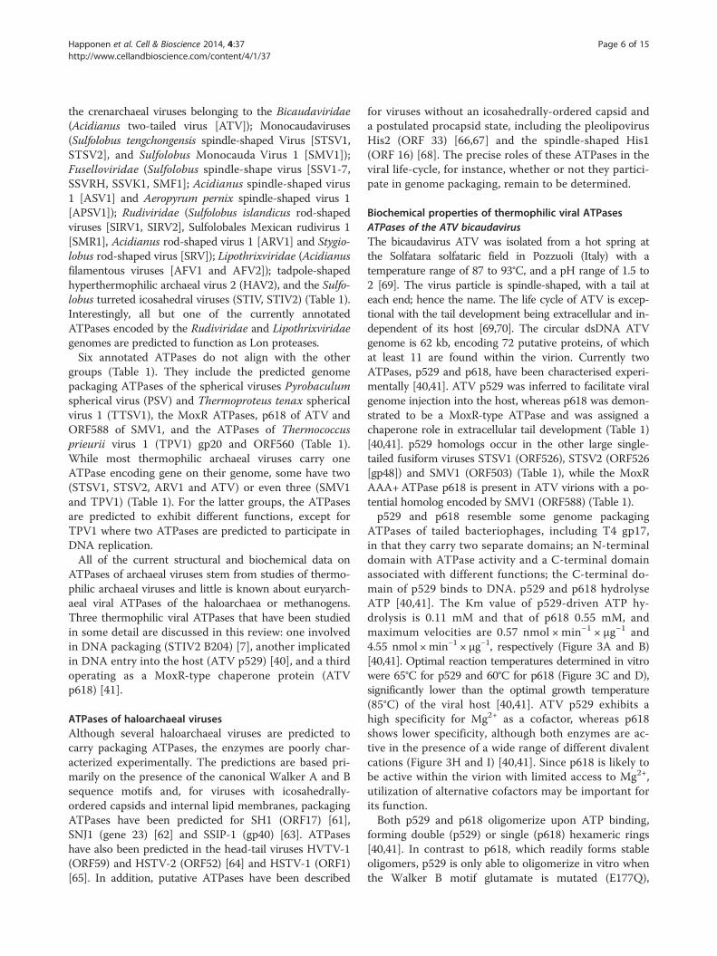
the crenarchaeal viruses belonging to the Bicaudaviridae(Acidianus two-tailed virus [ATV]); Monocaudaviruses(Sulfolobus tengchongensis spindle-shaped Virus [STSV1,STSV2], and Sulfolobus Monocauda Virus 1 [SMV1]);Fuselloviridae (Sulfolobus spindle-shape virus [SSV1-7,SSVRH, SSVK1, SMF1]; Acidianus spindle-shaped virus1 [ASV1] and Aeropyrum pernix spindle-shaped virus 1[APSV1]); Rudiviridae (Sulfolobus islandicus rod-shapedviruses [SIRV1, SIRV2], Sulfolobales Mexican rudivirus 1[SMR1], Acidianus rod-shaped virus 1 [ARV1] and Stygio-lobus rod-shaped virus [SRV]); Lipothrixviridae (Acidianusfilamentous viruses [AFV1 and AFV2]); tadpole-shapedhyperthermophilic archaeal virus 2 (HAV2), and the Sulfo-lobus turreted icosahedral viruses (STIV, STIV2) (Table 1).Interestingly, all but one of the currently annotatedATPases encoded by the Rudiviridae and Lipothrixviridaegenomes are predicted to function as Lon proteases.Six annotated ATPases do not align with the other
groups (Table 1). They include the predicted genomepackaging ATPases of the spherical viruses Pyrobaculumspherical virus (PSV) and Thermoproteus tenax sphericalvirus 1 (TTSV1), the MoxR ATPases, p618 of ATV andORF588 of SMV1, and the ATPases of Thermococcusprieurii virus 1 (TPV1) gp20 and ORF560 (Table 1).While most thermophilic archaeal viruses carry oneATPase encoding gene on their genome, some have two(STSV1, STSV2, ARV1 and ATV) or even three (SMV1and TPV1) (Table 1). For the latter groups, the ATPasesare predicted to exhibit different functions, except forTPV1 where two ATPases are predicted to participate inDNA replication.All of the current structural and biochemical data on
ATPases of archaeal viruses stem from studies of thermo-philic archaeal viruses and little is known about euryarch-aeal viral ATPases of the haloarchaea or methanogens.Three thermophilic viral ATPases that have been studiedin some detail are discussed in this review: one involvedin DNA packaging (STIV2 B204) [7], another implicatedin DNA entry into the host (ATV p529) [40], and a thirdoperating as a MoxR-type chaperone protein (ATVp618) [41].
ATPases of haloarchaeal virusesAlthough several haloarchaeal viruses are predicted tocarry packaging ATPases, the enzymes are poorly char-acterized experimentally. The predictions are based pri-marily on the presence of the canonical Walker A and Bsequence motifs and, for viruses with icosahedrally-ordered capsids and internal lipid membranes, packagingATPases have been predicted for SH1 (ORF17) [61],SNJ1 (gene 23) [62] and SSIP-1 (gp40) [63]. ATPaseshave also been predicted in the head-tail viruses HVTV-1(ORF59) and HSTV-2 (ORF52) [64] and HSTV-1 (ORF1)[65]. In addition, putative ATPases have been described
for viruses without an icosahedrally-ordered capsid anda postulated procapsid state, including the pleolipovirusHis2 (ORF 33) [66,67] and the spindle-shaped His1(ORF 16) [68]. The precise roles of these ATPases in theviral life-cycle, for instance, whether or not they partici-pate in genome packaging, remain to be determined.
Biochemical properties of thermophilic viral ATPasesATPases of the ATV bicaudavirusThe bicaudavirus ATV was isolated from a hot spring atthe Solfatara solfataric field in Pozzuoli (Italy) with atemperature range of 87 to 93°C, and a pH range of 1.5 to2 [69]. The virus particle is spindle-shaped, with a tail ateach end; hence the name. The life cycle of ATV is excep-tional with the tail development being extracellular and in-dependent of its host [69,70]. The circular dsDNA ATVgenome is 62 kb, encoding 72 putative proteins, of whichat least 11 are found within the virion. Currently twoATPases, p529 and p618, have been characterised experi-mentally [40,41]. ATV p529 was inferred to facilitate viralgenome injection into the host, whereas p618 was demon-strated to be a MoxR-type ATPase and was assigned achaperone role in extracellular tail development (Table 1)[40,41]. p529 homologs occur in the other large single-tailed fusiform viruses STSV1 (ORF526), STSV2 (ORF526[gp48]) and SMV1 (ORF503) (Table 1), while the MoxRAAA+ATPase p618 is present in ATV virions with a po-tential homolog encoded by SMV1 (ORF588) (Table 1).p529 and p618 resemble some genome packaging
ATPases of tailed bacteriophages, including T4 gp17,in that they carry two separate domains; an N-terminaldomain with ATPase activity and a C-terminal domainassociated with different functions; the C-terminal do-main of p529 binds to DNA. p529 and p618 hydrolyseATP [40,41]. The Km value of p529-driven ATP hy-drolysis is 0.11 mM and that of p618 0.55 mM, andmaximum velocities are 0.57 nmol × min−1 × μg−1 and4.55 nmol ×min−1 × μg−1, respectively (Figure 3A and B)[40,41]. Optimal reaction temperatures determined in vitrowere 65°C for p529 and 60°C for p618 (Figure 3C and D),significantly lower than the optimal growth temperature(85°C) of the viral host [40,41]. ATV p529 exhibits ahigh specificity for Mg2+ as a cofactor, whereas p618shows lower specificity, although both enzymes are ac-tive in the presence of a wide range of different divalentcations (Figure 3H and I) [40,41]. Since p618 is likely tobe active within the virion with limited access to Mg2+,utilization of alternative cofactors may be important forits function.Both p529 and p618 oligomerize upon ATP binding,
forming double (p529) or single (p618) hexameric rings[40,41]. In contrast to p618, which readily forms stableoligomers, p529 is only able to oligomerize in vitro whenthe Walker B motif glutamate is mutated (E177Q),
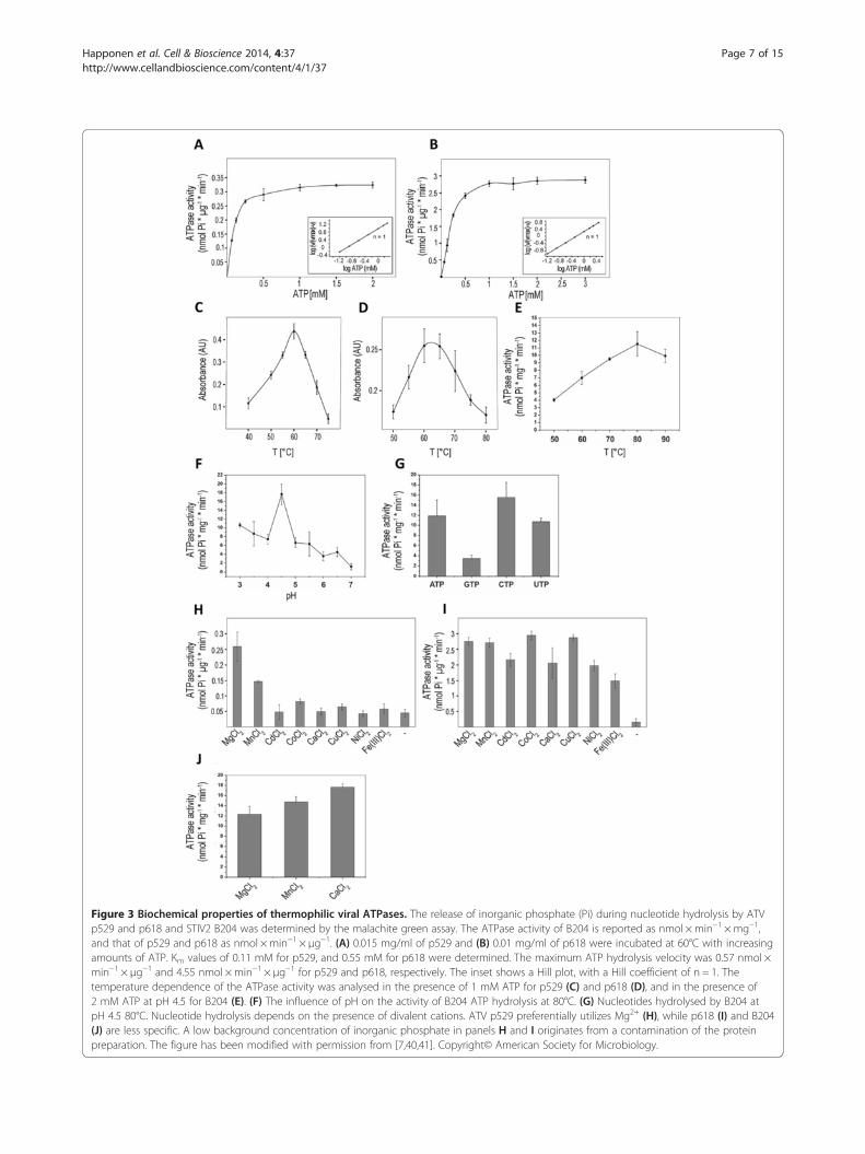
Figure 3 Biochemical properties of thermophilic viral ATPases. The release of inorganic phosphate (Pi) during nucleotide hydrolysis by ATVp529 and p618 and STIV2 B204 was determined by the malachite green assay. The ATPase activity of B204 is reported as nmol × min−1 × mg−1,and that of p529 and p618 as nmol ×min−1 × μg−1. (A) 0.015 mg/ml of p529 and (B) 0.01 mg/ml of p618 were incubated at 60°C with increasingamounts of ATP. Km values of 0.11 mM for p529, and 0.55 mM for p618 were determined. The maximum ATP hydrolysis velocity was 0.57 nmol ×min−1 × μg−1 and 4.55 nmol ×min−1 × μg−1 for p529 and p618, respectively. The inset shows a Hill plot, with a Hill coefficient of n = 1. Thetemperature dependence of the ATPase activity was analysed in the presence of 1 mM ATP for p529 (C) and p618 (D), and in the presence of2 mM ATP at pH 4.5 for B204 (E). (F) The influence of pH on the activity of B204 ATP hydrolysis at 80°C. (G) Nucleotides hydrolysed by B204 atpH 4.5 80°C. Nucleotide hydrolysis depends on the presence of divalent cations. ATV p529 preferentially utilizes Mg2+ (H), while p618 (I) and B204(J) are less specific. A low background concentration of inorganic phosphate in panels H and I originates from a contamination of the proteinpreparation. The figure has been modified with permission from [7,40,41]. Copyright© American Society for Microbiology.
Happonen et al. Cell & Bioscience 2014, 4:37 Page 7 of 15http://www.cellandbioscience.com/content/4/1/37

Happonen et al. Cell & Bioscience 2014, 4:37 Page 8 of 15http://www.cellandbioscience.com/content/4/1/37
suggesting that dissociation of the complex occurs afterATP hydrolysis. Oligomerization and ATPase activity ofthe isolated ATPase domain (amino acids 1–315) wereonly observed for p529 [40].
The genome packaging ATPase B204 of the icosahedralSTIV2STIV2 infects Sulfolobus islandicus and is an icosahedrally-symmetric virus with an internal membrane and verticesdecorated by large turrets thought to be involved inhost-cell recognition and attachment [71]. It was iso-lated from an acidic hot spring (88.3°C, pH 3.5), andhas a circular 16.6 kb dsDNA genome encoding 34predicted proteins, nine of which encode structuralproteins [7,71]. An STIV2-encoded ATPase – B204was predicted on the basis of sequence similarity toother P-loop ATPases [71]. This ATPase is most activeat pH 4.5 and 80°C, close to the optimal physiologicalconditions of the host [7,71] (Figure 3E and F). Thetemperature optimum of B204 is 20°C to 25°C higherthan that of ATV p618 and p529 and B204 can alsohydrolyse GTP, UTP, and CTP [7] (Figure 3G). More-over, as shown for ATV p618, B204 can also utilizeother cofactors in addition to Mg2+ [7] (Figure 3J).In contrast to ATV p529, B204 does not separate NTP
hydrolysis and DNA binding into distinct domains. Ithas been demonstrated via electrophoretic mobility shiftassays that B204 binds both ds and ss, linear and circularDNA, with no apparent sequence specificity, but lineardsDNA stimulated ATPase activity [7]. In this respect itresembles the phi29 genome packaging ATPase gp16,which also binds DNA in a non-specific manner, and its
Figure 4 B204 structure. (A) Ribbon diagram of a B204 monomer boundhighlighted in red indicate the conserved P9-motif described for the PRD1rainbow hues (blue to red) generated by aligning six monomers of chain Amonomers in the FtsK hexamer (PDB: 2IUU) using the program UCSF Chim(orange to red) lies on the capsid distal side. A short stretch of dsDNA (blamonomers. (C) A radially depth-cued isosurface representation (at 2 σ abov1679) [71] with one of the turrets replaced by our hexameric model of B20was rendered in UCSF Chimera [76].
ATPase activity is stimulated by DNA [21,72]. The min-imal length of a B204-bound DNA was estimated at20 bp [7], similar to the minimum length of a DNA mol-ecule bound to T4 gp17 [73]. However, B204 also bindsRNA in addition to DNA [7].
Structural properties of thermophilic viral ATPasesNTPase B204 is currently the only thermophilic viraldsDNA packaging NTPase for which the structure hasbeen determined. B204 is a 24.8 kDa monomer exhibit-ing nine β-strands and eight α-helices organised in thetopology of the FtsK-HerA superfamily (Figure 4A)[3,6,7]. The sequence shows similarity to that of genomepackaging ATPases of membrane-containing dsDNAviruses of the PRD1-like family [34,74,75].Based on the atomic models, the residues interacting
with the hydrolysable NTP are the Walker A motif resi-dues K13, K14, G16, K17 and S18, and residues Y19, Y186and I204 (Figure 5A) [7]. The nucleoside moiety stacks be-tween Y19 and Y186, but the sugar moiety does not inter-act with B204 [7]. The catalytic Mg2+-ion is coordinatedby the Walker A motif residue S18 [7]. Two active siteconformations were observed depending on the presenceor absence of a nucleotide in the P-loop [7]. The activesite pockets lacking a nucleotide are in the closed con-formation, whereas the active site pockets occupied by anucleotide are in the open conformation [7]. The max-imum difference between these two conformers is 1.8 Å[7]. The major differences between the closed and openconformers are as follows: in the absence of a nucleo-tide, i) the gap between Y19 and Y186 opens up; ii) K13and K14 bend away from the nucleotide binding site;
to AMPPCP and Mg2+ (green sphere) (PBD: 4KFU) [7]. The regions-like lineage of viruses [34]. (B) A hexamer model of B204 shown inof the AMPPCP B204 structure (PDB: 4KFU) onto each of the FtsKera [76]. According to our hypothesis the C-terminal part of B204ck and blue helix) is modelled into the channel formed by thee the mean) of the 20-Å resolution reconstruction of STIV2 (EMDB:4 (surface representation in rainbow hues as in panel B). The figure
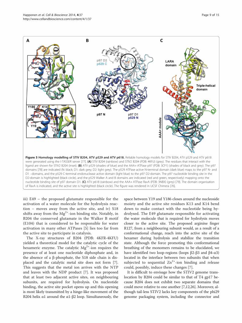
Figure 5 Homology modelling of STIV B204, ATV p529 and ATV p618. Reliable homology models for STIV B204, ATV p529 and ATV p618were generated using the I-TASSER server [77]. (A) STIV B204 (rainbow) and STIV2 B204 (PDB: 4KFU) (grey). The residues that interact with theligand are shown for STIV2 B204 (inset). (B) ATV p529 (shades of blue) and the AAA+ ATPase p97 (PDB: 3CF1) (shades of black and grey). The p97domains [78] are indicated (N: black; D1: dark grey; D2: light grey). The p529 ATPase active N-terminal domain (dark blue) maps to the p97 N- andD1 –domains, and the p529 C-terminal endonuclease active domain (light blue) to the p97 D2-domain. The p97 nucleotide binding site in theD2-domain is highlighted (black circle), and the p529 Walker A and B domains are indicated (red and green, respectively) mapping onto thenucleotide binding site of p97 domain D1. (C) ATV p618 (rainbow) and the AAA+ ATPase RavA (PDB: 3NBX) (grey) [79]. The domain organisationof RavA is indicated, and the active site is highlighted (black circle). The figure was rendered in UCSF Chimera [76].
Happonen et al. Cell & Bioscience 2014, 4:37 Page 9 of 15http://www.cellandbioscience.com/content/4/1/37
iii) E49 – the proposed glutamate responsible for theactivation of a water molecule for the hydrolysis reac-tion – moves away from the active site, and iv) S18shifts away from the Mg2+-ion binding site. Notably, inB204 the conserved glutamate in the Walker B motif(E104) that is considered to be responsible for wateractivation in many other ATPases [5] lies too far fromthe active site to participate in catalysis.The X-ray structures of B204 (PDB: 4KFR-4KFU)
yielded a theoretical model for the catalytic cycle of thehexameric enzyme. The catalytic Mg2+-ion requires thepresence of at least one nucleotide diphosphate and, inthe absence of a β-phosphate, the S18 side chain is dis-placed and the catalytic metal site does not form [7].This suggests that the metal ion arrives with the NTPand leaves with the NDP product [7]. It was proposedthat at least two adjacent active sites, on neighbouringsubunits, are required for hydrolysis. On nucleotidebinding, the active site pocket opens up and this openingis most likely transmitted by a hinge-like movement of theB204 helix α1 around the α1-β2 loop. Simultaneously, the
space between Y19 and Y186 closes around the nucleosidemoiety and the active site residues K13 and K14 benddown to make contact with the nucleotide being hy-drolysed. The E49 glutamate responsible for activatingthe water molecule that is required for hydrolysis movescloser to the active site. The proposed arginine fingerR127, from a neighbouring subunit would, as a result of aconformational change, reach into the active site of thehexamer during hydrolysis and stabilize the transitionstate. Although the force promoting this conformationalbreathing of the monomers remains to be elucidated, wehave identified two loop-regions (loops β2-β3 and β4-α3)located in the interface between two subunits that whensubjected to sequential Zn2+-ion binding and releasecould, possibly, induce these changes [7].It is difficult to envisage how the STIV2 genome trans-
location by B204 could be similar to that of T4 gp17 be-cause B204 does not exhibit two separate domains thatcould move relative to one another [7,12,26]. Moreover, al-though tail-less STIV2 lacks key components of the phi29genome packaging system, including the connector and

Happonen et al. Cell & Bioscience 2014, 4:37 Page 10 of 15http://www.cellandbioscience.com/content/4/1/37
pRNA, a similar genome packaging mechanism by revolu-tion rather than rotation could take place. In viruses suchas the PRD1-like viruses (including STIV2), with an or-dered icosahedral capsid and an internal lipid membrane,small membrane proteins have been suggested to generatea pore in the membrane at the packaging vertex and theymay function like the connectors of head-tail phages [80].There are several such proteins encoded by STIV2 thatcould fulfil this function [7,71]. Possibly the translocationmechanism of B204 is similar to that of phi29 gp16 in thatone ATP molecule is first bound to one subunit of theproposed hexameric structure. This would lead to a con-formational change rendering B204 susceptible to DNAbinding. Subsequent ATP hydrolysis-induced conform-ational changes, possibly aided by the B204 loops β2-β3and β4-α3, might then shift the DNA molecule to theadjacent ATP bound subunit. The current drawbacks tothis model are: i) STIV2 lacks some key components ofthe phi29 packaging system and ii) B204 binds DNA inthe absence of ATP under our assay conditions [7],The exact role of each of the residues in the Walker A
and B motifs in the catalysis remains to be elucidated al-though it is clear that residue S18 coordinates the metalion. Mutagenesis of K17 in the Walker A domain, and ofE104 in the Walker B motif, into alanine residues didnot affect B204 ATP hydrolysis and, therefore, they arenot essential for hydrolysis by monomeric B204 (L.J.H.,unpublished). Attempts at producing a biological hex-amer of STIV2 B204 have so far been unsuccessfuland our conclusions are based on a hexameric model(Figure 4B and C). An active hexameric form of B204would, in combination with mutagenesis studies, pro-vide valuable insights into the distinct roles of eachresidue during the catalytic cycle.
Homology modelling of the thermophilic viral ATPasesThe closest known relative to STIV2 is STIV [71,81].Originally, the predicted STIV genome packaging ATPasewas described as a 164 amino acid protein, but it was laterconfirmed that the original annotation was too short dueto a frame-shift sequencing error (M. Young, personalcommunication). We predicted a revised gene product forthe STIV ATPase, with a length of 204 amino acids, andan amino acid sequence identity of 90.2% to STIV2 B204.Therefore, we modelled the STIV B204 using I-TASSER(Table 2) [77] in order to explore the tertiary structure
Table 2 Homology modelling of thermophilic viral ATPases
Protein Length (AA) TM-score [0, 1]* RMSD (Å) Mo
STIV B204 204 0.92 ± 0.06 2.5 ± 1.9 Me
ATV P529 529 0.61 ± 0.14 9.4 ± 4.6 Me
ATV P618 618 0.57 ± 0.15 10.6 ± 4.6 Me
*A TM value > 0.5 implies that the model has the correct topology [82].**The STIV2 B204 structure in complex with AMPPCP (PDB: 4KFU) was used as a use
conservation between these two thermophilic ATPases(Figure 5A). The RMSD (root-mean square deviation)between our B204 structure complexed with AMPPCP(PDB: 4KFU) and the STIV B204 model is 2.5 ± 1.9 Å(Table 2), with well-conserved active site topology(Figure 5A, inset). Based on the high sequence identityand the apparent conserved active site topology, wepropose that the STIV B204 residues interacting withthe ATP are conserved between the two viruses (eventhough STIV2 B204 K14 is replaced by R14 in STIV)(Figure 1). Furthermore, it is highly likely that thecatalytic cycles of the two ATPases are similar.We also generated homology models for ATV p529
[40] and p618 [41] using I-TASSER [77] (Table 2) inorder to gain insight into their possible structural andfunctional conservation. They were inferred to be hom-ologous with the AAA+ ATPases p97 and MoxR, re-spectively [40,41]. The main parent model for p529used in our homology modelling with I-TASSER wasthe hexameric type II AAA+ ATPase p97 containingtwo nucleotide-binding domains D1 and D2, where theformer is inactive under physiological conditions [78,83](Figure 5B). Two distinct domains were described forATV p529: the N-terminal domain carrying ATPase activ-ity (Figure 5B, dark blue), and the C-terminal domainexhibiting endonuclease activity (Figure 5B, light blue)[40]. This correlates with descriptions of terminase-proteins of head-tail viruses, with one domain for DNA-binding and another with ATPase activity [1,18]. In ourmodel, the p529 N-terminal ATPase active site maps ontop of the p97 D1 nucleotide binding site (Figure 5B).The main parent model for p618 used in our homology
modelling was the hexameric RavA MoxR AAA+ATPase[79]. RavA consists of three domains: i) the N-terminalAAA+module (residues 1–306); ii) the triple-helical do-main (residues 307–330 and 442–497); and the iii) LARAdomain [79]. The overall topology of p618 follows that ofRavA, except for the three C-terminal helices present onlyin p618 (orange to red helices in Figure 5C). The overalltopology at the active site is highly conserved for bothproteins.
Role in virus life cycle and relationship to other virusesViruses with icosahedrally-ordered capsids package theirgenomes into a preformed procapsid with the help of atranslocating ATPase. One such ATPase is STIV2 B204
delled amino acids Main parent model (PDB id) Reference
t1-Ile204 4KFU** [7]
t1-Tyr529 3CF1 (p97) [78]
t1-Ser618 3NBX (RavA) [79]
r-defined restraint.

Happonen et al. Cell & Bioscience 2014, 4:37 Page 11 of 15http://www.cellandbioscience.com/content/4/1/37
and, based on our biochemical and structural datacombined with the knowledge of the assembly of othermembrane-containing and non-tailed viruses such asSTIV and PRD1, we have proposed a model for the roleof B204 in STIV2 (and STIV) assembly (Figure 6A) [7].B204 binds DNA unspecifically [7] which suggests thatit does not bind to a specific packaging sequence onthe STIV2 genome. This led us to the proposal that an-other STIV2 DNA-binding protein is responsible forthe recognition and recruitment of the genome to theputative packaging vertex. Currently a single DNA-binding protein B27 has been detected in the virion[71] and other possible co-functional DNA-bindingproteins could be STIV2 B116 or F98, for which theSTIV counterparts B116 and F93 have been shown tobind DNA but their functional roles remain unclear[84,85]. According to our model, procapsid assemblywould begin at the postulated packaging vertex con-currently with the STIV2 genome being recognized byB72. Small membrane proteins, most likely E51 andE76b, would form a pore in the viral membrane forgenome translocation. Recruitment of STIV2 DNA byB72 to the packaging vertex would generate the activemotor complex that, on ATP hydrolysis, would translo-cate the DNA into the procapsid. We propose furtherthat the STIV2 DNA is packaged as a linear moleculewhich is circularized within the capsid although no lig-ase gene has yet been identified. STIV B204 remains
Figure 6 Model for STIV2 genome packaging. Schematic model for genthe major capsid protein (light blue hexagon). The host-attachment structumembrane protein A55 (black spheres), as described for STIV [81]. The capsmembrane proteins (orange spheres) attached to it. The membrane proteinSTIV2 genome translocation at the packaging vertex [7]. STIV2 assembly inipostulated STIV2 genome recognition protein B72 (green sphere) would siminitiation complex thus recruiting the viral genome for packaging [7]. Accogenome packaging ATPase B204 that has assembled as a hexamer (red) atprocapsid via ATP powered hydrolysis. The figure is redrawn from [7].
associated with the virion and is not dissociating afterpackaging [86].
ATV p529 might be involved in genome injectionATV p529 has been proposed to participate in viralDNA delivery into the host [40]. To date, most of ourknowledge on viral DNA injection into the host and itssubsequent release is derived from studies on tailed bac-teriophages, although a few archaeal viral studies havebeen performed [87]. Viral DNA is packaged at a veryhigh density within virions, and several in vitro studieshave shown that this pressurized DNA can provide theforce for DNA ejection [88]. In contrast, DNA injectionhas been shown to be an active process involving bothpush and pull forces that are mediated by viral and hostproteins and may require ATP. For example, phi29DNA is injected via a two-step “push-pull” mechanism.Initially about 65% of the phi29 DNA in pushed into thehost cell, probably as a result of the pressure inside theviral capsid. Subsequently the remaining DNA is pulledinto the host cell facilitated by viral protein gp17 in anenergy-dependent process, although gp17 has not beenassociated with ATPase activity [89,90]. To date, DNAejection from archaeal virions has received little atten-tion. One of the few viruses that has been studied is thespindle-shaped haloarchaeal virus His1 [87] and, as forphi29, it was shown that His1 DNA ejection is only par-tial, and most likely requires host factors for completion
ome encapsidation of STIV2. The capsid of STIV2 is composed of A345,res – the turrets – are composed of B631 (dark blue triangles) and theid encloses the viral lipid membrane (yellow), with several predicteds E51 and E76b are proposed to form a pore in the membrane fortiates by the formation of the procapsid. We have proposed that theultaneously recognize and bind to the STIV2 genome, forming the
rding to our hypothesis, the initiation complex is recognised by thethe packaging vertex. The STIV2 genome is translocated into the

Happonen et al. Cell & Bioscience 2014, 4:37 Page 12 of 15http://www.cellandbioscience.com/content/4/1/37
[87]. Moreover, apart from ATV p529 described here,we are not aware of other archaeal ATPases postulatedto participate in viral DNA entry into a host.To date, four, large, tailed, fusiform viruses infecting
the Sulfolobales have been isolated and characterised.These viruses include the two-tailed bicaudavirus ATVand the three single-tailed monocaudaviruses STSV1,STSV2 and SMV1. At present, little is known abouttheir mechanism of host attachment, DNA injection,DNA packaging and viral release. ATPases have beenshown to be involved in some of these processes ineukaryal, bacterial and archaeal viruses indicating thatsimilar ATPase-driven mechanisms are likely to apply tothese thermophilic archaeal viruses. All four fusiformvirus genomes encode at least one ATPase. There is con-servation between ATV p529, ORF526 (STSV1), ORF526(STSV2), and ORF503 (SMV1) as well as ORF506 of thetadpole-shaped HAV2 (Table 1). However, only ATV p529has been characterized biochemically. It has a two-domainstructure and the domains exhibit ATPase activity as wellas DNA-binding and endonuclease activity. The endo-nuclease activity suggests that p529 may be important inprocessing the viral genome during encapsidation, whereit can be likened to, for example, T4 gp17, which cleavesthe viral genome upon headful packaging.ATV p529 is exceptional in the sense that both its N-
terminal ATPase domain and its C-terminal domain bindDNA [40]. DNA binding of the N-terminal domain isnegatively influenced by the presence of ATP, suggestingthat a conformational change induced by ATP bindinginhibits DNA binding. This contrasts with the proper-ties of the phi29 gp16 DNA packaging ATPase, whereDNA affinity is induced by a conformational changecaused by ATP binding [30]. The isolated C-terminaldomain of p529 also exhibits endonuclease activity thatis not observed for the wild-type protein, again indicatingthat activity may be dependent on conformation [40].Immobilized p529 pulls down the transmembrane
oligopeptide-binding host protein SSO1273 consideredto be a potential virus receptor [40,91]. Initial DNA ejec-tion into the host could be driven by pressure as describedfor bacterial, eukaryal and archaeal viruses [87-89,92].During DNA injection, the double hexameric structure ofp529 could provide a pore through which the remainingviral DNA is pulled into the host cell. This would implythat the extra energy needed to finalise viral DNA entrymay be provided by the virus independently of host fac-tors. ATV p529 was initially not identified within the vir-ion using SDS-PAGE and Edman degradation of extractedprotein bands [69], perhaps due to low abundance. How-ever, a very low concentration of the protein would be suf-ficient to build a functional complex. Additional studieson p529 are needed to verify its role in viral DNA trans-location into the host. Further, it is possible that p529
interacts with SSO1273 on the host’s inner membraneduring DNA packaging in STSV2, but this hypothesis hasyet to be explored experimentally.
ATV p618 is a MoxR-type ATPaseATV p618 was classified as a MoxR-type ATPase [41].MoxR-type ATPases are found amongst archaea and bac-teria, and they have been classified into seven subfamilies[93]. They are proposed to exhibit chaperone-like activitiesand are involved in various stress response pathways [94].The role of p618 in the ATV life cycle was investigated byassaying its interactions with nucleic acids and other ATVproteins. ATV p618 is a major virion component [69,70]and it interacts with at least four other virus proteins butnot with viral DNA [41]. Three of these proteins p387,p653, and p800 have been found in the virion and p800can generate long filaments [69,70]. The fourth protein,p892, which carries a von Willebrand factor type A(VWA) domain, interacts strongly with p618 [41]. A hom-ologous protein has been detected in SMV1 virions [44]although not yet in ATV virions. Proteins exhibiting aVWA domain are known to interact with MoxR-likeATPases, as shown for the E.coli RavA ATPase [93]. Bothp387 and p892 enhance p618 ATPase activity. From thisset of observations, it has been concluded that p618 par-ticipates in extracellular ATV tail development [41]. Thishypothesis was further strengthened by the observationthat the fourth interacting ATV protein, p653, is localizedin the virion tails [41]. Further studies are needed to local-ise p618 within the virion tail, and to elucidate its role intail development.
ConclusionsSeveral ATPases are encoded in archaeal viral genomes –both crenarchaeal and euryarchaeal – and these enzymeshave been implicated in a variety of functions during theviral life cycle. They include DNA entry into the host,genome packaging into viral capsids, modulation of DNAreplication, and as proteases and chaperones, but minimalexperimental evidence exists of their precise structures orspecific functions. To date, functional studies have onlybeen reported for three ATPases of thermophilic cre-narchaeal viruses, and none for the ATPases associatedwith euryarchaeal viruses. These studies provide a mereglimpse into the functional diversity of these archaealATPases. More research is needed in order to answer keyquestions, such as: i) How does genome packaging occurfor the bottle-shaped, droplet-shaped, spindle-shaped andpleomorphic viruses of the archaeal domain? Do theseviruses assemble their capsid proteins around the viralgenome, or do they utilise an active packaging mechan-ism involving ATP hydrolysis? ii) To what extent are thefunctional roles of archaeal viral ATPases inferred frombioinformatical analyses and homology predictions correct?
Happonen et al. Cell & Bioscience 2014, 4:37 Page 13 of 15http://www.cellandbioscience.com/content/4/1/37
iii) Can the extremophilic viral ATPases yield novel in-sights into how energy metabolism functions under ex-treme environmental conditions? iv) Since many archaealviruses reside in extreme environments, and their virionsare stable under extreme conditions, are they generallyamenable to important biotechnological and nanotechno-logical applications? Clearly the field is set for extensivein-depth studies of the diverse ATPases of the archaeal vi-ruses with the results having relevance both for a varietyof different viral life cycles and of strong potential interestfor biotechnology and nanotechnology.
Competing interestsThe authors declare that they have no competing interest.
Authors’ contributionsLJH performed the homology modelling of STIV B204, ATV p529 and p618,LJH and SE made the figures, LJH and RAG made the sequence alignmentsand annotations, LJH, SE, RAG, and SJB wrote the manuscript. All authorsread and approved the final manuscript.
AcknowledgementsThis work was supported by Academy of Finland grant 139178 (to SJB) andgrants from the Danish Natural Science Research Council (to RAG).
Author details1Department of Clinical Sciences, Division of Infection Medicine, LundUniversity, SE-221 84 Lund, Sweden. 2Archaea Centre, Department of Biology,University of Copenhagen, Ole Maaløes Vej 5, DK-2200 Copenhagen N,Denmark. 3Institute of Biotechnology, University of Helsinki, (Viikinkaari 1),P.O. Box 65, FI-00014 Helsinki, Finland.
Received: 4 September 2013 Accepted: 13 June 2014Published: 23 July 2014
References1. Guo P, Lee TJ: Viral nanomotors for packaging of dsDNA and dsRNA.
Mol Microbiol 2007, 64(4):886–903.2. Hanson PI, Whiteheart SW: AAA+ proteins: have engine, will work. Nat Rev
Mol Cell Biol 2005, 6(7):519–529.3. Iyer LM, Makarova KS, Koonin EV, Aravind L: Comparative genomics of the
FtsK-HerA superfamily of pumping ATPases: implications for the originsof chromosome segregation, cell division and viral capsid packaging.Nucleic Acids Res 2004, 32(17):5260–5279.
4. Walker JE, Saraste M, Runswick MJ, Gay NJ: Distantly related sequences inthe alpha- and beta-subunits of ATP synthase, myosin, kinases and otherATP-requiring enzymes and a common nucleotide binding fold. EMBO J1982, 1(8):945–951.
5. Goetzinger KR, Rao VB: Defining the ATPase center of bacteriophage T4DNA packaging machine: requirement for a catalytic glutamate residuein the large terminase protein gp17. J Mol Biol 2003, 331(1):139–154.
6. Iyer LM, Leipe DD, Koonin EV, Aravind L: Evolutionary history and higherorder classification of AAA+ ATPases. J Struct Biol 2004, 146(1–2):11–31.
7. Happonen LJ, Oksanen E, Liljeroos L, Goldman A, Kajander T, Butcher SJ:The Structure of the NTPase That Powers DNA Packaging into SulfolobusTurreted Icosahedral Virus 2. J Virol 2013, 87(15):8388–8398.
8. Massey TH, Mercogliano CP, Yates J, Sherratt DJ, Lowe J: Double-strandedDNA translocation: structure and mechanism of hexameric FtsK. Mol Cell2006, 23(4):457–469.
9. Subramanya HS, Bird LE, Brannigan JA, Wigley DB: Crystal structure of aDExx box DNA helicase. Nature 1996, 384(6607):379–383.
10. Mancini EJ, Kainov DE, Grimes JM, Tuma R, Bamford DH, Stuart DI: Atomicsnapshots of an RNA packaging motor reveal conformational changeslinking ATP hydrolysis to RNA translocation. Cell 2004, 118(6):743–755.
11. Nadanaciva S, Weber J, Wilke-Mounts S, Senior AE: Importance ofF1-ATPase residue alpha-Arg-376 for catalytic transition statestabilization. Biochemistry 1999, 38(47):15493–15499.
12. Sun S, Kondabagil K, Gentz PM, Rossmann MG, Rao VB: The structure of theATPase that powers DNA packaging into bacteriophage T4 procapsids.Mol Cell 2007, 25(6):943–949.
13. Guenther B, Onrust R, Sali A, O’Donnell M, Kuriyan J: Crystal structure ofthe delta’ subunit of the clamp-loader complex of E. coli DNApolymerase III. Cell 1997, 91(3):335–345.
14. Bochtler M, Hartmann C, Song HK, Bourenkov GP, Bartunik HD, Huber R:The structures of HsIU and the ATP-dependent protease HsIU-HsIV.Nature 2000, 403(6771):800–805.
15. Liu J, Smith CL, DeRyckere D, DeAngelis K, Martin GS, Berger JM: Structureand function of Cdc6/Cdc18: implications for origin recognition andcheckpoint control. Mol Cell 2000, 6(3):637–648.
16. Sievers F, Wilm A, Dineen D, Gibson TJ, Karplus K, Li W, Lopez R, McWilliam H,Remmert M, Soding J, Thompson JD, Higgins DG: Fast, scalable generation ofhigh-quality protein multiple sequence alignments using Clustal Omega.Mol Syst Biol 2011, 7:539.
17. Gouet P, Courcelle E, Stuart DI, Metoz F: ESPript: analysis of multiplesequence alignments in PostScript. Bioinformatics 1999, 15(4):305–308.
18. Rao VB, Feiss M: The bacteriophage DNA packaging motor. Annu RevGenet 2008, 42:647–681.
19. Smith DE, Tans SJ, Smith SB, Grimes S, Anderson DL, Bustamante C: Thebacteriophage straight phi29 portal motor can package DNA against alarge internal force. Nature 2001, 413(6857):748–752.
20. Simpson AA, Tao Y, Leiman PG, Badasso MO, He Y, Jardine PJ, Olson NH, MoraisMC, Grimes S, Anderson DL, Rossmann MG: Structure of the bacteriophagephi29 DNA packaging motor. Nature 2000, 408(6813):745–750.
21. Guo P, Peterson C, Anderson D: Prohead and DNA-gp3-dependent ATPaseactivity of the DNA packaging protein gp16 of bacteriophage phi 29.J Mol Biol 1987, 197(2):229–236.
22. Schwartz C, De Donatis GM, Fang H, Guo P: The ATPase of the phi29 DNApackaging motor is a member of the hexameric AAA+ superfamily.Virology 2013, 443(1):20–27.
23. Guo P, Schwartz C, Haak J, Zhao Z: Discovery of a new motion mechanismof biomotors similar to the earth revolving around the sun withoutrotation. Virology 2013, 446(1–2):133–143.
24. Schwartz C, De Donatis GM, Zhang H, Fang H, Guo P: Revolution ratherthan rotation of AAA+ hexameric phi29 nanomotor for viral dsDNApackaging without coiling. Virology 2013, 443(1):28–39.
25. Driedonks RA, Engel A, tenHeggeler B, van D: Gene 20 product ofbacteriophage T4 its purification and structure. J Mol Biol 1981,152(4):641–662.
26. Sun S, Kondabagil K, Draper B, Alam TI, Bowman VD, Zhang Z, Hegde S,Fokine A, Rossmann MG, Rao VB: The structure of the phage T4 DNApackaging motor suggests a mechanism dependent on electrostaticforces. Cell 2008, 135(7):1251–1262.
27. Sun S, Gao S, Kondabagil K, Xiang Y, Rossmann MG, Rao VB: Structure andfunction of the small terminase component of the DNA packagingmachine in T4-like bacteriophages. Proc Natl Acad Sci U S A 2012,109(3):817–822.
28. Fuller DN, Raymer DM, Kottadiel VI, Rao VB, Smith DE: Single phage T4DNA packaging motors exhibit large force generation, high velocity, anddynamic variability. Proc Natl Acad Sci U S A 2007, 104(43):16868–16873.
29. Kanamaru S, Kondabagil K, Rossmann MG, Rao VB: The functional domainsof bacteriophage t4 terminase. J Biol Chem 2004, 279(39):40795–40801.
30. Schwartz C, Guo P: Ultrastable pRNA hexameric ring gearing hexamericphi29 DNA-packaging motor by revolving without rotating and coiling.Curr Opin Biotechnol 2013, 24(4):581–590.
31. Zhao Z, Khisamutdinov E, Schwartz C, Guo P: Mechanism of one-way traffic ofhexameric phi29 DNA packaging motor with four electropositive relayinglayers facilitating antiparallel revolution. ACS Nano 2013, 7(5):4082–4092.
32. Kainov DE, Mancini EJ, Telenius J, Lisal J, Grimes JM, Bamford DH, Stuart DI,Tuma R: Structural basis of mechanochemical coupling in a hexamericmolecular motor. J Biol Chem 2008, 283(6):3607–3617.
33. Lisal J, Tuma R: Cooperative mechanism of RNA packaging motor. J BiolChem 2005, 280(24):23157–23164.
34. Strömsten NJ, Bamford DH, Bamford JK: In vitro DNA packaging of PRD1: acommon mechanism for internal-membrane viruses. J Mol Biol 2005,348(3):617–629.
35. Ziedaite G, Kivelä HM, Bamford JK, Bamford DH: Purified membrane-containing procapsids of bacteriophage PRD1 package the viral genome.J Mol Biol 2009, 386(3):637–647.

Happonen et al. Cell & Bioscience 2014, 4:37 Page 14 of 15http://www.cellandbioscience.com/content/4/1/37
36. Strömsten NJ, Bamford DH, Bamford JK: The unique vertex of bacterialvirus PRD1 is connected to the viral internal membrane. J Virol 2003,77(11):6314–6321.
37. Rice G, Tang L, Stedman K, Roberto F, Spuhler J, Gillitzer E, Johnson JE,Douglas T, Young M: The structure of a thermophilic archaeal virus showsa double-stranded DNA viral capsid type that spans all domains of life.Proc Natl Acad Sci U S A 2004, 101(20):7716–7720.
38. Häring M, Peng X, Brügger K, Rachel R, Stetter KO, Garrett RA, Prangishvili D:Morphology and genome organization of the virus PSV of thehyperthermophilic archaeal genera Pyrobaculum and Thermoproteus: anovel virus family, the Globuloviridae. Virology 2004, 323(2):233–242.
39. Ahn DG, Kim SI, Rhee JK, Kim KP, Pan JG, Oh JW: TTSV1, a new virus-likeparticle isolated from the hyperthermophilic crenarchaeoteThermoproteus tenax. Virology 2006, 351(2):280–290.
40. Erdmann S, Scheele U, Garrett RA: AAA ATPase p529 of Acidianustwo-tailed virus ATV and host receptor recognition. Virology 2011,421(1):61–66.
41. Scheele U, Erdmann S, Ungewickell EJ, Felisberto-Rodrigues C, Ortiz-Lombardia M, Garrett RA: Chaperone role for proteins p618 and p892 inthe extracellular tail development of Acidianus two-tailed virus. J Virol2011, 85(10):4812–4821.
42. Xiang X, Chen L, Huang X, Luo Y, She Q, Huang L: Sulfolobustengchongensis spindle-shaped virus STSV1: virus-host interactions andgenomic features. J Virol 2005, 79(14):8677–8686.
43. Erdmann S, Chen B, Huang X, Deng L, Liu C, Shah SA, Le Moine BS, Sobrino CL,Wang H, Wei Y, She Q, Garrett RA, Huang L, Lin L: A novel single-tailed fusi-form Sulfolobus virus STSV2 infecting model Sulfolobus species.Extremophiles 2014, 18(1):51–60.
44. Erdmann S, Le Moine BS, Garrett RA: Inter-viral conflicts that exploit hostCRISPR immune systems of Sulfolobus. Mol Microbiol 2014, 91(5):900–917.
45. Redder P, Peng X, Brügger K, Shah SA, Roesch F, Greve B, She Q, Schleper C,Forterre P, Garrett RA, Prangishvili D: Four newly isolated fusellovirusesfrom extreme geothermal environments reveal unusual morphologiesand a possible interviral recombination mechanism. Environ Microbiol2009, 11(11):2849–2862.
46. Palm P, Schleper C, Grampp B, Yeats S, McWilliam P, Reiter WD, Zillig W:Complete nucleotide sequence of the virus SSV1 of the archaebacteriumSulfolobus shibatae. Virology 1991, 185(1):242–250.
47. Stedman KM, She Q, Phan H, Arnold HP, Holz I, Garrett RA, Zillig W:Relationships between fuselloviruses infecting the extremelythermophilic archaeon Sulfolobus: SSV1 and SSV2. Res Microbiol 2003,154(4):295–302.
48. Peng X: Evidence for the horizontal transfer of an integrase gene from afusellovirus to a pRN-like plasmid within a single strain of Sulfolobusand the implications for plasmid survival. Microbiology 2008,154(Pt 2):383–391.
49. Wiedenheft B, Stedman K, Roberto F, Willits D, Gleske AK, Zoeller L, Snyder J,Douglas T, Young M: Comparative genomic analysis of hyperthermophilicarchaeal Fuselloviridae viruses. J Virol 2004,78(4):1954–1961.
50. Rice G, Stedman K, Snyder J, Wiedenheft B, Willits D, Brumfield S,McDermott T, Young MJ: Viruses from extreme thermal environments.Proc Natl Acad Sci U S A 2001, 98(23):13341–13345.
51. Servin-Garciduenas LE, Peng X, Garrett RA, Martinez-Romero E: Genomesequence of a novel archaeal fusellovirus assembled from themetagenome of a mexican hot spring. Genome Announcements 2013,1(2):e0016413.
52. Mochizuki T, Sako Y, Prangishvili D: Provirus induction inhyperthermophilic archaea: characterization of Aeropyrum pernixspindle-shaped virus 1 and Aeropyrum pernix ovoid virus 1. J Bacteriol2011, 193(19):5412–5419.
53. Bettstetter M, Peng X, Garrett RA, Prangishvili D: AFV1, a novel virusinfecting hyperthermophilic archaea of the genus acidianus. Virology2003, 315(1):68–79.
54. Häring M, Vestergaard G, Brügger K, Rachel R, Garrett RA, Prangishvili D:Structure and genome organization of AFV2, a novel archaeallipothrixvirus with unusual terminal and core structures. J Bacteriol 2005,187(11):3855–3858.
55. Vestergaard G, Häring M, Peng X, Rachel R, Garrett RA, Prangishvili D:A novel rudivirus, ARV1, of the hyperthermophilic archaeal genusAcidianus. Virology 2005, 336(1):83–92.
56. Peng X, Blum H, She Q, Mallok S, Brügger K, Garrett RA, Zillig W, Prangishvili D:Sequences and replication of genomes of the archaeal rudiviruses SIRV1and SIRV2: relationships to the archaeal lipothrixvirus SIFV and someeukaryal viruses. Virology 2001, 291(2):226–234.
57. Vestergaard G, Shah SA, Bize A, Reitberger W, Reuter M, Phan H, Briegel A,Rachel R, Garrett RA, Prangishvili D: Stygiolobus rod-shaped virus and theinterplay of crenarchaeal rudiviruses with the CRISPR antiviral system.J Bacteriol 2008, 190(20):6837–6845.
58. Servin-Garciduenas LE, Peng X, Garrett RA, Martinez-Romero E: Genomesequence of a novel archaeal rudivirus recovered from a mexican hotspring. Genome Announcements 2013, 1(2):e0016413.
59. Garrett RA, Prangishvili D, Shah SA, Reuter M, Stetter KO, Peng X:Metagenomic analyses of novel viruses and plasmids from a culturedenvironmental sample of hyperthermophilic neutrophiles. EnvironMicrobiol 2010, 12(11):2918–2930.
60. Gorlas A, Koonin EV, Bienvenu N, Prieur D, Geslin C: TPV1, the first virusisolated from the hyperthermophilic genus Thermococcus. EnvironMicrobiol 2012, 14(2):503–516.
61. Bamford DH, Ravantti JJ, Rönnholm G, Laurinavicius S, Kukkaro P, Dyall-SmithM, Somerharju P, Kalkkinen N, Bamford JK: Constituents of SH1, a novellipid-containing virus infecting the halophilic euryarchaeon Haloarculahispanica. J Virol 2005, 79(14):9097–9107.
62. Zhang Z, Liu Y, Wang S, Yang D, Cheng Y, Hu J, Chen J, Mei Y, Shen P,Bamford DH, Chen X: Temperate membrane-containing halophilicarchaeal virus SNJ1 has a circular dsDNA genome identical to that ofplasmid pHH205. Virology 2012, 434(2):233–241.
63. Aalto AP, Bitto D, Ravantti JJ, Bamford DH, Huiskonen JT, Oksanen HM:Snapshot of virus evolution in hypersaline environments from thecharacterization of a membrane-containing Salisaeta icosahedral phage1. Proc Natl Acad Sci U S A 2012, 109(18):7079–7084.
64. Pietilä MK, Laurinmäki P, Russell DA, Ko CC, Jacobs-Sera D, Butcher SJ,Bamford DH, Hendrix RW: Insights into head-tailed viruses infectingextremely halophilic archaea. J Virol 2013, 87(6):3248–3260.
65. Pietilä MK, Laurinmäki P, Russell DA, Ko CC, Jacobs-Sera D, Hendrix RW, BamfordDH, Butcher SJ: Structure of the archaeal head-tailed virus HSTV-1 completesthe HK97 fold story. Proc Natl Acad Sci U S A 2013, 110(26):10604–10609.
66. Bath C, Cukalac T, Porter K, Dyall-Smith ML: His1 and His2 are distantlyrelated, spindle-shaped haloviruses belonging to the novel virus group.Salterprovirus. Virology 2006, 350(1):228–239.
67. Pietilä MK, Atanasova NS, Manole V, Liljeroos L, Butcher SJ, Oksanen HM,Bamford DH: Virion architecture unifies globally distributedpleolipoviruses infecting halophilic archaea. J Virol 2012, 86(9):5067–5079.
68. Pietilä MK, Atanasova NS, Oksanen HM, Bamford DH: Modified coat proteinforms the flexible spindle-shaped virion of haloarchaeal virus His1.Environ Microbiol 2013, 15(6):1674–1686.
69. Häring M, Vestergaard G, Rachel R, Chen L, Garrett RA, Prangishvili D:Virology: independent virus development outside a host. Nature 2005,436(7054):1101–1102.
70. Prangishvili D, Vestergaard G, Häring M, Aramayo R, Basta T, Rachel R,Garrett RA: Structural and genomic properties of the hyperthermophilicarchaeal virus ATV with an extracellular stage of the reproductive cycle.J Mol Biol 2006, 359(5):1203–1216.
71. Happonen LJ, Redder P, Peng X, Reigstad LJ, Prangishvili D, Butcher SJ:Familial relationships in hyperthermo- and acidophilic archaeal viruses.J Virol 2010, 84(9):4747–4754.
72. Lee TJ, Guo P: Interaction of gp16 with pRNA and DNA for genomepackaging by the motor of bacterial virus phi29. J Mol Biol 2006,356(3):589–599.
73. Alam TI, Rao VB: The ATPase domain of the large terminase protein,gp17, from bacteriophage T4 binds DNA: implications to the DNApackaging mechanism. J Mol Biol 2008, 376(5):1272–1281.
74. Abrescia NG, Bamford DH, Grimes JM, Stuart DI: Structure unifies the viraluniverse. Annu Rev Biochem 2012, 81:795–822.
75. Benson SD, Bamford JK, Bamford DH, Burnett RM: Does commonarchitecture reveal a viral lineage spanning all three domains of life?Mol Cell 2004, 16(5):673–685.
76. Pettersen EF, Goddard TD, Huang CC, Couch GS, Greenblatt DM, Meng EC,Ferrin TE: UCSF Chimera–a visualization system for exploratory researchand analysis. J Comput Chem 2004, 25(13):1605–1612.
77. Zhang Y: I-TASSER server for protein 3D structure prediction. BMCBioinformatics 2008, 9:40.

Happonen et al. Cell & Bioscience 2014, 4:37 Page 15 of 15http://www.cellandbioscience.com/content/4/1/37
78. Davies JM, Brunger AT, Weis WI: Improved structures of full-length p97, anAAA ATPase: implications for mechanisms of nucleotide-dependentconformational change. Structure 2008, 16(5):715–726.
79. El Bakkouri M, Gutsche I, Kanjee U, Zhao B, Yu M, Goret G, Schoehn G,Burmeister WP, Houry WA: Structure of RavA MoxR AAA+protein reveals thedesign principles of a molecular cage modulating the inducible lysinedecarboxylase activity. Proc Natl Acad Sci U S A 2010, 107(52):22499–22504.
80. Butcher SJ, Manole V, Karhu NJ: Lipid-containing viruses: bacteriophagePRD1 assembly. Adv Exp Med Biol 2012, 726:365–377.
81. Veesler D, Ng TS, Sendamarai AK, Eilers BJ, Lawrence CM, Lok SM, Young MJ,Johnson JE, Fu CY: Atomic structure of the 75 MDa extremophile Sulfolobusturreted icosahedral virus determined by CryoEM and X-ray crystallography.Proc Natl Acad Sci U S A 2013, 110(14):5504–5509.
82. Zhang Y, Skolnick J: Scoring function for automated assessment ofprotein structure template quality. Proteins 2004, 57(4):702–710.
83. Pye VE, Dreveny I, Briggs LC, Sands C, Beuron F, Zhang X, Freemont PS:Going through the motions: the ATPase cycle of p97. J Struct Biol 2006,156(1):12–28.
84. Larson ET, Eilers B, Menon S, Reiter D, Ortmann A, Young MJ, Lawrence CM:A winged-helix protein from Sulfolobus turreted icosahedral virus pointstoward stabilizing disulfide bonds in the intracellular proteins of ahyperthermophilic virus. Virology 2007, 368(2):249–261.
85. Larson ET, Eilers BJ, Reiter D, Ortmann AC, Young MJ, Lawrence CM: A newDNA binding protein highly conserved in diverse crenarchaeal viruses.Virology 2007, 363(2):387–396.
86. Maaty WS, Ortmann AC, Dlakic M, Schulstad K, Hilmer JK, Liepold L,Weidenheft B, Khayat R, Douglas T, Young MJ, Bothner B: Characterizationof the archaeal thermophile Sulfolobus turreted icosahedral virusvalidates an evolutionary link among double-stranded DNA viruses fromall domains of life. J Virol 2006, 80(15):7625–7635.
87. Hanhijärvi KJ, Ziedaite G, Pietilä MK, Haeggstrom E, Bamford DH: DNAejection from an archaeal virus-a single-molecule approach. Biophys J2013, 104(10):2264–2272.
88. Molineux IJ: Fifty-three years since Hershey and Chase; much ado aboutpressure but which pressure is it? Virology 2006, 344(1):221–229.
89. Gonzalez-Huici V, Salas M, Hermoso JM: The push-pull mechanism ofbacteriophage O29 DNA injection. Mol Microbiol 2004, 52(2):529–540.
90. Gonzalez-Huici V, Salas M, Hermoso JM: Requirements for Bacillus subtilisbacteriophage phi29 DNA ejection. Gene 2006, 374:19–25.
91. Gogliettino M, Balestrieri M, Pocsfalvi G, Fiume I, Natale L, Rossi M, Palmieri G:A highly selective oligopeptide binding protein from the archaeonSulfolobus solfataricus. J Bacteriol 2010, 192(12):3123–3131.
92. Bauer DW, Huffman JB, Homa FL, Evilevitch A: Herpes virus genome, thepressure is on. J Am Chem Soc 2013, 135(30):11216–11221.
93. Snider J, Houry WA: MoxR AAA+ ATPases: a novel family of molecularchaperones? J Struct Biol 2006, 156(1):200–209.
94. Wong KS, Houry WA: Novel structural and functional insights into theMoxR family of AAA+ ATPases. J Struct Biol 2012, 179(2):211–221.
doi:10.1186/2045-3701-4-37Cite this article as: Happonen et al.: Adenosine triphosphatases ofthermophilic archaeal double-stranded DNA viruses. Cell & Bioscience2014 4:37.
Submit your next manuscript to BioMed Centraland take full advantage of:
• Convenient online submission
• Thorough peer review
• No space constraints or color figure charges
• Immediate publication on acceptance
• Inclusion in PubMed, CAS, Scopus and Google Scholar
• Research which is freely available for redistribution
Submit your manuscript at www.biomedcentral.com/submit