actin polymerization - cytoskeleton · see figure 1 for a typical positive control pyrene actin...
TRANSCRIPT

Manual
Cytoskeleton, Inc.
The Protein
Experts
cytoskeleton.com Phone: (303) 322.2254 Fax: (303) 322.2257
Customer Service: [email protected]
Technical Support: [email protected]
V 18.0
Actin Polymerization
Biochem Kit™
(Muscle Actin)
Cat. # BK003

cytoskeleton.com Page 2

cytoskeleton.com Page 3 cytoskeleton.com
Section I: Introduction
Overview ........................................................................................................... 5
Kit Uses ............................................................................................................. 5
Section II: Important Technical Notes
Fluorimeter settings ........................................................................................... 6
Section III: Kit Contents .................................................................................................. 8
Section IV: Reconstitution and Storage of Components .............................................. 9
Section V: Things to do Prior to Beginning the Assay ................................................. 10
Section VI: Actin Polymerization/Depolymerization Assays
Actin Polymerization Assay .............................................................................. 11
Actin Depolymerization Assay .......................................................................... 13
Section VII: Troubleshooting .......................................................................................... 14
Appendix 1 ....................................................................................................................... 16
Appendix 2 ....................................................................................................................... 17
Manual Contents

cytoskeleton.com Page 4

cytoskeleton.com Page 5 cytoskeleton.com
The Actin Polymerization Biochem Kit is an extremely quick and economical way to obtain
an answer concerning effectors of actin polymerization and/or depolymerization. If you
are new to the field you may not know that actin requires ATP and divalent cations for
stability, without this knowledge it is easy to obtain incorrect data, which would result in
inappropriate experimental interpretation. This kit is designed to guide you through the
process of studying actin polymerization/depolymerization.
Overview
The Actin Polymerization Biochem Kit is based on the enhanced fluorescence of pyrene conjugated actin that occurs during polymerization. The enhanced fluorescence that occurs when pyrene G-actin (monomer) forms pyrene F-actin can be used to follow polymerization over time. Also, by using preformed pyrene F-actin, it is possible to follow depolymerization. Both cell/tissue extracts and purified proteins can be added to the reaction mixture to identify their effect on actin polymerization. The components of the kit can also be used separately for other actin based assays such as a spin-down assays to detect F-actin binding proteins (see Cat. # BK001) or size exclusion chromatography to identify G-actin binding proteins.
Kit uses:
1. To show quantitative / qualitative effects on actin polymerization by the addition of a tissue extract, an actin binding protein, or compound.
2. To show quantitative / qualitative effects on actin polymerization by addition of
an F-actin nucleating protein, compound, or extract.
3. To show quantitative / qualitative effects on steady-state F-actin levels by addition of an F-actin severing protein, compound, or tissue extract.
4. To show quantitative / qualitative effects on actin depolymerization by addition of an actin binding protein, compound, or tissue extract.
5. To determine the critical concentration of actin polymerization under various experimental conditions
I: Introduction

cytoskeleton.com Page 6
The following technical notes should be read carefully prior to beginning the assay.
Equipment Required
1. Fluorimeter with an excitation wavelength of 350 or 360 +/- 20 nm and an emission wavelength of 407 or 410 +/- 10 nm or 420 +/- 20 nm (see below for instrument settings).
2. Optional: Small capacity fluorescence spectrophotometer cuvette (100-1000 μl)
3. Optional: Ultracentrifuge capable of centrifuging 200 μl volumes at 100,000 x g at 4°C and 24°C.
Examples are: 1. Beckman Airfuge with Ultraclear tubes (Beckman, Cat. # 344718).
2. SW50 ultracentrifuge rotor with adapters for Ultraclear tubes (Beckman, Cat. # 344718).
3. Tabletop ultracentrifuge (Beckman) with TLA-100 rotor.
Note: If a fluorimeter reader is not available, a small capacity cuvette and a fluorescent spectrophotometer can be used for all pyrene-actin polymerization and depolymerization measurements. The excitation beam should be set on low power and the gain of emission signal set at 100-1000x. The excitation beam must be shuttered between reads to prevent bleaching of the pyrene fluorescence.
II: Important Technical Notes
cytoskeleton.com Page 7 cytoskeleton.com
Fluorimeter settings
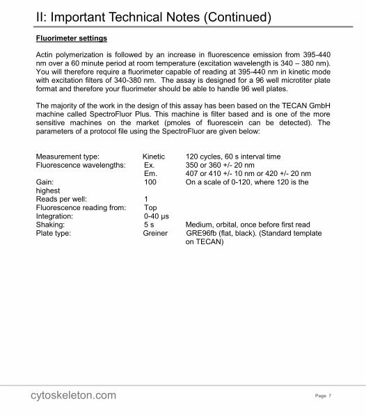
Actin polymerization is followed by an increase in fluorescence emission from 395-440 nm over a 60 minute period at room temperature (excitation wavelength is 340 – 380 nm). You will therefore require a fluorimeter capable of reading at 395-440 nm in kinetic mode with excitation filters of 340-380 nm. The assay is designed for a 96 well microtiter plate format and therefore your fluorimeter should be able to handle 96 well plates.
The majority of the work in the design of this assay has been based on the TECAN GmbH machine called SpectroFluor Plus. This machine is filter based and is one of the more sensitive machines on the market (pmoles of fluorescein can be detected). The parameters of a protocol file using the SpectroFluor are given below:
Measurement type: Kinetic 120 cycles, 60 s interval time Fluorescence wavelengths: Ex. 350 or 360 +/- 20 nm Em. 407 or 410 +/- 10 nm or 420 +/- 20 nm Gain: 100 On a scale of 0-120, where 120 is the highest Reads per well: 1 Fluorescence reading from: Top Integration: 0-40 μs Shaking: 5 s Medium, orbital, once before first read Plate type: Greiner GRE96fb (flat, black). (Standard template on TECAN)
II: Important Technical Notes (Continued)

cytoskeleton.com Page 8
This kit contains sufficient reagents for approximately 60-120 assays of 200 μl
volume depending on choice of polymerization concentration.
III: Kit Contents
Reagents
Cat. # Quantity Description
Pyrene Labeled
Muscle Actin AP05 5 x 1 mg Lyophilized. >99% pure labeled actin, 1
mg per tube.
General Actin Buffer
BSA01-010 1 x 100 ml Lyophilized. 5 mM Tris-HCl pH 8.0 and 0.2 mM CaCl2 when resuspended .
Actin Polymerization Buffer
BSA02-001 2 x 2 ml Lyophilized. 500 mM KCl, 20 mM MgCl2, 0.05M guanidine carbonate and 10 mM ATP when resuspended. 10X strength.
ATP stock BSA04-001 1x 1 ml Lyophilized. 100 mM ATP when
resuspended.
Tris-HCl pH 7.5 BSTR-01 1 x 5 ml Lyophilized. 100 mM when reconstituted.
96 well plate
Corning Costar
# 3915
1 plate Black polystyrene assay plate.

cytoskeleton.com Page 9 cytoskeleton.com
Many of the components of this kit have been provided in lyophilized form. Prior to beginning the assay it is necessary to reconstitute several components as described in Table 3. Components that are reconstituted and stored as directed will have a shelf life of 6 months.
Table 3. Kit Component Reconstitution and Storage.
IV: Reconstitution and Storage of Components
Kit Component Reconstitution Storage Conditions
Pyrene Muscle
Actin
Resuspend one 1 mg aliquot when needed with 50 μl of ice cold sterile de-ionized water. Aliquot into 5 μl volumes and snap freeze in liquid nitrogen. Store at -70°C. Note: These frozen aliquots will be 20% less active than unfrozen protein.
Store lyophilized
protein desiccated at 4°C.
Store resuspended protein at -70°C.
General Actin Buffer
Reconstitute with 100 ml of sterile de-ionized water.
Store lyophilized buffer at 4°C
Store resuspended buffer at 4°C
Actin Polymerization Buffer
Reconstitute with 2 ml of 100 mM Tris-HCl pH 7.5.
Aliquot into 200 μl volumes, snap freeze in liquid nitrogen and store at -70°C.
Store lyophilized buffer desiccated at 4°C.
Store resuspended buffer at -70°C.
ATP Reconstitute with 1.0 ml of 100 mM Tris-HCl pH 7.5.
Aliquot into 100 μl volumes and store at -70°C.
Store lyophilized buffer desiccated at 4°C.
Store resuspended buffer at -70°C.
Tris-HCl Reconstitute with 5 ml of sterile de-ionized water to give a final buffer composition of 100 mM Tris-HCl pH 7.5 Required for ATP and Actin Polymerization Buffer resuspension.
Store lyophilized buffer desiccated at 4°C.
Store resuspended buffer at 4°C.

cytoskeleton.com Page 10
Buffer preparation:
1. Rapidly defrost one aliquot of Actin Polymerization Buffer and ATP by placing each tube in a room temperature water bath and then place on ice. After thawing, spin each tube for 5 s in a microfuge to collect the liquid at the bottom of the tube. Place on ice.
2. Prepare G-buffer by adding 2 μl of ATP (Cat. # BSA04) for every 1 ml of General Actin Buffer (Cat. # BSA01).
Pyrene G-actin preparation (Polymerization assay only):
1. Prepare G-actin stock by adding 225 μl of ice cold G-buffer to one 5 μl frozen aliquot of pyrene actin (final conc. is 0.4 mg/ml). Pipet up and own to mix and leave on ice for 1 h to depolymerize actin oligomers that have formed during storage.
2. Centrifuge the actin at 14k rpm at 4°C for 30 min.
3. Remove the supernatant to a new tube on ice.
Pyrene F- actin preparation (Depolymerization assay only):
1. Quickly thaw four frozen pyrene actin aliquots on ice. Combine into a single tube and keep on ice.
2. Dilute the actin to 1 mg/ml by adding 400 μl of G-buffer.
3. Polymerize the actin by adding 10 μl of 10x Actin Polymerization Buffer (0.25x final strength) and incubate at room temperature for 1 h.
4. This is the F-actin stock, it is stable for 1 h after preparation.
Test protein preparation:
1. Make the test protein at the highest possible concentration in an actin compatible buffer (see Appendix 1). The high concentration (preferably >20 mM) is necessary to give you every chance of detecting actin interactions. Centrifuge the test protein at 150,000 x g for 1 h at 4°C.
2. Remove supernatant and place on ice, this is your test protein stock.
We recommend using the buffers and protein stocks described above as a starting point for polymerization/ depolymerization assays. As you become familiar with the experimental system the final actin stock solutions can be varied as required.
V: Things to do Prior to Beginning the Assay

cytoskeleton.com Page 11 cytoskeleton.com
A simple actin polymerization assay using the 96 well plate (provided) is described below. A good reference for more detailed analysis can be found in Chapter 3 of The Cytoskeleton: A Practical Approach (J. Cooper, Eds Carraway and Carraway, IRL Press, 1992).
NOTE: When not being measured in the fluorimeter all samples should be shuttered to prevent the excitation beam from bleaching the pyrene fluorescence leading to erroneous results. When measuring the fluorescence the shutter should be opened for no longer than 7 s every 30 s for 1 h.
Actin Polymerization Assay
1. A total of EIGHT wells (eg. A1-H1) are required for a typical polymerization reaction with appropriate controls. Only six of these wells will contain pyrene actin. A typical polymerization test is setup as follows:
Wells A1/B1 are G-buffer baseline controls
Wells C1/D1 are G-buffer-pyrene actin positive controls
Wells E1/F1 are test buffer-pyrene actin controls
Wells G1/H1 are test buffer and protein (drug)-pyrene actin reactions
2. Prepare SIX G-actin stock samples as described in Section V, Pyrene G-actin preparation. Combine into a single tube on ice.
3. Pipet 200 μl of G-buffer into wells A1-B1.
4. Pipet 200 μl of G-actin stock into wells C1-H1.
5. Place the 96 well plate into the fluorimeter, shake for 5 s and read the samples once every 60 s for a total of 3 min to establish a baseline fluorescent measurement for all samples.
6. After 3 min, pause the read, and add (1). 20 μl of test buffer to wells E1 and F1, and (2). 20 μl of the test protein/drug into wells G1 and H1. Return the plate to the fluorimeter, shake for 5 s and continue reading for 20 min. This will indicate if the
test buffer or test protein/drug sample alone can enhance actin polymerization.
7. After 20 min, pause the read and add 20 μl of 10x Actin Polymerization Buffer to all EIGHT wells. Return the plate to the fluorimeter, shake for 5 s and continue the read for 1 h or until the fluorescent signal plateaus.
8. Compare the final polymerization profiles to identify any polymerization/inhibitory activities associated with the test protein/drug. Contact technical service ([email protected]) for a Microsoft Excel data template.
9. See Figure 1 for a typical positive control pyrene actin polymerization reaction.
VI: Actin Polymerization/Depolymerization Studies

cytoskeleton.com Page 12
NOTE: Some actin binding proteins require alternative conditions for different activities (e.g. cofilin requires pH 8.0 for F-actin binding and full severing activity and pH 6.8 for F-actin binding activity only). Several parameters may have to be tested in this assay in order to characterize a particular protein or compound (eg. pH titration (6.5-8.5). A particularly useful parameter to vary is the amount of Actin Polymerization Buffer added. At 0.25x strength Actin Polymerization Buffer pH 7.0, cofilin is clearly seen to alter actin polymerization. Whereas the reaction performed with 1.0x Actin Polymerization Buffer would not show a difference. Cofilin should be used at 20 μg/ml final concentration in this assay.
Figure 1. Pyrene actin polymerization assay.
Actin polymerization was carried out as described in Section VI. Duplicate samples of pyrene actin and General Actin Buffer alone were assayed for 3 min to establish a baseline fluorescence value. At 3 min 20 µl of 10x Actin Polymerization buffer was added to all the wells and fluorescence was assayed every 30 s for 1 h. NOTE: Actin polymerization resulted in a 7 fold increase in fluorescence compared to monomeric actin levels. Arrowhead indicates the fluorescence signal from pyrene G-actin. Arrow shows the increases fluorescence associated with pyrene F-actin.
VI: Actin Polymerization/Depolymerization Studies

cytoskeleton.com Page 13 cytoskeleton.com
Actin Depolymerization Assay
1. A total of EIGHT wells (eg. A2-H2) are required for a typical depolymerization reaction with appropriate controls. Only six of these wells will contain pyrene F-actin. A typical depolymerization test is setup as follows:
Wells A2/B2 are G-buffer baseline controls
Wells C2/D2 are G-buffer-pyrene F-actin positive controls
Wells E2/F2 are test buffer-pyrene F-actin controls
Wells G2/H2 are test buffer and protein (drug)-pyrene F-actin reactions
2. Dilute the F-actin stock (Section V) to 0.2 mg/ml by adding 1.6 ml of G-buffer.
3. Pipet 200 μl of G-buffer into wells A2-B2.
4. Pipet 200 μl of F-actin stock into wells C2-H2.
5. Place the 96 well plate into the fluorimeter, shake for 5 s and read the samples once every 60 s for a total of 3 min to establish a peak fluorescent measurement for all samples.
6. After 3 min, pause the read and add (1): 20 μl of test buffer to wells E2 and F2 and (2). 20 μl of the test protein/drug into wells G2 and H2. Return the plate to the fluorimeter, shake for 5 s and continue reading for 1 h.
7. The depolymerization of Pyrene F-actin will be apparent by a reduction in the fluorescent signal over time.
8. Compare the final depolymerization profiles to identify any depolymerizing activities associated with the test protein/drug. Contact technical service ([email protected]) for a Microsoft Excel data template.
VI: Actin Polymerization/Depolymerization Studies

cytoskeleton.com Page 14
VII: Troubleshooting
Observation Possible cause Correction
1. No increase in fluorescence between G-actin control and F-actin test.
1. Sensitivity of fluorimeter is set too low.
2. Denatured actin.
1. Increase emission gain or increase bandwidth of emission channel, or increase intensity of excitation wavelength.
2. Follow correct freeze/thaw instructions.
2. During polymerization increase in fluorescence is too slow.
1. Actin concentration is too
low.
2. Excitation light is too intense.
1. Increase actin concentration
greater than 0.2 mg/ml.
2. Reduce light intensity reaching the sample. Increase gain to compensate if necessary.
3. During polymerization increase in fluorescence is too quick.
1. Actin polymerization buffer is too concentrated.
2. Actin concentration is too
high.
3. Nucleation is too great.
1. Use less Actin Polymerization Buffer.
2. Decrease actin
concentration.
3. Centrifuge samples (100,000 x g for 1 h at 4oC) prior to adding test solution or Actin Polymerization Buffer.
4. During polymerization increase in fluorescence is not reproducible.
1. Inconsistent preparation of pyrene actin solutions.
2. Fluorescence spectrophotometer is sensitive to changes in cuvette.
3. Actin filaments are sheared differentially during pipetting.
1. More consistent technique.
2. Change from single cuvette. monochromatic machine to 96-well plate reader.
3. Use equal force and mixing technique to all samples, especially important for assays requiring multiple pipetting steps.

cytoskeleton.com Page 15 cytoskeleton.com
VIII: Troubleshooting (Continued)
Observation Possible cause Correction
5. Test sample has no effect on polymerization.
Buffer components of the reaction interfere with the activity of the test protein or compound.
1. CaCl2 too high.
2. ATP too high.
3. MgCl2 too high.
4. Tris-HCl.
5. pH is incorrect.
6. Sodium azide present.
7. Protein binds abnormally to pyrene actin.
8. Test substance does not affect actin polymerization.
1. Add EGTA to 2 mM.
2. Do not add ATP to General Actin Buffer or Actin Polymerization Buffer. Actin
is stable for 1 h after dilution, after this point actin denaturation will be significant.
3. Dilute polymerization buffer to 0.1 or 0.25x strength.
4. Prepare Hepes or PIPES containing Actin Buffer.
5. Actin can be polymerized at pH 6.0-8.5.
6. Do not add sodium azide to buffer; make General Actin Buffer each day that you perform the experiment.
7. Use different ratios of unlabeled to pyrene labeled actin to see the effect.

cytoskeleton.com Page 16
G-actin polymerizes to form F-actin
Globular-actin (G-actin) readily polymerizes under physiological conditions to form Filamentous-actin (F-actin) with the concomitant hydrolysis of ATP. F-actin is a double-helical filament as shown below:
Figure 2. Double-helical structure of actin filaments.
Actin can polymerize from both ends in vitro, however, the rate of polymerization is not equal. This results in an intrinsic polarity in the actin filament. It has therefore become the convention to term the rapidly polymerizing end the plus-end
(see Figure 1) or barbed-end while the slow growing end is called the minus-end or pointed-end.
The propensity of actin to polymerize is dependent upon the affinity of actin monomers for filament ends. Thus, there is an actin monomer concentration below which actin will not polymerize; this value has been termed the Critical Concentration (CC). At monomer concentrations above the CC, the actin will polymerize until the free monomer concentration is equal to the CC. When one is working with actin in vitro the extent of actin polymerization depends upon the conditions used. For example, at 4°C muscle actin has a CC of 0.03 mg/ml in the presence of Mg2+ (2 mM) and KCl (50 mM), but when these ions are absent, the CC is approximately 3.0 mg/ml. Thus, by altering the ionic type and strength one can alter the amount of polymer formed. Non-muscle actin has its own CC values, for example, at 4oC in the presence of Mg2+ (2 mM) and KCl (50 mM) the CC is approximately 0.04 mg/ml. If Mg2+ and KCl are replaced with Ca2+, the CC will increase to nearly 4 mg/ml. Finally, the CC of non-muscle actin can be reduced to 0.03 mg/ml by increasing the temperature to 30°C (Gordon D.J., Boyer J.L. and Korn E.D. 1977. Comparative biochemistry of non-muscle actins (Journal of Biological Chemistry,
252, 8300-8309).
Conditions in which actin is stable
G-actin is stable upon thawing for two days at 4°C. F-actin is not stable to freezing but can be stored at 4°C for one month. F-actin can be transferred to a variety of buffers (e.g. HEPES, phosphate, etc) without detrimental effects. Actin requires a divalent cation, pH
6.5 - 8.0 and ATP for stability. F-actin also requires Mg2+ for stability. Assays for Actin
Appendix 1

cytoskeleton.com Page 17 cytoskeleton.com
Actin Polymerization Assay:
The most versatile, sensitive and easiest actin polymerization assay consists of pyrene conjugated actin (Cat. # AP05) and a fluorescence spectrophotometer. Fluorescence of pyrene actin is enhanced two to twenty fold by the association of actin monomer into the polymer form (Kouyama and Mihashi, 1981. Fluorimetry study of N-(1-pyrenyl)
iodoacetamide-labeled F-actin. (Eur. Jo. Biochem, 114, 33-38, see Figure 2).
Figure 3. Polymerization of actin as measured by pyrene actin fluorescence.
Actin polymerization follows three phases, similar to microtubule assembly; these are lag phase, growth and steady state as depicted in Figure 2. The extent of polymerization is indicated by the steady state level of fluorescence, an upper limit can be measured by adding phalloidin (an actin stabilizing compound) which pushes the CC down to <0.01 mg/ml. This assay is greater than 95% accurate and the sample is not disturbed during the assay. Further details of setting up this assay are supplied with pyrene actin (Cat. # AP05).
Actin Monomer Assay:
DNase inhibition assays are based on the high affinity interaction between G-actin and DNase 1 which results in inhibition of DNase activity (Blikstad et al. 1978. Selective assay for monomeric and filamentous actin in cell extracts, using inhibition of deoxyribonuclease 1. Cell, 15, 935-943). The value of this assay is that it can distinguish between G- and F-actin. In this regard, it has been used to selectively assay the amounts of monomeric and filamentous actin in cell extracts. This assay has also been successfully adapted to study actin binding proteins that generate G- actin from actin filaments; an example of this is gelsolin protein.
Appendix 2
Flu
ore
sce
nce
Time
Steady state
phase
Grow th
phase
Lag
phase

cytoskeleton.com Phone: (303) 322.2254 Fax: (303) 322.2257
Customer Service: [email protected]
Technical Support: [email protected]