acta medica okayama - connecting repositories · for the study of megakaryocytic functions, the...
TRANSCRIPT
Acta Medica OkayamaVolume 17, Issue 1 1963 Article 1
FEBRUARY 1963
Tissue culture of spleen : Studies on thegrowing pattern and its evaluation for clinicaldiagnosis of leukemias and other hematologic
disorders
Kiyoshi Hiraki∗ Tadashi Ofuji† Hiroshi Sunami‡
Zensuke Ota∗∗ Koichi Kitajima†† Koji Shinagawa‡‡
Masayoshi Kibata§ Isao Miyoshi¶ Atsumasa Hanazawa‖
∗Okayama University,†Okayama University,‡Okayama University,∗∗Okayama University,††Okayama University,‡‡Okayama University,§Okayama University,¶Okayama University,‖Okayama University,
Copyright c©1999 OKAYAMA UNIVERSITY MEDICAL SCHOOL. All rights reserved.
Tissue culture of spleen : Studies on thegrowing pattern and its evaluation for clinicaldiagnosis of leukemias and other hematologic
disorders∗
Kiyoshi Hiraki, Tadashi Ofuji, Hiroshi Sunami, Zensuke Ota, Koichi Kitajima,Koji Shinagawa, Masayoshi Kibata, Isao Miyoshi, and Atsumasa Hanazawa
Abstract
It has been found that the clinical tissue culture method devised in our laboratory for bonemarrow is satisfactorily applicable to the in vitro study of human and animal splenic tissues. Thepresent experiments have shown that the growth pattern, migration mode or cellular population ofthe cultured splenic cells is fairly characteristic of each disease condition, and the technic appearsto be a valuable diagnostic aid in the practice of hematology. Direct vision biopsy with the la-paroscope is safe and unattended by untoward side effects, enabling us to obtain excellent biopsymaterial from the enlarged spleen in various blood dyscrasias.
∗PMID: 14078635 [PubMed - OLDMEDLINE] Copyright c©OKAYAMA UNIVERSITY MEDI-CAL SCHOOL
Acta Med. Okayama 17, 1-17 (1963)
TISSUE CULTURE OF SPLEEN: STUDIES ON THE GROWINGPATTERN AND ITS EVALUATION FOR CLINICAL
DIAGNOSIS OF LEUKEMIAS AND OTHERHEMATOLOGIC DISORDERS
Kiyoshi HIRAKI, Tadashi OFUJI, Hiroshi SUNAMI, Zensuke OTA,
Koichi KITAJIMA, Koji SHINAGAWA, Masayoshi KIBATA,
Isao MIYOSHI and Atsumasa HANZAWA
Department of Internal Medicine, Okayama University Medical School,Okayama (Director: Prof. K. Hiraki)
Received for publication, January 23, 1963
Using the tissue culture method devised by HIRAKI et al./-s we have suc
ceeded in elucidating the mechanism of platelet formation from megakaryocytes
and in establishing a new method valuable for the differential diagnosis of
granulocytic, lymphocytic and monocytic leukemias. These results were reported
at the VI,4 VIlli and VIlI6 International Congress of Hematology.
Application of the same tissue culture technic has enabled us to observe
splenic cells in vitro which were obtained by biopsy from patients suffering from
leukemias and other various blood dyscrasias.
The present paper deals with the growth pattern of cultured splenic tissue
and cell morphology as studied mainly under the phase contrast microscope.
MATERIALS AND METHODS
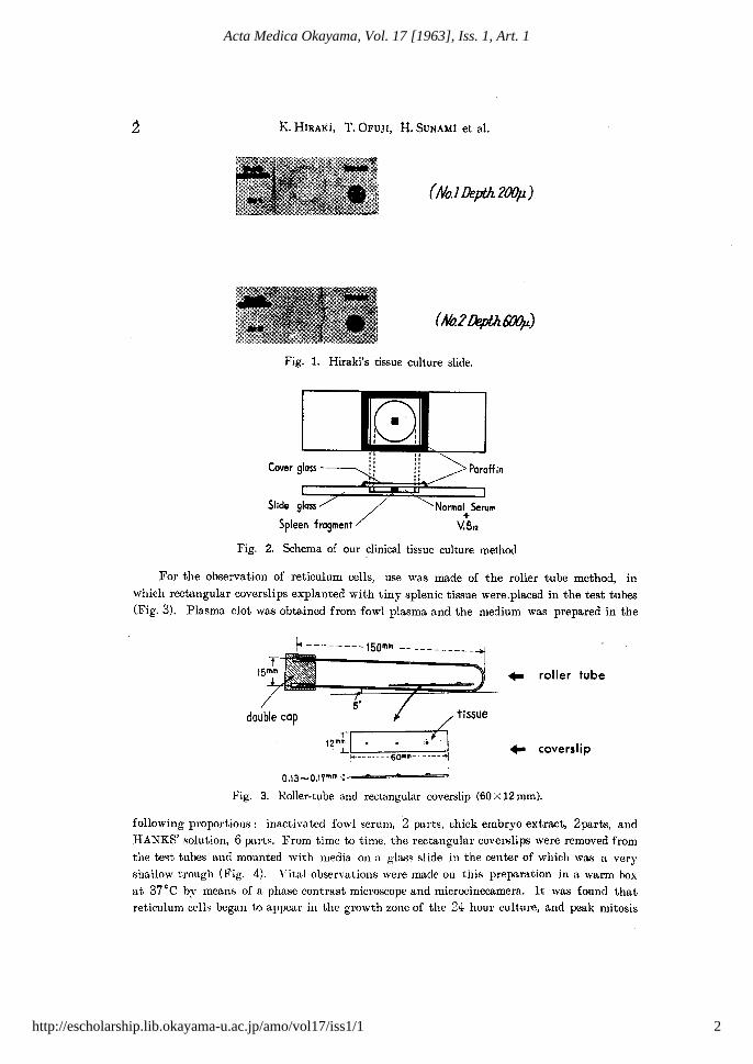
Splenic tissue obtained from experimental animals (mouse, rat, guinea pig, rabbit,dog, and cat) and patients with various blood disorders were used. Only the hospitalizedpatients were subjected to laparoscopy. They were brought to an operating room and underlocal anesthesia splenic tissue was removed with the aid of a laparoscope. In addition, exploration of the liver and other abdominal organs was fully performed and liver biopsy wasalso done whenever indicated. The fresh tissues were cut into small pieces with sterileknives and cultured in the HIRAKI's tissue culture slide No. 1 (Fig. 1). As described inthe previous papers,I-3 a small piece of the tissue, about 1 mm. in size, was placed in thecenter of circle marked on the culture slide which was perviously mounted with one drop ofhomologous blood serum and one drop of vitamin BJ2 (100 r per m!.), and sealed with thecoverslip as illustrated in Fig. 2. For the study of megakaryocytic functions, the HIRAKI'stissue culture slide No. 2 was used (Fig. 1).7.8
Aft.el' incubation at 37°C, the ohservation was carried out under the phase contrast '1ndfluorescence microscope for 48 hours at various intervals of 6, 12, and 24 hours. In someinstances, microcillematogrq,phy was taken with or without vital staining with J-anus greenand neutral red.9
1
1
Hiraki et al.: Tissue culture of spleen : Studies on the growing pattern and its
Produced by The Berkeley Electronic Press, 1963
K. HIRAKi, '1'.OFUJI, H. SUNAMl et al.
(No.l Depth 200p)
(No.21Jepth6OOp)
Fig. 1. Hiraki's tissue culture slide.
(:)',I ,I I
Cover gloss
Slide glass
Spleen fragment
"III.'.I."
""I I.1::.,
Paroffin
Normal Seru",+
Y.B12
.. coverslip
Fig. 2. Schema of our clinical tissue culture method
For the observation of reticulum cells, use was made of the roller tube method, in
which rectangular coverslips explanted with tiny splenic tissue were.placed in the test tubes
(Fig. 3). Plasma clot was obtained from fowl plasma and the me:lium was prepared in the
.. roller tube
-----7;:" tissue12mrl J
L..r -·_-·-·.-.'--:6:-:-0==lIIm=--.•-.--:-+1
0.13-0.17"'''' }=_......_ ...-=~==-
Fig. 3. Roller-tube and rectangular coverslip (60 X 12 mm).
following proportions: inactivated fowl serum, 2 parts, chick embryo extract, 2parts, andHANKS' solution, 6 partH. From time to time, the rectangular coverslips were removed from
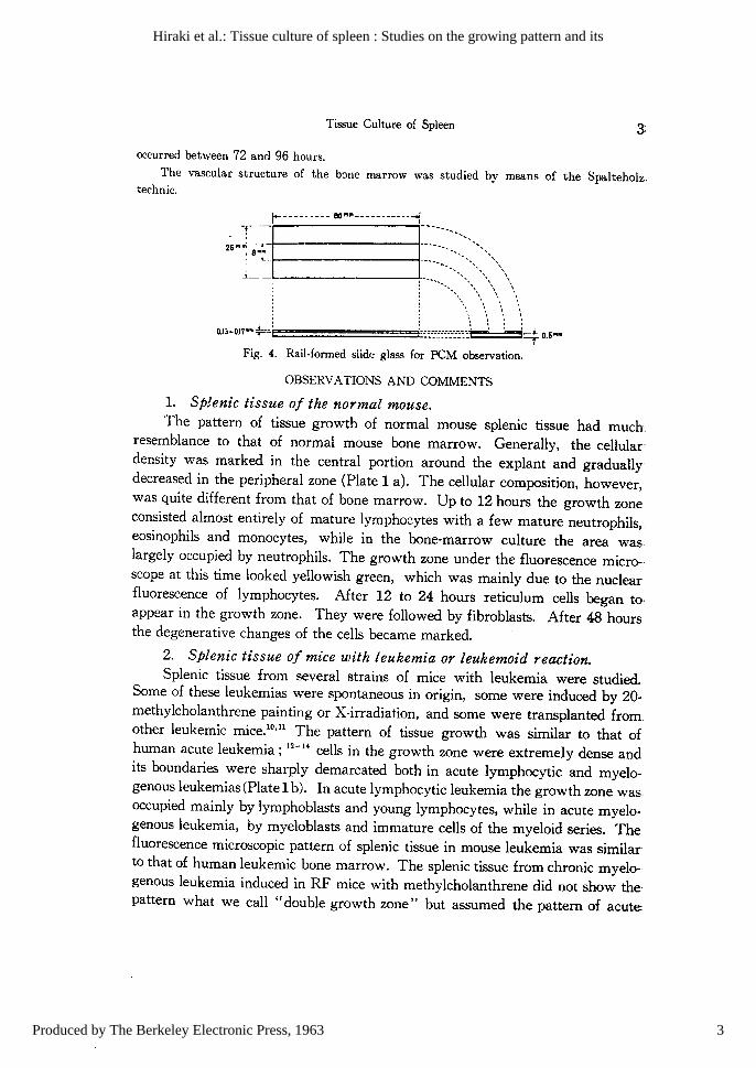
the te<;t tubes and mounted with media on a glass slide in the center of which was a veryshallow trough (Fig. 4). Vital observations were made on this prep..'tration in a warm box
at 37°0 by means of a phase contrast microscope and microcinecamera. It was found thatreticulum cells began to appear in the growth zone of the 24 hour culture, and peak mitosis
2
Acta Medica Okayama, Vol. 17 [1963], Iss. 1, Art. 1
http://escholarship.lib.okayama-u.ac.jp/amo/vol17/iss1/1
Tissue Culture of Spleen
occurred between 72 and 96 hours.The vascular structure of the bane marrow was studied by means of the Spalteholz.
technic.
. -r- r-
I
--------- 80.".------------1
1
_
26 In'" 8·.t.------- ...... ... ... , ...: _.t.._.-- :--•••, "\
,- • ,,,..... \:.\,;\\ \\
0.13-0.17"" +--: j::::::.:::-:-::::..H H4 o.s....
Fig. 4. Rail-formed slid(~ glass for PCM observation.
OBSERVAnONS AND COMMENTS
1. Splenic tissue of the normal mouse.The pattern of tissue growth of normal mouse splenic tissue had much
resemblance to that of normal mouse bone marrow. Generally, the cellular'density was marked in the central portion around the explant and graduallydecreased in the peripheral zone (Plate 1 a). The cellular composition, however,was quite different from that of bone marrow. Up to 12 hours the growth zoneconsisted almost entirely of mature lymphocytes with a few mature neutrophils,eosinophils and monocytes, while in the bone-marrow culture the area waslargely occupied by neutrophils. The growth zone under the fluorescence microscope at this time looked yellowish green, which was mainly due to the nuclearfluorescence of lymphocytes. After 12 to 24 hours reticulum cells began to·appear in the growth zone. They were followed by fibroblasts. After 48 hoursthe degenerative changeE of the cells became marked.
2. Splenic tissue of mice with leukemia or leukemoid reaction.Splenic tissue from several strains of mice with leukemia were studied.
Some of these leukemias were spontaneous in origin, some were induced by 20methylcholanthrene painting or X-irradiation, and some were transplanted fromother leukemic mice.10on The pattern of tissue growth was similar to that ofhuman acute leukemia ; 12-14 cells in the growth zone were extremely dense andits boundaries were sharply demarcated both in acute lymphocytic and myelogenous leukemias (Plate 1b). In acute lymphocytic leukemia the growth zone wasoccupied mainly by lymphoblasts and young lymphocytes, while in acute myelogenous leukemia, by myeloblasts and immature cells of the myeloid series. Thefluorescence microscopic pattern of splenic tissue in mouse leukemia was similarto that of human leukemic bone marrow. The splenic tissue from chronic myelogenous leukemia induced in RF mice with methylcholanthrene did not show the·pattern what we call "double growth zone" but assumed the pattern of acute
3
Hiraki et al.: Tissue culture of spleen : Studies on the growing pattern and its
Produced by The Berkeley Electronic Press, 1963

4 K. HIRAKI, T.OFUJI, H. SUNAMI et al.
leukemia. This might have been due to the fact that in chronic myelogenousleukemia maturation of leukemic cells did not proceed in the spleen as in thebone marrow, which in culture exhibited the "double growth zone". Differential.cell counts of splenic and bone marrow imprints gave support to this view.
In leukemoid reactions induced in pure strain mice by either saponin poisoning or M-Y sarcoma transplantation, the cell composition of the growth zone wasquite different from that of normal spleen, although the pattern of tissue growthwas rather similar (Fig. 5). Young granulocytes and megakaryocytes predomi-
Fig. 5. Spleen of mouse intoxicated by saponin. 6 hour culture.
nated. Fluorescence micr03~:::>py revealed the growth zone of diffusely reddish·orange color, which was due to the fluorescence of granules of young granulocyticcells that appeared in a large number in the growth zone.
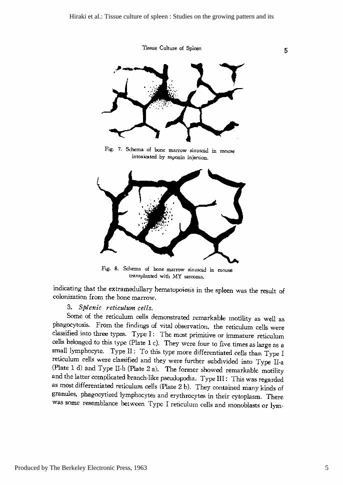
In the leukemoid reactions, the vascular beds of the bone marrow demonstrated some destruction of the walls of the venous sinusoids (Figs. 6, 7 and 8),
Fig. 6. Schema of bone marrow sinusoid in mouse.
4
Acta Medica Okayama, Vol. 17 [1963], Iss. 1, Art. 1
http://escholarship.lib.okayama-u.ac.jp/amo/vol17/iss1/1
Tissue Cuiture of Spleen
,#...,............." ---~-'-
Fig. 7. Schema of bone marrow sinusoid In mouseintoxicated by saponin injection.
Fig. 8. Schema of bone marrow sinusoid in mousetransplanted with MY sarcoma.
5
indicating that the extramedullary hematopoiesis in the spleen was the result ofcolonization from the bone marrow.
3. Splenic reticulum cells.Some of the reticulum cells demonstrated remarkable motility as well as
phagocytosis. From the findings of vital observation, the reticulum cells wereclassified into three types. Type I: The most primitive or immature reticulumcells belonged to this type (Plate 1 c). They were four to five times as large as asmall lymphocyte. Type II: To this type more differentiated cells than Type Ireticulum cells were classified and they were further subdivided into Type II-a(Plate 1 d) and Type II-b (Plate 2 a). The former showed remarkable motilityand the latter complicated branch-like pseudopodia. Type Ill: This was regardedas most differentiated reticulum cells (Plate 2 b). They contained many kinds ofgranules, phagocytized lymphocytes and erythrocytes in their cytoplasm. Therewas some resemblance between Type I reticulum cells and monoblasts or lym-
5
Hiraki et al.: Tissue culture of spleen : Studies on the growing pattern and its
Produced by The Berkeley Electronic Press, 1963
K. HIRAKI, T.OFUJI, H. SUNAMl et al.
phogonias (AMANO)/o between Type Il-a reticulum cells and monocytes, betweenType Il-b reticulum cells and fibroblasts (Plate 2 c), and between Type III reti·culum cells and ascitic phagocytes or histiocytes. But judging from their migratory from, mitochondrial distribution, appearance of granules and vacuoles withvital stains, and findings with fluorescence microscopy, we were able to differentiate one from another.
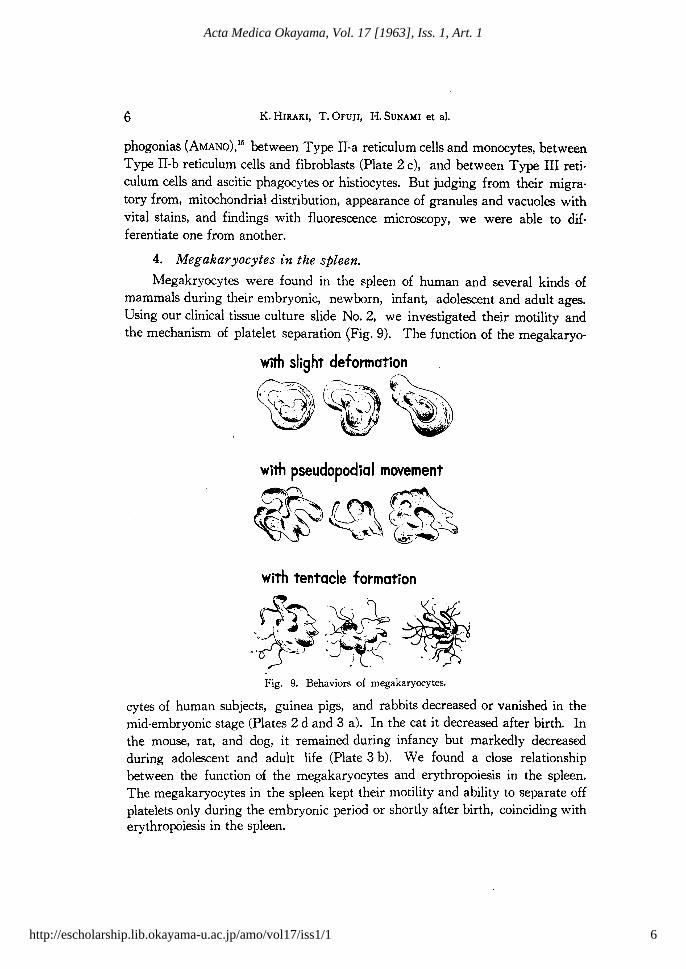
4. Megakaryocytes in the spleen.
Megakryocytes were found in the spleen of human and several kinds ofmammals during their embryonic, newborn, infant, adolescent and adult ages.Using our clinical tissue culture slide No. 2, we investigated their motility andthe mechanism of platelet separation (Fig. 9). The function of the megakaryo·
with pseudopodiaI movement
~.~'.'.'..D fl~~, ~-~.. ,-: '..:;~.. , (,#,- .
::\. ' ,.' ~ ."-.' '.:~~ (.;~.
~'
with tentacle formation
~..,\. . "l
. ~". ~.~.. ,~SS ~.... ~"~~.. J7t"""~ .~
~:-...... ···-r· '
0' .~ • J~. • .. . .
Fig, 9. Behaviors of megakaryocytes.
cytes of human subjects, guinea pigs, and rabbits decreased or vanished in themid-embryonic stage (Plates 2 d and 3 a). In the cat it decreased after birth. Inthe mouse, rat, and dog, it remained during infancy but markedly decreasedduring adolescent and adult life (Plate 3 b). We found a close relationshipbetween the function of the megakaryocytes and erythropoiesis in the spleen.The megakaryocytes in the spleen kept their motility and ability to separate offplatelets only during the embryonic period or shortly after birth, coinciding witherythropoiesis in the spleen.
6
Acta Medica Okayama, Vol. 17 [1963], Iss. 1, Art. 1
http://escholarship.lib.okayama-u.ac.jp/amo/vol17/iss1/1
Tissue Cuitnre of SplC'eIl 7
13
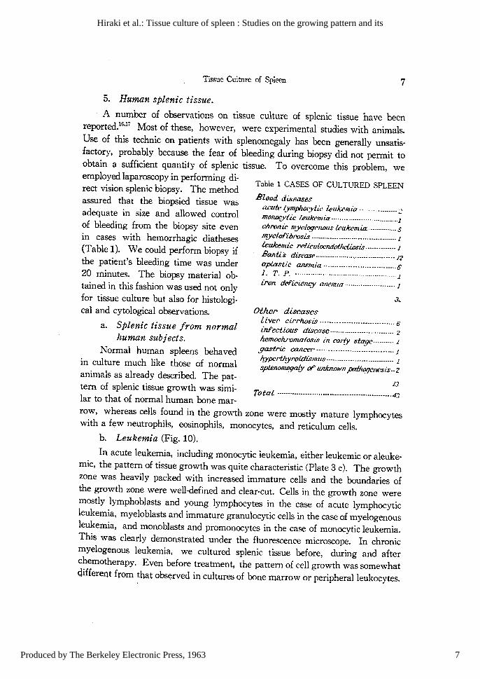
Tabie 1 CASES OF CULTURED SPLEEN
Total ···················································4:1
Other> diseaseslive,. cirrhosis 6
infectious disease 2
h.em.ochrolllatosis in COlry stage· 1
gastric caJlCCr ·································1q.ypcrU'Yf'Oidismlls 1
splenom.egaLy 01' ILRJcrwwn pntJll!lJenesis·· 2
Blood diseasesil.<~utt~ lymplwryiit' l.eukelltia.· ::'monlXJ'tie I.eukcmia······················· lchronic myrl.ogf'llollS lrulcemia··· ..· ,sD{JIclof'ibrosis J
lculcemic rcti.culocndofheliosis·············· J
Banti's discasc··············· ..··.··..··· ·.··.··12aplastic anemia ·· 0I. T. P. ···················································1iron deficiency alU'mia )
5. Human splenic tissue.
A number of observations on tissue culture of splenic tissue have beenreported.1G
,17 Most of these, however, were experimental studies with animals.Use of this technic on patients with splenomegaly has been generally unsatisfactory, probably because the fear of bleeding during biopsy did not permit toobtain a sufficient quantity of splenic tissue. To overcome this problem, weemployed laparoscopy in performing direct vision splenic biopsy. The methodassured that the biopsied tissue wasadequate in size and allowed controlof bleeding from the biopsy site evenin cases with hemorrhagic diatheses(Table 1). We could perform biopsy ifthe patient's bleeding time was under20 minutes. The biopsy material ob·tained in this fashion was used not onlyfor tissue culture but also for histological and cytological observations.
a. Splenic tissue from normalhuman subjects.
Normal human spleens behavedin culture much like those of normalanimals as already described. The pattern of splenic tissue growth was similar to that of normal human bone mar·row, whereas cells found in the growth zone were mostly mature lymphocyteswith a few neutrophils, eosinophils, monocytes, and reticulum cells.
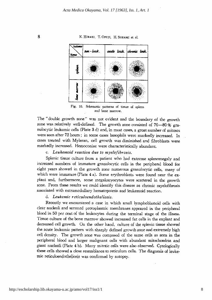
b. Leukemia (Fig. 10).
In acute leukemia, including monocytic leukemia, either leukemic or aleuke·mic, the pattern of tissue growth was quite characteristic (Plate 3 c). The growthzone was heavily packed with increased immature cells and the boundaries ofthe growth zone were well-defined and clear-cut. Cells in the growth zone weremostly lymphoblasts and young lymphocytes in the case of acute lymphocyticleukemia, myeloblasts and immature granulocytic cells in the case of myelogenousleukemia, and monoblasts and promonocytes in the case of monocytic leukemia.This was clearly demonstrated under the fluorescence microscope. In chronicmyelogenous leukemia, we cultured splenic tissue before, during and afterchemotherapy. Even before treatment, the pattern of cell growth was somewhatdifferent frorn that observed in cultures of bone marrow or peripheralleukocytes.
7
Hiraki et al.: Tissue culture of spleen : Studies on the growing pattern and its
Produced by The Berkeley Electronic Press, 1963
8 K. HIRAKI, T.OFUJI, H. SUNAMI et al.
0':'·"
,-~,t,~Ii''.
Fig. 10. Schematic patterns of tissue of spleenand bone marrow.
The "double growth zone" was not evident and the boundary of the growthzone was relatively well-defined. The growth zone consisted of 70-8096 granulocytic leukemic cells (Plate 3 d) and, in most cases, a great number of mitoseswere seen after 72 hours; in some cases basophils were markedly increased. Incases treated with Myleran, cell growth was diminished and fibroblasts weremarkedly increased. Hemoconiae were characteristically abundant.
c. Leukemoid reaction due to myelofibrosis.Splenic tissue culture from a patient who had extreme splenomegaly and
increased numbers of immature granulocytic cells in the peripheral blood foreight years showed in the growth zone numerous granulocytic cells, many ofwhich were immature (Plate 4 a). Some erythroblasts were found near the explant and, furthermore, some megakaryocytes were scattered in the growthzone. From these results we could identify this disease as chronic myelofibrosisassociated with extramedullary hematopoiesis and leukemoid reaction.
d. Leukemic reticuloendotheliosis.Recently we encountered a case in which small lymphoblastoid cells with
clear nucleoli and serrated protoplasmic membranes appeared in the peripheralblood in 50 per cent of the leukocytes during the terminal stage of the illness.Tissue culture of the bone marrow showed increased fat cells in the explant anddecreased cell growth. On the other hand, culture of the splenic tissue showedthe acute leukemic pattern with sharply defined growth zone and extremely highcell density. The growth zone was composed of the same cells as seen in theperipheral blood and larger malignant cells with abundant mitochondria andgiant nucleoli (Plate 4 b). Many mitotic cells were also observed. Cytologicallythese cells showed a close resemblance to reticulum cells. The diagnosis of leukemic reticqloendotheliosis was confirmed by autopsy.
8
Acta Medica Okayama, Vol. 17 [1963], Iss. 1, Art. 1
http://escholarship.lib.okayama-u.ac.jp/amo/vol17/iss1/1

Tissue Culture of Spleen 9
e. Aplastic anemia.
In aplastic anemia the growth pattern was almost normal in contrast tohypoplastic bone marrow. A few promyelocytes and myelocytes were alwaysfound in the growth zone. However, in some cases, plasma cells and eosinophilswere definitely increased; in one case, we observed many wandering reticulumcells which phagocytized erythrocytes (Plate 4 c).
f. Banti's disease.
Compared with normal controls, most cases showed reduced growth rateand cell density. The growth zone consisted predominantly of mature lymphocytes and a few neutrophils. In the terminal stage, splenic reticulum cells andfibroblasts were relatively increased.
g. Idiopathic thrombocytopenic purpura.
In idiopathic thrombocytopenic purpura, it was of interest to note thatmany megakayocytes with diminished motility and platelet separation appearedin the growth zone as in the bone marrow culture.
h. Splenomegaly due to infection.
The pattern of tissue growth resembled the normal but its growth rate wasvery high and mature neutrophils were increased up to 30--40 per cent of thetotal cells in the growth zone, while immature granulocytic cells did not appearin contrast to myelogenous leukemia and leukemoid reaction (Plate 4 d).
SUMMARY
It has been found that the clinical tissue culture method devised in ourlaboratory for bone marrow is satisfactorily applicable to the in vitro study ofhuman and animal splenic tissues. The present experiments have shown thatthe growth pattern, migration mode or cellular population of the cultured spleniccells is fairly characteristic of each disease condition, and the technic appears tobe a valuable diagnostic aid in the practice of hematology.
Direct vision biopsy with the laparoscope is safe and unattended by untowardside effects, enabling us to obtain excellent biopsy material from the enlargedspleen in various blood dyscrasias.
REFERENCES
1. HIRAKI, K., OFUJI, T. and SUNAMI, H.: The method of tissue culture (mainly of the bonemarrow) and a simple method of observing tissue. Acta Med. Okayama 10, 99, 1956
2. HIRAKI, K., OFUJI, T. and WATARI, Z. : Observation of various living blood cells by tissueculture of the bone marrow. ibid. 10, 110, 1956
3. HIRAKI, K., SUNAMI, H. and SOGAW A, T. : Fundamental studies on the peripheralleukocyteCll!tl,lre and its clinical application. ibi4. 10, 139, 1958
9
Hiraki et al.: Tissue culture of spleen : Studies on the growing pattern and its
Produced by The Berkeley Electronic Press, 1963
10 K. HIRAKI, T.OFUJI, H. SUNAMI et al.
4. HIRAKI, K.: The function of the megakaryocyte (motility, Reparation of the platelet andphaghcytosis): observations both in idiopathic thrombocytopenic purpura and in normaladults. Proc. 6th Intern. Congr. Intern. Soc. Hemat. 537, 1958
5. HIRAKI, K: Studies on diagnosis of leukemin by tissue culture. Proc. 7th Intern. Congr.Intern. Soc. Hemat. 307, 1960
6. HIRAKI, K: Studies on chemotherapy of leukemia by a new bone-marrow tissue culturetechnic. Proc. 8th Intern. Congr. Intern. Soc. Hemat. 371, 1962
7. HIRAKI, K, OFUJI, T., KOBAYASHI, T., SUNAMI, H. and AWAI,K.: On'the function of themegakaryocyte (motility, separation of the platelet, phagocytosis). Observations both inidiopathic thrombocytopenic purpura and normal adult. Acta Med. Okayama 10, 57, 1950
8. HIRAKI, K, SUNAMI, I;I. and OGAWARA, K: Studies on the megarycoytes, platelets separationand degeneration. ibid. 12, 187, 1958
9. OTA, Z.: A study on the cytomorphologic structure of blood cells by vital staining. Part 1.Normal human blood cells in the bone marrow. Part 2. Leukemic cells in the bone marrow.ibid. 14, 22, 1960
10. OKADA, K., MIYOSHI, 1., KAWAMURA, N. and SATO, M.: Studies on leukemogenesis bycell-free filtrates of AKR mouse leukemia, Acta Haemat. fap. 25, 1, 1962
11. HIRAKI, K, IRINO, S. and SOTA, S.: Studies on cytogenesis of RF mouse leukemia inducer!by X-ray irradiation. Proc. 8th Congr. Europ. Soc. Haemat. 347, 1962
12. HIRAKI, K., OFUJI, T. and HATTORI, Y.: Diagnosis of leukemia, and aplastic anemia bythe bone marrow culture. Acta Med. Okayama 10, 130, 1956
13. HIRAKI, K.: Studies on diagnosis of leukemia by tissue culture. ibid. 12, 84, 1958
14. HIRAKI, K.: Etude de la chemotherapie des leucemies par une nouve11e technique de culturede moelle osseuse. N. R. F. Hemat. 1, 410, 1961
15. AMANo, S.. UNNO, G., HANAOKA, M. and TAMAKI, Y.: Studies on the discrimination oflymphocytes and plasma cells. Supplements on advocation of the Iymphogonia-theory. ActaPath. fap. \, 117, 1951
16. FAZZARI, J.: Culture "in vitro" di milza embryonale ed adulta Arch. expr. Zel/jorschg.2. 307, 1926
17. ERDMANN, R, EIsNER, H. und LAsEo, H.: Das Verhalten der foetalen. postfoetalen undaU3gewachsenen Rattenmilz unter verschiedene Bedingungen in vitro. Arch. exper. Zenjorschg. 2. 361. 1926
EXPLANATION FOR PLATE 1
a. Normal mouse spleen. 6 hour culture.b. RF mouse spleen with acute lymphocytic leukemia. 6 hour culture.c. Splenic reticulum cell (Type 1) in normal mouse. 72 hour culture.d. Splenic reticulum cell (Type Ha) showing active movement in normal mouse.
10
Acta Medica Okayama, Vol. 17 [1963], Iss. 1, Art. 1
http://escholarship.lib.okayama-u.ac.jp/amo/vol17/iss1/1

Tissue Culture of Spleen
11
Hiraki et al.: Tissue culture of spleen : Studies on the growing pattern and its
Produced by The Berkeley Electronic Press, 1963

12 K. HIRAKI, T. OFUJI, H. SUNAMI et al.
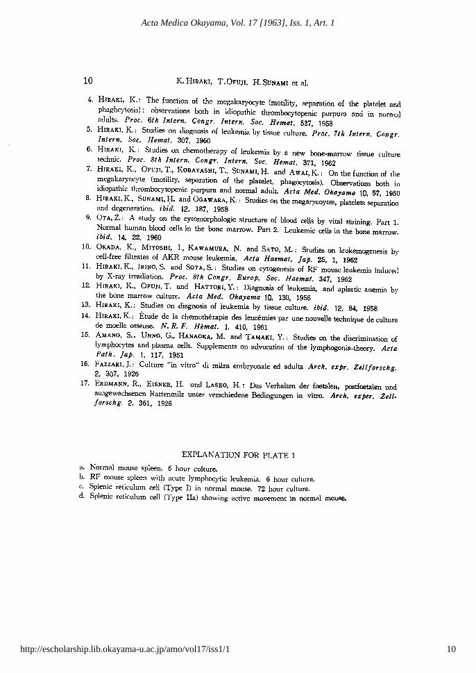
EXPLANATlON FOR PLATE 2
a. Splenic reticulum cell (Type !I-b) in normal mouse.b. Splenic reticulum cell (Type Ill) with many phagocytized granules in norrral m:luse.c. Fibroblast in normal mouse spleen.d. Human splenic megakaryocyte in 4-month·old fetus with tentacle formation. 18 hour
culture.
12
Acta Medica Okayama, Vol. 17 [1963], Iss. 1, Art. 1
http://escholarship.lib.okayama-u.ac.jp/amo/vol17/iss1/1
Tissue Culture of Spleen
Plate 2
13
13
Hiraki et al.: Tissue culture of spleen : Studies on the growing pattern and its
Produced by The Berkeley Electronic Press, 1963

14 K. HIRAKI, T. OFUJI. H. SUNAMI et al.
EXPLANATION FOR PLATE 3
a. Human splenic megakaryocyte in 5-month-old fetus with pseudopodial m~vement. 18hour culture.
b. Megarkyocyte with tentacle formation in young mouse. 18 hour culture.c. Human spleen in acute lymphocytic leukemia. 6 hour culture.d. Human spleen in chronic myelogenous leukemia. 48 hour culture.
14
Acta Medica Okayama, Vol. 17 [1963], Iss. 1, Art. 1
http://escholarship.lib.okayama-u.ac.jp/amo/vol17/iss1/1

· Culture of SpleenTIssue 15
15
Hiraki et al.: Tissue culture of spleen : Studies on the growing pattern and its
Produced by The Berkeley Electronic Press, 1963

16 Tissue Culture of Spleen
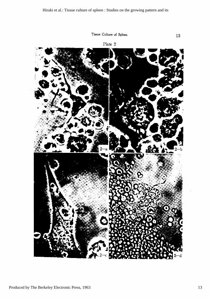
EXPLANATION FOR PLATE 4
a. Human spleen in leukemoid reaction due to myelofibrosis. 6 hour culture.b. Human spleen in leukemic rdiculoendotheliosis. 48 hour culture.c. Human reticulum cell with p'ngocytized erythrocytes in aplastic anemia. 48 hour culture.d. Human spleen in infectious disease. 6 hour culture.
16
Acta Medica Okayama, Vol. 17 [1963], Iss. 1, Art. 1
http://escholarship.lib.okayama-u.ac.jp/amo/vol17/iss1/1
K. HIRAKI, T. OFUJI, H. SUNAMI, et al.
Plate 4
17
17
Hiraki et al.: Tissue culture of spleen : Studies on the growing pattern and its
Produced by The Berkeley Electronic Press, 1963