acral lentiginous melanoma, indolent subtype diagnosed by ...acral lentiginous melanoma, indolent...
TRANSCRIPT
En Bloc Excision in ALM
Vol. 29, No. 3, 2017 327
Received March 28, 2016, Revised July 29, 2016, Accepted for publication August 7, 2016
Corresponding author: Hyun-sun Park, Department of Dermatology, SMG-SNUBoramae Medical Center, 20 Boramae-ro 5-gil, Dongjak-gu, Seoul 07061, Korea. Tel: 82-2-870-2880, Fax: 82-2-831-0714, E-mail: [email protected]
This is an Open Access article distributed under the terms of the Creative Commons Attribution Non-Commercial License (http://creativecommons.org/licenses/by-nc/4.0) which permits unrestricted non-commercial use, distribution, and reproduction in any medium, provided the original work is properly cited.
Copyright © The Korean Dermatological Association and The Korean Society for Investigative Dermatology
pISSN 1013-9087ㆍeISSN 2005-3894Ann Dermatol Vol. 29, No. 3, 2017 https://doi.org/10.5021/ad.2017.29.3.327
CASE REPORT
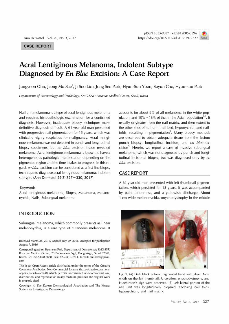
Fig. 1. (A) Dark black colored pigmented band with about 1-cm width on the left thumbnail. Ulceration, onychodystrophy, and Hutchinson’s sign were observed. (B) Left lateral portion of the nail unit was longitudinally biopsied, enclosing nail folds, hyponychium, and nail matrix.
Acral Lentiginous Melanoma, Indolent Subtype Diagnosed by En Bloc Excision: A Case Report
Jungyoon Ohn, Jeong Mo Bae1, Ji Soo Lim, Jong Seo Park, Hyun-Sun Yoon, Soyun Cho, Hyun-sun Park
Departments of Dermatology and 1Pathology, SMG-SNU Boramae Medical Center, Seoul, Korea
Nail unit melanoma is a type of acral lentiginous melanoma and requires histopathologic examination for a confirmed diagnosis. However, inadequate biopsy techniques make definitive diagnosis difficult. A 61-year-old man presented with progressive nail pigmentation for 15 years, which was clinically highly suspicious for malignancy. Acral lentigi-nous melanoma was not detected in punch and longitudinal biopsy specimens, but en bloc excision tissue revealed melanoma. Acral lentiginous melanoma is known to have a heterogeneous pathologic manifestation depending on the pigmented region and the time it takes to progress. In this re-gard, en bloc excision can be considered as a first-line biopsy technique to diagnose acral lentiginous melanoma, indolent subtype. (Ann Dermatol 29(3) 327∼330, 2017)
-Keywords-Acral lentiginous melanoma, Biopsy, Melanoma, Melano-nychia, Nails, Subungual melanoma
INTRODUCTION
Subungual melanoma, which commonly presents as linear melanonychia, is a rare type of cutaneous melanoma. It
accounts for about 2% of all melanoma in the white pop-ulation, and 10%∼18% of that in the Asian population1-3. It usually originates from the nail matrix, and then extent to the other sites of nail unit: nail bed, hyponychial, and nail-folds, resulting in pigmentation4. Many biopsy methods are described to obtain adequate tissue from the lesion: punch biopsy, longitudinal incision, and en bloc ex-cision5. Herein, we report a case of invasive subungual melanoma, which was not diagnosed by punch and longi-tudinal incisional biopsy, but was diagnosed only by en bloc excision.
CASE REPORT
A 61-year-old man presented with left thumbnail pigmen-tation, which persisted for 15 years. It was accompanied by pain, tenderness, and a yellowish discharge. About 1-cm wide melanonychia, onychodystrophy in the middle
J Ohn, et al
328 Ann Dermatol
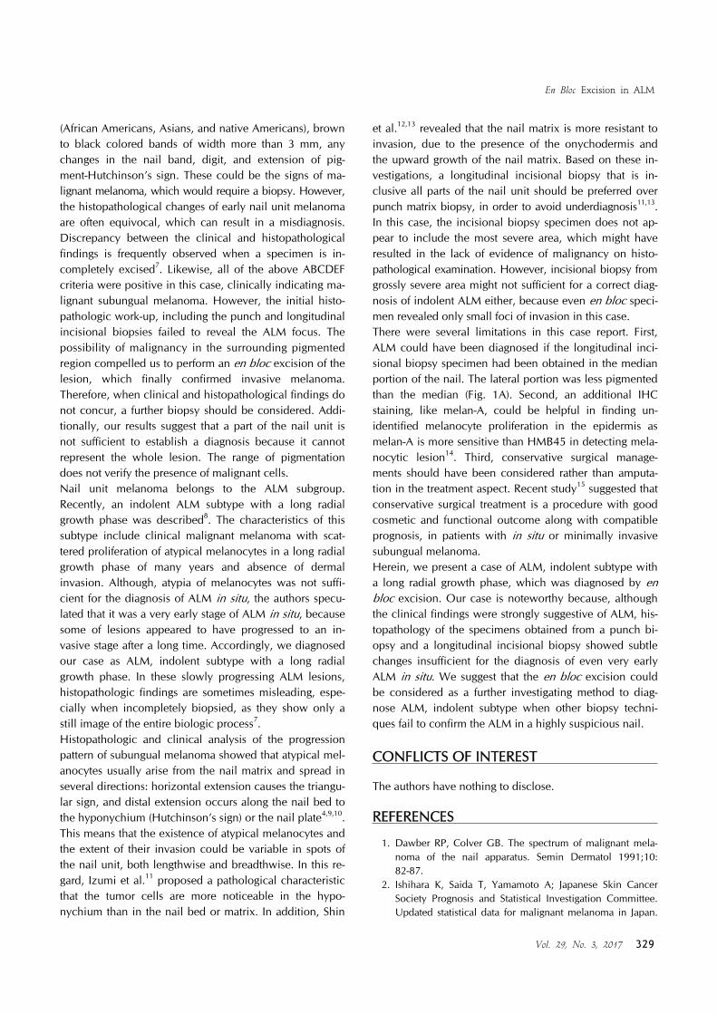
Fig. 3. (A) Two months later, the nail unit presented with plate destruction and intermittent bleeding from the nailbed. (B) En blocexcisional specimen of the nail unit. (C) Scattered pigmentation was observed in the hyponychium and the nail bed with lymphocytic infiltration in the onychodermis layer (H&E, ×10). (D) Atypical melanocytes with hyperchromatic nuclei invaded the dermis layer (H&E, ×100). (E, F) Immunohistochemical stains for human melanoma black 45 and Ki-67 were positive (×100).
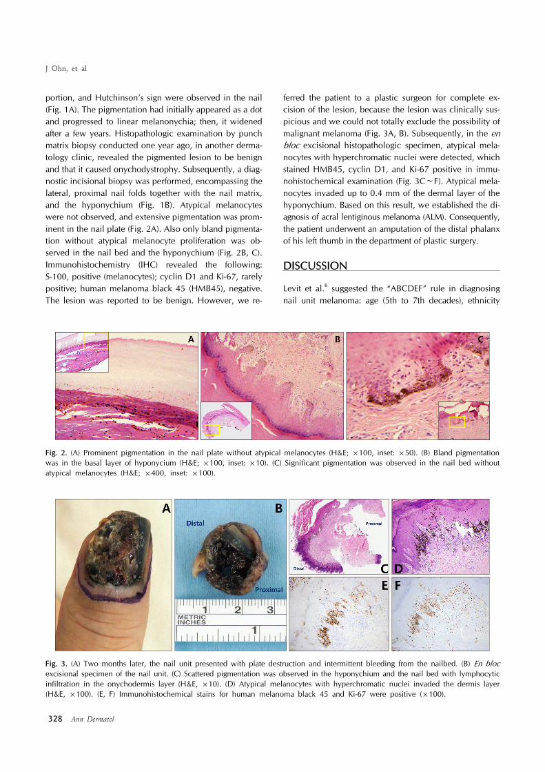
Fig. 2. (A) Prominent pigmentation in the nail plate without atypical melanocytes (H&E; ×100, inset: ×50). (B) Bland pigmentation was in the basal layer of hyponycium (H&E; ×100, inset: ×10). (C) Significant pigmentation was observed in the nail bed without atypical melanocytes (H&E; ×400, inset: ×100).
portion, and Hutchinson’s sign were observed in the nail (Fig. 1A). The pigmentation had initially appeared as a dot and progressed to linear melanonychia; then, it widened after a few years. Histopathologic examination by punch matrix biopsy conducted one year ago, in another derma-tology clinic, revealed the pigmented lesion to be benign and that it caused onychodystrophy. Subsequently, a diag-nostic incisional biopsy was performed, encompassing the lateral, proximal nail folds together with the nail matrix, and the hyponychium (Fig. 1B). Atypical melanocytes were not observed, and extensive pigmentation was prom-inent in the nail plate (Fig. 2A). Also only bland pigmenta-tion without atypical melanocyte proliferation was ob-served in the nail bed and the hyponychium (Fig. 2B, C). Immunohistochemistry (IHC) revealed the following: S-100, positive (melanocytes); cyclin D1 and Ki-67, rarely positive; human melanoma black 45 (HMB45), negative. The lesion was reported to be benign. However, we re-
ferred the patient to a plastic surgeon for complete ex-cision of the lesion, because the lesion was clinically sus-picious and we could not totally exclude the possibility of malignant melanoma (Fig. 3A, B). Subsequently, in the en bloc excisional histopathologic specimen, atypical mela-nocytes with hyperchromatic nuclei were detected, which stained HMB45, cyclin D1, and Ki-67 positive in immu-nohistochemical examination (Fig. 3C∼F). Atypical mela-nocytes invaded up to 0.4 mm of the dermal layer of the hyponychium. Based on this result, we established the di-agnosis of acral lentiginous melanoma (ALM). Consequently, the patient underwent an amputation of the distal phalanx of his left thumb in the department of plastic surgery.
DISCUSSION
Levit et al.6 suggested the “ABCDEF” rule in diagnosing nail unit melanoma: age (5th to 7th decades), ethnicity
En Bloc Excision in ALM
Vol. 29, No. 3, 2017 329
(African Americans, Asians, and native Americans), brown to black colored bands of width more than 3 mm, any changes in the nail band, digit, and extension of pig-ment-Hutchinson’s sign. These could be the signs of ma-lignant melanoma, which would require a biopsy. However, the histopathological changes of early nail unit melanoma are often equivocal, which can result in a misdiagnosis. Discrepancy between the clinical and histopathological findings is frequently observed when a specimen is in-completely excised7. Likewise, all of the above ABCDEF criteria were positive in this case, clinically indicating ma-lignant subungual melanoma. However, the initial histo-pathologic work-up, including the punch and longitudinal incisional biopsies failed to reveal the ALM focus. The possibility of malignancy in the surrounding pigmented region compelled us to perform an en bloc excision of the lesion, which finally confirmed invasive melanoma. Therefore, when clinical and histopathological findings do not concur, a further biopsy should be considered. Addi-tionally, our results suggest that a part of the nail unit is not sufficient to establish a diagnosis because it cannot represent the whole lesion. The range of pigmentation does not verify the presence of malignant cells. Nail unit melanoma belongs to the ALM subgroup. Recently, an indolent ALM subtype with a long radial growth phase was described8. The characteristics of this subtype include clinical malignant melanoma with scat-tered proliferation of atypical melanocytes in a long radial growth phase of many years and absence of dermal invasion. Although, atypia of melanocytes was not suffi-cient for the diagnosis of ALM in situ, the authors specu-lated that it was a very early stage of ALM in situ, because some of lesions appeared to have progressed to an in-vasive stage after a long time. Accordingly, we diagnosed our case as ALM, indolent subtype with a long radial growth phase. In these slowly progressing ALM lesions, histopathologic findings are sometimes misleading, espe-cially when incompletely biopsied, as they show only a still image of the entire biologic process7. Histopathologic and clinical analysis of the progression pattern of subungual melanoma showed that atypical mel-anocytes usually arise from the nail matrix and spread in several directions: horizontal extension causes the triangu-lar sign, and distal extension occurs along the nail bed to the hyponychium (Hutchinson’s sign) or the nail plate4,9,10. This means that the existence of atypical melanocytes and the extent of their invasion could be variable in spots of the nail unit, both lengthwise and breadthwise. In this re-gard, Izumi et al.11 proposed a pathological characteristic that the tumor cells are more noticeable in the hypo-nychium than in the nail bed or matrix. In addition, Shin
et al.12,13 revealed that the nail matrix is more resistant to invasion, due to the presence of the onychodermis and the upward growth of the nail matrix. Based on these in-vestigations, a longitudinal incisional biopsy that is in-clusive all parts of the nail unit should be preferred over punch matrix biopsy, in order to avoid underdiagnosis11,13. In this case, the incisional biopsy specimen does not ap-pear to include the most severe area, which might have resulted in the lack of evidence of malignancy on histo-pathological examination. However, incisional biopsy from grossly severe area might not sufficient for a correct diag-nosis of indolent ALM either, because even en bloc speci-men revealed only small foci of invasion in this case.There were several limitations in this case report. First, ALM could have been diagnosed if the longitudinal inci-sional biopsy specimen had been obtained in the median portion of the nail. The lateral portion was less pigmented than the median (Fig. 1A). Second, an additional IHC staining, like melan-A, could be helpful in finding un-identified melanocyte proliferation in the epidermis as melan-A is more sensitive than HMB45 in detecting mela-nocytic lesion14. Third, conservative surgical manage-ments should have been considered rather than amputa-tion in the treatment aspect. Recent study15 suggested that conservative surgical treatment is a procedure with good cosmetic and functional outcome along with compatible prognosis, in patients with in situ or minimally invasive subungual melanoma.Herein, we present a case of ALM, indolent subtype with a long radial growth phase, which was diagnosed by en bloc excision. Our case is noteworthy because, although the clinical findings were strongly suggestive of ALM, his-topathology of the specimens obtained from a punch bi-opsy and a longitudinal incisional biopsy showed subtle changes insufficient for the diagnosis of even very early ALM in situ. We suggest that the en bloc excision could be considered as a further investigating method to diag-nose ALM, indolent subtype when other biopsy techni-ques fail to confirm the ALM in a highly suspicious nail.
CONFLICTS OF INTEREST
The authors have nothing to disclose.
REFERENCES
1. Dawber RP, Colver GB. The spectrum of malignant mela-noma of the nail apparatus. Semin Dermatol 1991;10: 82-87.
2. Ishihara K, Saida T, Yamamoto A; Japanese Skin Cancer Society Prognosis and Statistical Investigation Committee. Updated statistical data for malignant melanoma in Japan.
J Ohn, et al
330 Ann Dermatol
Int J Clin Oncol 2001;6:109-116.3. Kim JH, Park JH, Lee DY. Site distribution of cutaneous
melanoma in South Korea: a retrospective study at a single tertiary institution. Int J Dermatol 2015;54:e38-e39.
4. Tan KB, Moncrieff M, Thompson JF, McCarthy SW, Shaw HM, Quinn MJ, et al. Subungual melanoma: a study of 124 cases highlighting features of early lesions, potential pitfalls in diagnosis, and guidelines for histologic reporting. Am J Surg Pathol 2007;31:1902-1912.
5. Baran R, Kechijian P. Longitudinal melanonychia (melano-nychia striata): diagnosis and management. J Am Acad Dermatol 1989;21:1165-1175.
6. Levit EK, Kagen MH, Scher RK, Grossman M, Altman E. The ABC rule for clinical detection of subungual melanoma. J Am Acad Dermatol 2000;42:269-274.
7. Park HS, Cho KH. Acral lentiginous melanoma in situ: a diagnostic and management challenge. Cancers (Basel) 2010;2:642-652.
8. Kim JY, Choi M, Jo SJ, Min HS, Cho KH. Acral lentiginous melanoma: indolent subtype with long radial growth phase. Am J Dermatopathol 2014;36:142-147.
9. Lee DY. Variable sized cellular remnants in the nail plate of longitudinal melanonychia: evidence of subungual mela-noma. Ann Dermatol 2015;27:328-329.
10. Phan A, Dalle S, Touzet S, Ronger-Savlé S, Balme B, Thomas L. Dermoscopic features of acral lentiginous melanoma in a large series of 110 cases in a white population. Br J Dermatol 2010;162:765-771.
11. Izumi M, Ohara K, Hoashi T, Nakayama H, Chiu CS, Nagai T, et al. Subungual melanoma: histological examination of 50 cases from early stage to bone invasion. J Dermatol 2008;35:695-703.
12. Shin HT, Park SW, Lee DY, Jang KT, Mun GH, Cheung L. A case of subungual melanoma with tumor invasion sparing the nail matrix dermis. Ann Dermatol 2014;26:655-657.
13. Shin HT, Jang KT, Mun GH, Lee DY, Lee JB. Histopa-thological analysis of the progression pattern of subungual melanoma: late tendency of dermal invasion in the nail matrix area. Mod Pathol 2014;27:1461-1467.
14. Blessing K, Sanders DS, Grant JJ. Comparison of immu-nohistochemical staining of the novel antibody melan-A with S100 protein and HMB-45 in malignant melanoma and melanoma variants. Histopathology 1998;32:139-146.
15. Sureda N, Phan A, Poulalhon N, Balme B, Dalle S, Thomas L. Conservative surgical management of subungual (matrix derived) melanoma: report of seven cases and literature review. Br J Dermatol 2011;165:852-858.