aberrant root canal anatomy: a review -...
TRANSCRIPT
50
Aberrant Root Canal Anatomy: A Review
ABERRANT ROOT CANAL ANATOMY: A REVIEW
Dr. Sasidhar Nallapati B.D.S.
Successful endodontic treatment involves accurate
diagnosis, good understanding of the biological
principles and excellent execution of the treatment. To
be able to execute an excellent treatment, it’s imperative
that the clinician has comprehensive knowledge of the
root canal anatomy and the know-how to locate and
treat this anatomy.
Many outcome studies conducted over the past few
decades showed incomplete debridement and
disinfection of root canal space as the most important
factor in endodontic treatment failure (1,2,3). Missed
canal in the initial treatment as a significant cause of
root canal failure was shown by Hoen et al (4). They
also showed a significant relation between an
asymmetrical obturation in a root space and the
incidence of a missed canal in an initial treatment.
Methodology:
Root canal anatomy is studied by both in vitro and in
vivo methods. In vivo methods include clinical treatment
of a tooth followed by radiographic evaluation of the
root canal anatomy. In vitro methods include
1. Direct observation
2. Microscopic observation
3. Macroscopic sectioning
4. Microscopic sectioning
5. Dyes
6. Filling and decalcification
7. Filling and clearing
8. Radiography
9. Contrasting media (Hypaque)
10. Cone beam Tomography.
Classification:
Weine classified root canal anatomy into 3 types.
Type I: One canal with one orifice and one apical
foramen (1-1)
Type II: Two canals that merge into one and exit as
one canal (2-1)
Type III: One canal that divides into two and exit as
two canals. (2-2)
Vertucci’s classification was more elaborate and it
covered 8 types.
Type I 1-1
Type II 2-1
Type III 1-2 -1
Type IV 2-2
Type V 1-2
Type VI 2-1-2
Type VII 1-2-1-2
Type VIII 3-3
Studies with Percentages:
The methodology employed and the criteria used to
describe root canal anatomy will decide the percentages
of canals found in any tooth. For example Neaverth et
al (5) have used “two separate canals could be
visualized on radiographic examination (two files or two
GP points to no less than a mm short of the length)” as
the criteria for two canals in a mesiobuccal root of a
maxillary first molar. On the other hand Sempira et al
(6) have used the presence of “two canals to at least 4
mm from the apex” in the mesiobuccal root of maxillary
first molar to determine the percentage of Mb2 canal.
These differences in criteria dictate the great variation
seen in the percentages of root canals seen in different
studies. However, if one reviews the published evidence
certain features of aberrant anatomy stand out in a tooth/
root.
Ethnicity has a significant influence on aberrant
anatomy,(26). Radix Entomolaris, an extra distal root in
a mandibular molar, is often seen in Oriental and Eskimo
populations (23). Similarly 2 and 3 canal premolars are
seen frequently in black populations (12,15,27).‘C’
shaped anatomy is seen more commonly in Chinese,
Korean and Indian populations (22,25).
Bilateral symmetry is a feature of aberrant anatomy.
Rarer the aberration, the more common is the bilateral
symmetry (28).
Clinical Management of Aberrant Anatomy:
Radiography: Angled views of teeth reveal aberrant
anatomy. Angled views allow us to visualize the root
anatomy in 3 dimensions so that better assessment of
the root canal anatomy is made. It is imperative that at
least 2 angled views shall be taken before attempting
endodontic treatment.
Dr. Sashi Nallapati obtained his dental
degree from the Govt. Dental College and
Hospital, Hyderabad, India. He completed
his post graduate training in the specialty
of Endodontics from Nova Southeastern
University (NSU), Davie, Florida, USA.
He maintains a practice limited to
Endodontics in Kingston, Jamaica. He serves on the
faculty of NSU in the dept. of Post Graduate Endodontics.
Dr. Nallapati authored several clinical articles and lectures
across the globe. His hobbies include digital photography,
swimming and reading. he can be reached at
www.endojamaica.com
50 - 62 Sasidhar Nallapati.pmd 12/5/2007, 12:50 PM50
51
Aberrant Root Canal Anatomy: A Review
Tooth Feature
Maxillary Central Incisor Over 60% lateral canals (7)
Maxillary Lateral Incisor Lingual curvatureLarge mid root canal diameter
Maxillary Canine Single canal (9)Lateral canals
Maxillary First Premolar Three canals with two canals in the buccal root (mesiobuccal, distobuccal
and palatal canals) (11,12)
Maxillary Second Premolar Three canals with two canals in the buccal root(mesiobuccal and distobuccal
and palatal canals) (12)
Maxillary First Molar Two mesiobuccal canals in majority of cases. Occasionally three mesiobuccal
canals, two distobucal and two palatal canals (16,17,18,19)
Maxillary Second Molar Two mesiobuccal canals in majority of the cases. ‘C’ shaped canals (30)
MandibularIncisors Two canals that join apically. Occasionally 2 separate canals (8)
Mandibular Canine Two canals. Buccal and lingual (9)
Mandibular First Premolar Two to three canals. Mesiobuccal, distobuccal and lingual.‘C’ shaped anatomy
occasionally (10,13,15)
Mandibular Second Premolar Two to three canals. Mesiobuccal, distobuccal and lingual.‘C’ shaped anatomy
occasionally (14,15)
Mandibular First Molar Four to six canals. Three mesial canals and three distal canals. Radix
entomolaris with a separate distolingual root (20,21,23,24)
MandibularSecond Molar Four to five canals. Three mesial canals. ‘C’ shaped anatomy(22,25)
Once a canal disappears mid-root, one shall always
suspect a bifurcation. Twin periodontal ligament outlines
of the root also indicate a broad root and therefore extra
anatomy. Guide files are placed in root canals and
radiographs are taken to reveal the symmetry of the
guide file to the external contours of the root. If there is
an asymmetry, it often means hidden anatomy. Also
guide files reveal any hidden curvatures that are in
multiple planes. (Fig 1-3)
Clinical Anatomy: Contour of the gingiva often
indicates aberrant anatomy. For example, broad buccal
gingiva in a maxillary or mandibular premolar may
suggest a broad buccal root. This in turn may suggest
Figure 2: Distobuccal canal located.
Figure 1: Off-centre appearance of guide file in the
buccal and palatal roots of maxillary premolar. Figure 3: All three canals obturated.
50 - 62 Sasidhar Nallapati.pmd 12/5/2007, 12:50 PM51

52
Aberrant Root Canal Anatomy: A Review
the presence of two canals in the buccal root. Gingival
recession some times may reveal a bifurcation of the
buccal root indicating two canals in maxillary and
mandibular premolar teeth (fig 4). Teeth that have an
extra cusp or an aberrant clinical crown may indicate
aberrant pulp chamber and root canal anatomy (fig 5-
8).
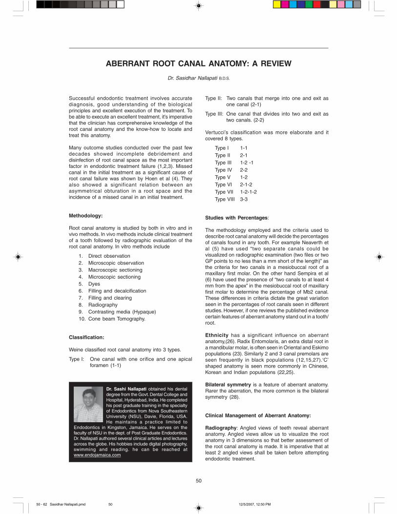
Magnification: A surgical operating microscope (S.O.M)
(www.globalsurgical.com) is highly recommended to
perform all endodontic therapy (fig 9). An S.O.M not
only magnifies the chamber anatomy in great detail but
also allows great amount of light to illuminate the pulp
chamber. This allows the operator to understand the
subtleties of pulp chamber anatomy, visualize the pulpal
floor and locate root canal orifices. This becomes even
more useful when there are pulpal floor calcifications
blocking canal orifices (31). Significant amount of
preparation of the pulpal floor is required to identify and
carve calcifications away to locate root canal orifices.
The use of an SOM greatly facilitates this process and
makes endodontic therapy more predictable and less
stressful to the operator.
Size and extension of access plays an important role
in the predictable location of all root canals. Modified
access extensions are required to identify canals like
Figure 4: Gingival recession allows to see the bifurcation
of the buccal root in a mandibular premolar.
Figure 5: Extra cusp on the distal in a maxillary second molar. Figure 8: Post-operative radiograph of the molar.
Figure 7: Preoperative radiograph of the molar.
Figure 6: Access reveals two palatal canals and
one mb canal that bifurcated.
Mb2 in a maxillary molar, Middle mesial canal in the
mesial root of a mandibular molar, second buccal canal
in a maxillary premolar and the radix entomolaris in a
mandibular molar. Initial access shape is determined
by the shape of the pulpal floor (32). Then, appropriate
extensions are made to facilitate the location and
straight line access into all canals. Complete unroofing
of the chamber roof is a basic step in access design
and yet many clinicians err by not removing the roof of
the chamber (fig10a,10b).
50 - 62 Sasidhar Nallapati.pmd 12/5/2007, 12:50 PM52
53
Aberrant Root Canal Anatomy: A Review
Subtle color changes are observed between the coronal
dentin and the pulpal floor dentin under the SOM. This
is useful in the location of the pulp chamber and canal
orifices. Pulpal floor is usually is dark gray in color and
contrasts from the light colored axial dentin. When
access preparations are made this color difference
allows the operator to be very precise in removing the
axial dentin to expose the pulpal floor. There is also a
very subtle difference in color between tertiary dentin
and axial dentin. This difference allows the operator to
be once again very precise in carving away this
irritational dentin to expose the pulpal floor and
subsequently canal orifices.
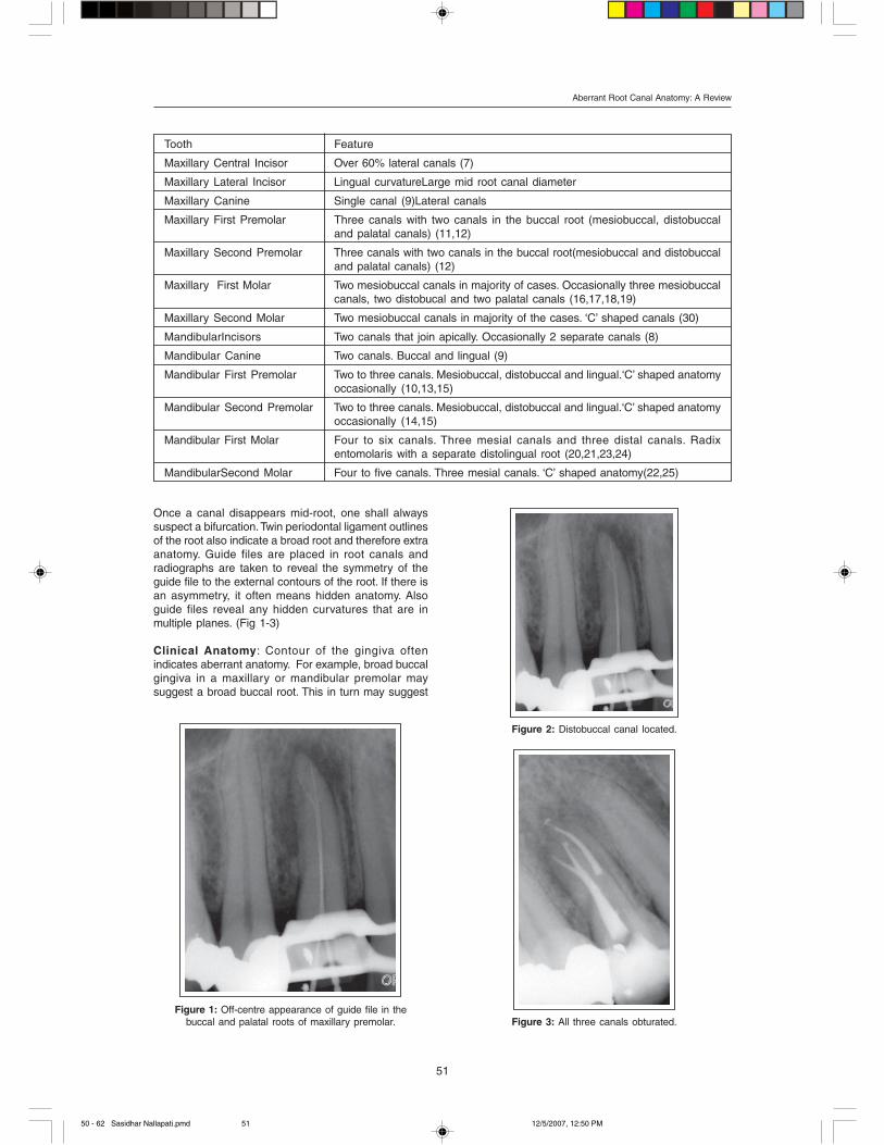
Isthmus: It is the space that connects two or more
canals that exist in the same root (29). Isthmus is the
frequent location of an aberrant canal (fig 11a-b).
Isthmus houses pulp tissue (12a-c). If the isthmus exists
only in the coronal third, it should be eliminated to
remove the pulp tissue. If the isthmus extends from the
coronal to the middle/apical third, it should be
Figure 9: Surgical operating microscope.
Figure 10a: Roof of the chamber not unroofed
in the original treatment.
Figure 10b: Complete removal of the roof reveals a
second distal canal in a mandibular molar.
Figure 11b: Three mesial canals and two distal canals.
Figure 11a: Isthmus explored between Mb and
ML canals in a mandibular molar.
Pulpal Floor: When access preparations are made,
coronal dentin is removed to make entry into the pulp
chamber space. Often times the pulp chamber space
is reduced in size or completely filled with either
secondary dentin or tertiary dentin.
50 - 62 Sasidhar Nallapati.pmd 12/5/2007, 12:50 PM53
54
Aberrant Root Canal Anatomy: A Review
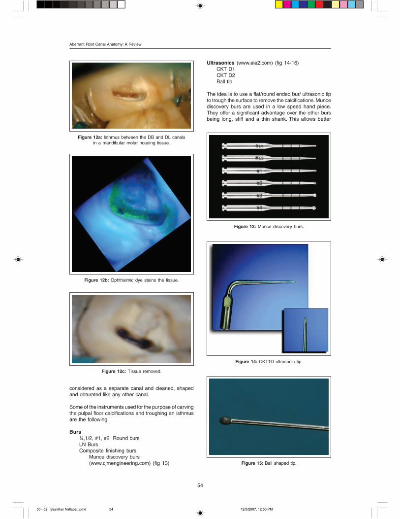
Ultrasonics (www.eie2.com) (fig 14-16)
CKT D1
CKT D2
Ball tip
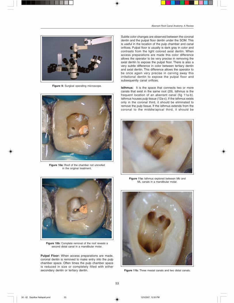
The idea is to use a flat/round ended bur/ ultrasonic tip
to trough the surface to remove the calcifications. Munce
discovery burs are used in a low speed hand piece.
They offer a significant advantage over the other burs
being long, stiff and a thin shank. This allows better
Figure 12a: Isthmus between the DB and DL canals
in a mandibular molar housing tissue.
Figure 12b: Ophthalmic dye stains the tissue.
Figure 12c: Tissue removed.
considered as a separate canal and cleaned, shaped
and obturated like any other canal.
Some of the instruments used for the purpose of carving
the pulpal floor calcifications and troughing an isthmus
are the following.
Burs
¼,1/2, #1, #2 Round burs
LN Burs
Composite finishing burs
Munce discovery burs
(www.cjmengineering.com) (fig 13) Figure 15: Ball shaped tip.
Figure 13: Munce discovery burs.
Figure 14: CKT1D ultrasonic tip.
50 - 62 Sasidhar Nallapati.pmd 12/5/2007, 12:50 PM54

55
Aberrant Root Canal Anatomy: A Review
Figure 16: CKT 2D ultrasonic tip.
Figure 17b: Final access after the canals are relocated.
Figure 17a: Initial access in a maxillary molar
revealing four canals.
visualization when working deep in the chamber or root.
Some of the limitations of this series of burs are in
posterior teeth in patients with limited mouth opening.
CKT series ultrasonic tips from Eie2 are excellent for
both chamber and deep root troughing. They are
especially useful in posterior teeth and in patients with
limited mouth opening.
Canal Relocation:
All canals in multirooted teeth, exit the pulp chamber at
an angle. Once a canal is located, this exit curvature
shall be minimized or eliminated. This will create a
straight line access to the mid root (fig 17a-b). This in
turn facilitates negotiation of the canal to the apex. The
following instruments can be used for this purpose.
Burs
¼, 1/2, #1, #2 Round burs
LN Burs
Composite finishing burs
Munce discovery burs
(www.cjmengineering.com)
Ultrasonics (www.eie2.com)
CKT D1
CKT D2
Ball tip
Gates Glidden drills
4, 3, 2 (sizes 90,70,50)
Niti files
Access shapers
Dyes can help locating pulp tissue in pulp chamber.
Sable seek (www.ultradent.com), methylene blue
(www.vista-dental.com) or fluorescein sodium
(www.haag-streit.com) are the appropriate choices (33).
Dyes stain any vital or dystrophic pulp tissue in the
chamber or root canal space. Under magnification this
staining guides the operator to the pathway to the pulp
canals.
Similarly, transillumination the pulp chamber with an
external fiber-optic light source allows differentiation of
sclerotic dentin from normal dentin. This in turn allows
precise removal of the sclerotic dentin to locate root
canal orifices (fig 18).
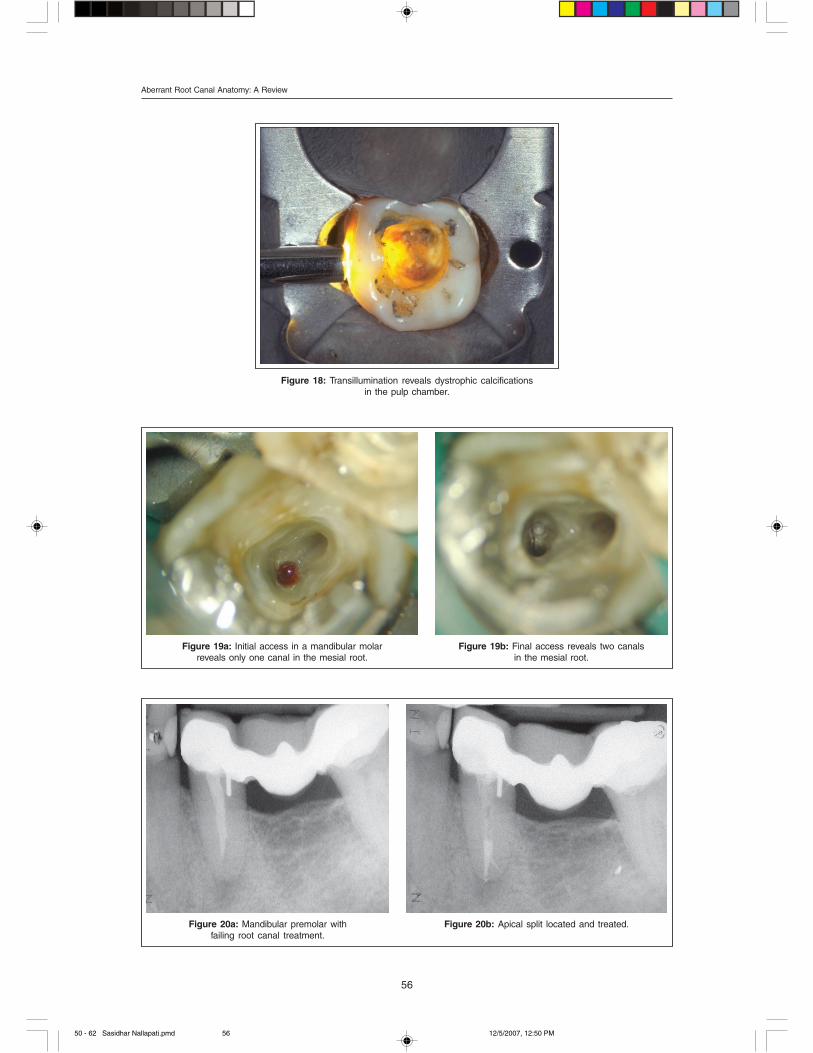
Sub chamber bifurcations. In mesial roots of
mandibular second molars, buccal roots of maxillary
second molars and buccal roots of maxillary premolars,
only one canal is seen on making the access. Upon
careful troughing of the canal orifice, this single canal
is found dividing into two canals apical to the chamber
level (fig 19a-b).
Apical bifurcations on the other hand can be seen in
any root of any tooth. Adequate coronal flaring and using
pre-bent hand files in an exploratory fashion to scout
the walls of the root canal often help in the location of
the apical bifurcations. Subsequently a straight line
access (with in the limits of safety) to the apical
bifurcation will help clean, shape and obturate the
splitting canals (20a-b).
50 - 62 Sasidhar Nallapati.pmd 12/5/2007, 12:50 PM55
56
Aberrant Root Canal Anatomy: A Review
Figure 19a: Initial access in a mandibular molar
reveals only one canal in the mesial root.
Figure 19b: Final access reveals two canals
in the mesial root.
Figure 18: Transillumination reveals dystrophic calcifications
in the pulp chamber.
Figure 20a: Mandibular premolar with
failing root canal treatment.
Figure 20b: Apical split located and treated.
50 - 62 Sasidhar Nallapati.pmd 12/5/2007, 12:50 PM56

57
Aberrant Root Canal Anatomy: A Review
‘C’ shaped canals are frequently seen in mandibular
second molars and less frequently in maxillary second
molars and mandibular premolars (fig 21a-c). The canal
shape resembles the letter ‘C’. There are three different
variations to this entity.
1. All canals are joined in the ‘C’
2. Two canals are joined in the ‘C’ and one canal
stays separate.
3. All canals stay separate with in the ‘C’
One of the most difficult aspects of treating this anatomy
is the predictable removal of pulp tissue in the isthmus
that connects all canals. Ultrasonic debridement, special
irrigation techniques and intra-canal medicaments have
a better chance in debriding and disinfecting these
canals (fig 22a-b).
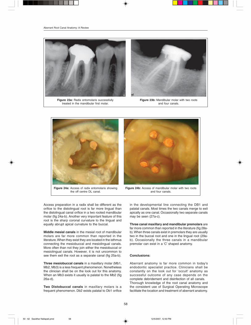
Radix Entomolaris is separate root that exists usually
to the distolingual in a mandibular molar. A mandibular
Figure 21c: Post op radiograph of the ‘C’ shaped
maxillary molar.
Figure 21b: DB and palatal canals are in ‘C’ shape.
Notice the two Mb canals.
molar with the radix shall have three roots with 4 canals,
not be confused with a mandibular molar with two roots
and four canals (fig. 23a-d).
Figure 21a: Maxillary molar with a ‘c’ shaped anatomy.
Figure 22a,b: Pre and Post op radiographs of retreatment
of ‘C’ shaped mandibular second molar.
50 - 62 Sasidhar Nallapati.pmd 12/5/2007, 12:50 PM57
58
Aberrant Root Canal Anatomy: A Review
Figure 23a: Radix entomolaris successfully
treated in the mandibular first molar.
Figure 23b: Mandibular molar with two roots
and four canals.
Figure 24a: Access of radix entomolaris showing
the off centre DL canal.
Figure 24b: Access of mandibular molar with two roots
and four canals.
Access preparation in a radix shall be different as the
orifice to the distolingual root is far more lingual than
the distolingual canal orifice in a two rooted mandibular
molar (fig 24a-b). Another very important feature of this
root is the sharp coronal curvature to the lingual and
equally abrupt apical curvature to the buccal.
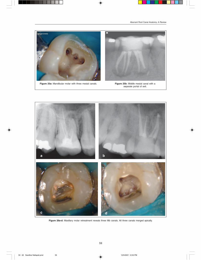
Middle mesial canals in the mesial root of mandibular
molars are far more common than reported in the
literature. When they exist they are located in the isthmus
connecting the mesiobuccal and mesiolingual canals.
More often than not they join either the mesiobuccal or
mesiolingual canals. However, it is not uncommon to
see them exit the root as a separate canal (fig 25a-b).
Three mesiobuccal canals in a maxillary molar (Mb1,
Mb2, Mb3) is a less frequent phenomenon. Nonetheless
the clinician shall be on the look out for this anatomy.
When an Mb3 exists it usually is palatal to the Mb2 (fig
26a-d).
Two Distobuccal canals in maxillary molars is a
frequent phenomenon. Db2 exists palatal to Db1 orifice
in the developmental line connecting the DB1 and
palatal canals. Most times the two canals merge to exit
apically as one canal. Occasionally two separate canals
may be seen (27a-c).
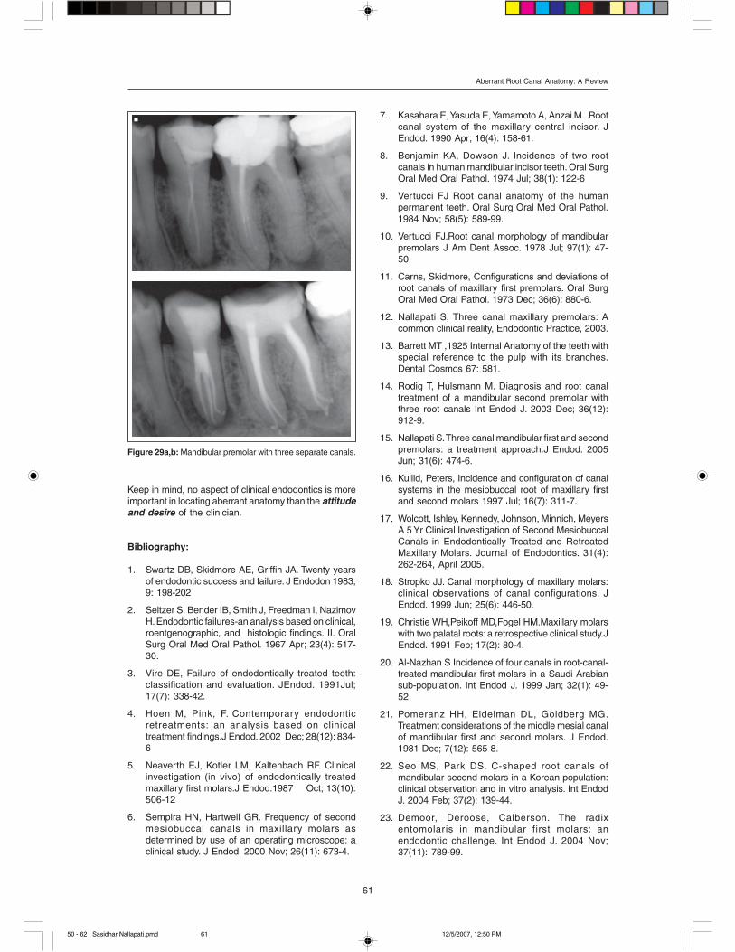
Three canal maxillary and mandibular premolars are
far more common than reported in the literature (fig 28a-
b). When three canals exist in premolars they are usually
two in the buccal root and one in the lingual root (29a-
b). Occasionally the three canals in a mandibular
premolar can exist in a ‘C’ shaped anatomy.
Conclusions:
Aberrant anatomy is far more common in today’s
endodontic specialist practice. Clinicians shall be
constantly on the look out for ‘occult’ anatomy as
successful outcome of any case depends on the
complete debridement and disinfection of all canals.
Thorough knowledge of the root canal anatomy and
the consistent use of Surgical Operating Microscope
facilitate the location and treatment of aberrant anatomy.
50 - 62 Sasidhar Nallapati.pmd 12/5/2007, 12:50 PM58
59
Aberrant Root Canal Anatomy: A Review
Figure 25a: Mandibular molar with three mesial canals. Figure 25b: Middle mesial canal with a
separate portal of exit.
Figure 26a-d: Maxillary molar retreatment reveals three Mb canals. All three canals merged apically.
a b
c d
50 - 62 Sasidhar Nallapati.pmd 12/5/2007, 12:50 PM59

60
Aberrant Root Canal Anatomy: A Review
a b
Figure 28a,b: Maxillary first premolar with three canals.
a b
c d
Figure 27a-d: A rare occurrence of two separate Db canals in a maxillary molar. Also notice three Mb canals.
50 - 62 Sasidhar Nallapati.pmd 12/5/2007, 12:50 PM60
61
Aberrant Root Canal Anatomy: A Review
Keep in mind, no aspect of clinical endodontics is more
important in locating aberrant anatomy than the attitudeand desire of the clinician.
Bibliography:
1. Swartz DB, Skidmore AE, Griffin JA. Twenty years
of endodontic success and failure. J Endodon 1983;
9: 198-202
2. Seltzer S, Bender IB, Smith J, Freedman I, Nazimov
H. Endodontic failures-an analysis based on clinical,
roentgenographic, and histologic findings. II. Oral
Surg Oral Med Oral Pathol. 1967 Apr; 23(4): 517-
30.
3. Vire DE, Failure of endodontically treated teeth:
classification and evaluation. JEndod. 1991Jul;
17(7): 338-42.
4. Hoen M, Pink, F. Contemporary endodontic
retreatments: an analysis based on clinical
treatment findings.J Endod. 2002 Dec; 28(12): 834-
6
5. Neaverth EJ, Kotler LM, Kaltenbach RF. Clinical
investigation (in vivo) of endodontically treated
maxillary first molars.J Endod.1987 Oct; 13(10):
506-12
6. Sempira HN, Hartwell GR. Frequency of second
mesiobuccal canals in maxillary molars as
determined by use of an operating microscope: a
clinical study. J Endod. 2000 Nov; 26(11): 673-4.
7. Kasahara E, Yasuda E, Yamamoto A, Anzai M.. Root
canal system of the maxillary central incisor. J
Endod. 1990 Apr; 16(4): 158-61.
8. Benjamin KA, Dowson J. Incidence of two root
canals in human mandibular incisor teeth. Oral Surg
Oral Med Oral Pathol. 1974 Jul; 38(1): 122-6
9. Vertucci FJ Root canal anatomy of the human
permanent teeth. Oral Surg Oral Med Oral Pathol.
1984 Nov; 58(5): 589-99.
10. Vertucci FJ.Root canal morphology of mandibular
premolars J Am Dent Assoc. 1978 Jul; 97(1): 47-
50.
11. Carns, Skidmore, Configurations and deviations of
root canals of maxillary first premolars. Oral Surg
Oral Med Oral Pathol. 1973 Dec; 36(6): 880-6.
12. Nallapati S, Three canal maxillary premolars: A
common clinical reality, Endodontic Practice, 2003.
13. Barrett MT ,1925 Internal Anatomy of the teeth with
special reference to the pulp with its branches.
Dental Cosmos 67: 581.
14. Rodig T, Hulsmann M. Diagnosis and root canal
treatment of a mandibular second premolar with
three root canals Int Endod J. 2003 Dec; 36(12):
912-9.
15. Nallapati S. Three canal mandibular first and second
premolars: a treatment approach.J Endod. 2005
Jun; 31(6): 474-6.
16. Kulild, Peters, Incidence and configuration of canal
systems in the mesiobuccal root of maxillary first
and second molars 1997 Jul; 16(7): 311-7.
17. Wolcott, Ishley, Kennedy, Johnson, Minnich, Meyers
A 5 Yr Clinical Investigation of Second Mesiobuccal
Canals in Endodontically Treated and Retreated
Maxillary Molars. Journal of Endodontics. 31(4):
262-264, April 2005.
18. Stropko JJ. Canal morphology of maxillary molars:
clinical observations of canal configurations. J
Endod. 1999 Jun; 25(6): 446-50.
19. Christie WH,Peikoff MD,Fogel HM.Maxillary molars
with two palatal roots: a retrospective clinical study.J
Endod. 1991 Feb; 17(2): 80-4.
20. Al-Nazhan S Incidence of four canals in root-canal-
treated mandibular first molars in a Saudi Arabian
sub-population. Int Endod J. 1999 Jan; 32(1): 49-
52.
21. Pomeranz HH, Eidelman DL, Goldberg MG.
Treatment considerations of the middle mesial canal
of mandibular first and second molars. J Endod.
1981 Dec; 7(12): 565-8.
22. Seo MS, Park DS. C-shaped root canals of
mandibular second molars in a Korean population:
clinical observation and in vitro analysis. Int Endod
J. 2004 Feb; 37(2): 139-44.
23. Demoor, Deroose, Calberson. The radix
entomolaris in mandibular first molars: an
endodontic challenge. Int Endod J. 2004 Nov;
37(11): 789-99.
Figure 29a,b: Mandibular premolar with three separate canals.
50 - 62 Sasidhar Nallapati.pmd 12/5/2007, 12:50 PM61
62
Aberrant Root Canal Anatomy: A Review
24. Fabra-Campos H.Three canals in the mesial root
of mandibular first permanent molars: a clinical
study. Int Endod J. 1989 Jan; 22(1): 39-43.
25. Ng YL, Aung TH, Alavi A, Gulabivala K. Root and
canal morphology of Burmese maxillary,
mandibular molars. Int Endod J. 2001 Dec; 34(8):
620-30.
26. Chopra P, Bal CS. Study of root canals and their
configuration in buccal roots of maxillary first
permanent molar. Indian J Dent Res. 1989 Jan-Mar;
1(1): 3-14.
27. Trope M, Elfenbein L, Tronstad L. Mandibular
premolars with more than one root canal in different
race groups J Endod. 1986 Aug; 12(8): 343-5.
28. Sabala CL,Benenati FW,Neas BR. Bilateral root or
root canal aberrations in a dental school patient
population. J Endod. 1994 Jan; 20(1): 38-42.
29. Weller NR, Niemczyk SP, Kim S (1995) Incidence
and Position of the canal Isthmus. Par t 1.
Mesiobuccal root of the maxillary first molar. Journal
of Endodontics.
30. Yang ZP, Yang SF, Lee G (1988) The root and root
canal anatomy of maxillary molars in a Chinese
population. Endodontics and Dental Traumatology
4, 215-8.
31. Carr GB. Microscopes in endodontics.J Calif Dent
Assoc. 1992 Nov; 20(11): 55-61.
32. Krasner P, Rankow HJ. Anatomy of the pulp-
chamber floor. J Endod. 2004 Jan; 30(1): 5-16.
33. Nallapati S, Glassman G. Use of ophthalmic dye in
root canal location, Endodontic practice 2004.
50 - 62 Sasidhar Nallapati.pmd 12/5/2007, 12:50 PM62