abdominal vascular catastrophes
TRANSCRIPT
Abdominal VascularCatastrophes
Manpreet Singh, MDa, Alex Koyfman, MDb, Joseph P. Martinez, MDc,*
KEYWORDS
� Mesenteric ischemia � Ruptured abdominal aortic aneurysm � Aorto-enteric fistula� Gastrointestinal bleeding
KEY POINTS
� Mesenteric ischemia (MI) has a variety of causes, each with its own historical clues toassist in diagnosis.
� Early CT angiography without waiting for administration of oral contrast should be pursuedin suspected cases of MI.
� Unexplained hypotension, syncope, or ecchymosis should prompt consideration ofruptured abdominal aortic aneurysm (AAA).
� Any amount of gastrointestinal (GI) bleeding in a patient with a history of AAA or AAA repairis an aortoenteric fistula (AEF) until proved otherwise.
INTRODUCTION
Abdominal vascular catastrophes are uncommon yet frequently fatal processes thatare of great interest to emergency physicians because rapid recognition and initiationof definitive treatment are essential to prevent long-term morbidity and mortality. Thelist of abdominal vascular catastrophes is broad, but the focus of this article is on MI,AAA, and AEF.
MESENTERIC ISCHEMIAIntroduction
Acute MI continues to remain an elusive disease to diagnose despite clinicians beingtaught in medical school and residency about the classic pain out of proportion withexamination presentation. Although a rare case of abdominal pain, with an annual inci-dence of 0.09% to 0.2% per year and approximately 1% of acute abdomen
Disclosure Statement: The authors have no financial relationships to disclose.a Department of Emergency Medicine, Harbor-UCLA Medical Center, 1000 W. Carson Boule-vard, Torrance, CA 90502, USA; b Department of Emergency Medicine, UT Southwestern Med-ical Center, 5323 Harry Hines Boulevard, Dallas, TX 75390, USA; c Department of EmergencyMedicine, University of Maryland School of Medicine, 110 South Paca Street, Sixth Floor,Suite 200, Baltimore, MD 21201, USA* Corresponding author.E-mail address: [email protected]
Emerg Med Clin N Am 34 (2016) 327–339http://dx.doi.org/10.1016/j.emc.2015.12.014 emed.theclinics.com0733-8627/16/$ – see front matter � 2016 Elsevier Inc. All rights reserved.
Singh et al328
hospitalizations,1,2 this is offset with a 60% to 80% mortality within the first 24 hours.3
It is imperative that there is no delay in diagnosis because delays in diagnosis lead toincreased mortality and morbidity in terms of amount of bowel requiring resection. Thepresentation of patients with MI is usually nonspecific with a falsely reassuring objec-tive abdominal examination, which can lead to a false sense of security because thelate findings of this disease process (absent bowel sounds, positive fecal occult bloodtest, focal/generalized peritonitis from visceral ischemia, elevated lactate, hypoten-sion, fever, and so forth) have not evolved. In general, a high degree of clinical suspi-cion should be based on a combination of history, examination, laboratory results, andimaging studies to arrive at the diagnosis of acute mesenteric ischemia.
Anatomy
The abdominal aorta gives off 3 major branches to the intestines (foregut, midgut, andhindgut), which are the celiac artery (CA), superior mesenteric artery (SMA), and inferiormesenteric artery (IMA).4 The CA perfuses the foregut (distal esophagus to secondportion of duodenum). Acute MI of the foregut is rare because the CA is a short, wideartery with good collateral flow. The SMA perfuses the midgut (duodenum to distaltransverse colon), which encompasses nearly the entire small bowel and two-thirdsof the large bowel. This is the most common embolic site of MI due to favorable takeoffangle (approximately 45�) from the aorta. The IMA perfuses the hindgut (transversecolon to rectum) and is rarely the sole vessel involved in MI. Collateral circulation fromthe CA or IMA generally allows sufficient perfusion in reduced SMA occlusion states.
Pathophysiology
In addition to the abdominal aortic anatomy, it is important to understand how thebowel layers are affected by MI, starting from the innermost to outermost layers (mu-cosa, submucosa, muscularis, and serosa). Early in the course of MI, the furthest layerfrom the blood supply (mucosa) is the first to become ischemic and is the reason forextreme pain, which is visceral in origin. Because the outer structures (muscle andserosa) have not become ischemic, however, there is minimal irritation of the parietalperitoneum when the examiner indents down against the serosa and the externallayers of the bowel. Hence, there is pain out of proportion with the examination earlyon in the disease process. Over a period of hours, the muscularis and serosal layersbecome ischemic and infarct, leading to peritoneal irritation and guarding with rigidity.At this point, the pain is in proportion with the examination. It is also important toconsider that between the early and late presentations (discussed previously), thereis a deceptive pain-free interval of approximately 3 to 6 hours caused by a declinein intramural pain receptors from hypoperfusion.5
Etiology
MI can be classified as acute versus chronic or as occlusive versus nonocclusive. Thefollowing are the major 4 causes of acute MI5:
� Acute arterial emboli – the most frequent cause of MI, accounting for 40% to50% of cases; the embolus usually lodges in the SMA.3 The proximal branchesof the SMA (jejunal and middle colic arteries) are usually preserved because theembolus lodges 3 cm to 10 cm distally from the SMA takeoff, where the arterytapers off and is just after the first major branch of the SMA (the middle colic ar-tery). As a result, the proximal small and large bowels are usually spared.6 Due topoorly developed collateral circulation, the onset of symptoms in cases of emboliis usually severe and dramatic pain.4 When the bowel becomes ischemic, it has a
Abdominal Vascular Catastrophes 329
propensity to empty itself, leading to vomiting or diarrhea, so-called gutemptying. This is one of the reasons that MI is often misdiagnosed as gastroen-teritis. Common predisposing factors include atrial fibrillation, cardiomyopathy,recent angiography, and valvular disorders, such as rheumatic valve disease.5
One-third of patients have had a previous embolic event, such as an embolicrenal infarct, embolic stroke, or peripheral arterial embolus.
� Acute arterial thrombosis – patients with long-standing atherosclerosis maydevelop plaque build-up at the origin of the SMA, a site of turbulent bloodflow. This subsequent stenosis may lead to long-standing postprandial pain(intestinal angina) and food fear with resultant weight loss. These symptoms ofchronic MI can be seen in up to 80% of patients who develop arterial thrombosis.If the plaque acutely ruptures or the stenosis reaches a critical level, patients maypresent with acute pain, similar to those with arterial emboli.3,6,7
� Mesenteric venous thrombosis (MVT) – generally found in patients with an under-lying hypercoagulable state; MVT accounts for 10% to 15% of cases. Patientstypically present with less severe and more insidious pain than those with arterialocclusion.5 A majority of patients present after more than 24 hours of symptoms.In 1 study, the mean symptom duration was 5 to 14 days, with many patientsexperiencing pain for 1 month prior to diagnosis.3 Predisposing risk factorsinclude malignancy, sepsis, liver disease or portal hypertension, sickle cell dis-ease, and pancreatitis.3,7 Many patients have heritable hematologic disorders,including protein C and protein S deficiency, antithrombin III deficiency, and fac-tor V Leiden mutation. One-half of patients with MVT have a personal or familyhistory of venous thromboembolism.
� Nonocclusive MI (NOMI) – this type of MI occurs in 20% of patients due to failureof autoregulation in low-flow states, such as hypovolemia, potent vasopressoruse, heart failure, or sepsis.3,6 The underlying ischemia from splanchnic vasocon-striction can further lead to hypotension from endogenous substances, perpetu-ating a vicious cycle.4,5 This accounts for the extremely highmortality rate, usuallydue to the poor health of the affected population, with multiple comorbidities,combined with the difficulty in treating the primary cause of diminished intestinalblood flow. Patients who present with abdominal pain postdialysis may haveNOMI secondary to intradialytic hypotension, leading to vasospasm.8
Clinical Findings
The presentation of MI is typically acute severe abdominal pain with a paucity of phys-ical examination findings. There is a widely variable range of performance character-istics of the history and physical examination, which underlines the diagnosticchallenge.7 History and physical examination findings, such as acute abdominalpain, pain out of proportion, peritoneal signs, guaiac-positive stool, acute abdominalpain, heart failure, and atrial fibrillation, have a wide range of sensitivities and arefrequently absent.9 Therefore, clinicians should be vigilant in considering MI in the dif-ferential diagnosis of abdominal pain of unclear etiology. Assessing a patient’s pretestprobability for disease, actively searching for known risk factors, and adding in cluesbased on a patient’s history and physical examination findings are an important pro-cess for wary clinicians. Early and aggressive imaging based on this process has beendemonstrated to decrease overall mortality from MI.10
Laboratory Studies
Numerous laboratory abnormalities have been described in MI, including elevatedamylase, lactate dehydrogenase, large base deficit, and metabolic acidosis. None
Singh et al330
of these findings is sensitive or specific for MI. Troponin I levels are often elevated. Thisfinding is not specific for MI and has been shown to lead to delays in definitive care ofthese patients and inappropriate cardiology consultations.11,12 Common laboratoryabnormalities, such as hemoconcentration, leukocytosis, and high anion-gap meta-bolic acidosis with elevated lactate (specifically D-lactate), are neither sensitive norspecific enough to be diagnostic and usually late findings.Diagnostic biomarkers are a tool that should bear a high sensitivity and specificity,
especially in MI, where early symptoms are nonspecific and mortality rises withdelayed or missed diagnosis. Many laboratory tests have been studied in AMI,including D-lactate, intestinal fatty acid–binding protein, glutathione S-transferase,ischemia-modified albumin, and D-dimer. Although some have shown promising earlyresults, none is sufficiently well established to either make or exclude the diagnosis ofAMI. The serum marker that practicing clinicians are most familiar with is lactate.Although MI mortality is associated with high lactate serum values, a normal serumlactate value does not exclude AMI.13 Early in the disease process, lactate is generallynormal as it travels through the portal venous system to the liver, where it is convertedinto glucose via the Cori cycle. As the ischemia load increases and the liver is not ableto keep up with the demand, lactate spills over into the systemic circulation, where iteventually increases in late stages.
Imaging Studies
Various imaging methods have been studied and used in the diagnosis of MI, includinglower GI endoscopy, radionuclide imaging, peritoneal fluid analysis, MRI, and perito-neoscopy. Imaging that is insensitive or low yield should be avoided, and any imagingthat is performed should be pursued in as expeditious a manner as possible, given thetime-sensitive nature of the disease. It has been shown that a multidisciplinaryapproach to suspected cases of AMI with streamlined protocols and early involvementof consultants can have an impact on overall mortality.14 The following are commondiagnostic modalities often described:
� Plain radiographs: the findings on a plain abdominal radiograph are usuallynonspecific (ie, small bowel distention with air-fluid levels or ileus), and 25% ofpatients may have normal findings.3 Patients with normal plain radiographshave a lower mortality rate, presumably because the findings that are visibleon plain radiographs are late findings seen in more advanced disease. Character-istic findings, such as thumb printing or thickening of bowel loops, occur in lessthan 40% of patients.3 Later findings, such as air in the bowel wall (pneumatosisintestinalis) and portal venous system, are ominous signs portending a poorprognosis.
� Ultrasound: the use of ultrasound to detect significant stenosis (>50%) in mesen-teric vessels has been shown and has a role in chronic MI, but the role of ultra-sound in making the diagnosis of acute ischemia is less well established. Thislikely is due to limited operator experience in AMI and the abnormality in patientbowel gas patterns that often accompanies AMI, which make visualization of themesenteric vessels more difficult.
� CT scan: CT is the most commonly used diagnostic tool in suspected MI; theinitial sensitivity was 64% but has now improved to 93%with the use of dynamiccontrast-enhanced CT.15,16 The addition of multidetector row CT (MDRCT) tech-nology has further improved results (Fig. 1).17,18 The use of MDRCT angiographydoes not require oral contrast,16 which has been shown to increase time to imageacquisition to 2 to 3 hours,19 a potentially lethal delay in cases of suspected MI.
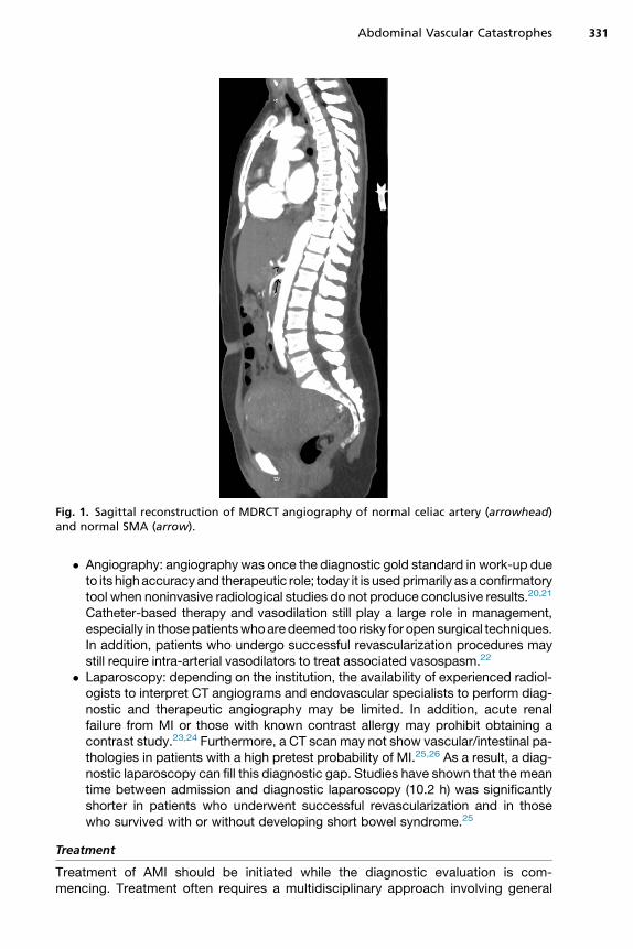
Fig. 1. Sagittal reconstruction of MDRCT angiography of normal celiac artery (arrowhead)and normal SMA (arrow).
Abdominal Vascular Catastrophes 331
� Angiography: angiography was once the diagnostic gold standard in work-up dueto its highaccuracyand therapeutic role; today it is usedprimarily asa confirmatorytool when noninvasive radiological studies do not produce conclusive results.20,21
Catheter-based therapy and vasodilation still play a large role in management,especially in thosepatientswhoaredeemed too risky for opensurgical techniques.In addition, patients who undergo successful revascularization procedures maystill require intra-arterial vasodilators to treat associated vasospasm.22
� Laparoscopy: depending on the institution, the availability of experienced radiol-ogists to interpret CT angiograms and endovascular specialists to perform diag-nostic and therapeutic angiography may be limited. In addition, acute renalfailure from MI or those with known contrast allergy may prohibit obtaining acontrast study.23,24 Furthermore, a CT scan may not show vascular/intestinal pa-thologies in patients with a high pretest probability of MI.25,26 As a result, a diag-nostic laparoscopy can fill this diagnostic gap. Studies have shown that the meantime between admission and diagnostic laparoscopy (10.2 h) was significantlyshorter in patients who underwent successful revascularization and in thosewho survived with or without developing short bowel syndrome.25
Treatment
Treatment of AMI should be initiated while the diagnostic evaluation is com-mencing. Treatment often requires a multidisciplinary approach involving general
Singh et al332
and vascular surgeons as well as interventional radiologists. Aggressive fluid resus-citation should be started to correct fluid deficit and metabolic derangements.Broad-spectrum antibiotics are generally given as well. Early surgical consultationis warranted, even before definitive testing is performed, especially in cases ofhigh pretest probability. The presence of peritoneal signs is usually an indicatorof late stages of the disease requiring emergency laparotomy and may obviateany confirmatory imaging.Once a diagnosis is established, surgical treatment of the underlying cause should
be performed (ie, embolectomy, thrombectomy, endarterectomy, or bypass). Antico-agulation should be started, in consultation with the treating surgeon. An importantpart of the postsurgical care involves reducing the profound vasospasm that accom-panies AMI. This is typically accomplished through intra-arterial papaverine infusionvia an indwelling catheter in the SMA. A growing area of research involves minimizingischemia-reperfusion injury.
Summary
MI is a vascular emergency, which all emergency physicians must consider early intheir abdominal pain differential. It continues to remain a diagnostic challenge, andany delay in diagnosis can contribute to the increases in the already high mortalityrate. Clues to the diagnosis should be sought for in the patient history (Table 1).Although the underlying cause varies, early diagnosis and prompt effective treatmentcan lead to improved clinical outcome. Time is bowel, so if there is a high clinical sus-picion for MI, surgical and interventional radiology consultants should be involvedearly and in parallel with an expeditious diagnostic evaluation.
ABDOMINAL AORTIC ANEURYSMIntroduction
An AAA in itself is a hallmark emergency medicine presentation, where it is a tickingtime bomb if not recognized because patients are asymptomatic until it becomespainful as it expands until it ruptures. Once ruptured, overall mortality is as high as90%; even those with treatment have a 40% to 50% survival.27 The classic presenta-tion consists of abdominal/flank pain, hypotension, and a pulsatile abdominal mass,but this is only present in 50% of all cases at best.
Anatomy/Pathophysiology
A true aneurysm is a dilatation of all 3 arterial layers (intima, media, and adventitia)through a degenerative process that remains unclear but involves the degradationof the media, where elastin is normally found. As a result, the aortic wall becomesmore susceptible to influences of high blood pressure. The aorta varies in size by
Table 1Historical clues in suspected causes of mesenteric ischemia
Etiology Historical Clue
SMA embolus One-third have prior embolic event
SMA thrombosis 80% Have history of intestinal angina
MVT One-half have personal or family history of deep vein thrombosis/pulmonary embolism
NOMI More commonly seen in dialysis patients
Abdominal Vascular Catastrophes 333
age, gender, and body habitus, with the average diameter less than 2.0 cm, but, ingeneral, anything above 3.0 cm is considered an aneurysm.28 The risk of rupture in-creases with the size and rate of expansion.29
Most AAAs originate in the infrarenal aorta, below the takeoff of the renal vessels. Inthis area, the diameter of the aorta is decreasing and contains a lesser proportion ofelastin. Although this is the most common location of AAAs, they may also occur inthe suprarenal, pararenal, and juxtarenal areas. These anatomic variations are impor-tant when discussing patient candidates for endovascular aortic repair (EVAR), dis-cussed later.
Causes/Risk Factors
Although the degenerative process remains unclear, the following are well-defined riskfactors30 that contribute to AAAs:
� Smoking� Hypertension� Male gender� Connective tissue disorder (ie, Marfan syndrome and Ehlers-Danlossyndrome)
� Atherosclerosis� Infection/arteritis
Clinical Findings
The clinical manifestation of an AAA varies considerably, depending on the location ofrupture (Box 1). Intraperitoneal rupture typically manifests as sudden death and rarelysurvives to reach medical attention. Retroperitoneal bleeds may tamponade offtemporarily, allowing a patient to present for medical care. In 75% of cases, acutesevere pain is the most common presentation of rupture, where the location of painvaries based on the site of rupture.31 Those close to renal vessels have flank pain lead-ing to possible renal colic mimicry, whereas those anterior cause abdominal pain andposterior cause back pain. Once a rupture stabilizes, the pain may subside, leading toa false sense of security for both patient and physician. Other uncommon presenta-tions of AAA rupture include radicular femoral/sciatic pain due to nerve compressionfrom the hematoma, acute inguinal hernia from sudden increases in intraperitonealpressure, and acute high-output heart failure or massive leg swelling from ruptureinto the inferior vena cava (aortocaval fistula). Unexplained hypotension or syncope,even transient, in a patient with risk factors for ruptured AAA should prompt con-sideration of the condition. This is evident because it is considered part of the differ-ential of a hypotensive patient when performing the Rapid Ultrasound for Shock andHypotension examination. Even without rupturing, AAAs can cause subacute flank,
Box 1
Location of rupture in abdominal aortic aneurysm (in decreasing frequency)
� Retroperitoneal
� Intraperitoneal
� Vena cava (aortocaval fistula)
� GI tract (AEF)

Singh et al334
abdominal, or back pain due to rapid enlargement and compression of the surround-ing structures.The physical examination on these patients may be unrevealing but can offer clues
to the diagnosis. Although insensitive, 50% of patients with an AAA have a pulsatilemass palpable in the epigastrium.32 Besides an abdominal examination, a fullvascular examination palpating major pulses (radial, carotid, femoral, and popliteal)should be done to look unequal pulses that can hint to acute aortic syndromes.Although ecchymotic signs, such as Grey Turner (flank), Cullen (periumbilical), Fox(inguinal ligament), and Bryant (scrotal) signs, are neither sensitive nor specific foran AAA, unexplained ecchymosis should always prompt consideration of thisvascular emergency.
Imaging Studies
Bedside ultrasound has emerged as the test of choice when screening patients forAAA (Fig. 2). Ultrasound has a 98% sensitivity in fasted patients undergoing screening,and although bowel gas and body habitus can hinder the examination, this is less of anissue in imaging the larger aneurysms that are likely to present ruptured. If a patientwith a known history of AAA arrives unstable with symptoms consistent with rupture,however, no confirmatory diagnostic tests are necessary and the patient should betransferred to an operating room expeditiously.Abdominal CT imaging is generally advised for hemodynamically stable patients,
although ultrasound should be performed immediately in patients with high clinicalsuspicion to aid in triage and to speed disposition. In addition to assessing for alter-native conditions, CT provides important anatomic information about the AAA thatmay be important in surgical planning for open and closed (ie, endovascular) surgi-cal approaches (Fig. 3). Although contrast is not required, its administration is help-ful to obtain more aortic detail for preoperative planning and to ascertain whetherthe patient is a candidate for EVAR. Signs of rupture on CT include retroperitonealhematoma, free intraperitoneal blood, an indistinct aortic wall, and loss of thenormal fat plane around the aorta. Signs of impending rupture or an unstable aneu-rysm also may be seen and include layering of hematoma within the aorta (crescentsign), breaks in the calcification of the wall, and blebs or other irregularity within thewall.
Fig. 2. Bedside ultrasound showing 4.1-cm � 4.5-cm AAA.

Fig. 3. CT of large AAA – axial slice of abdominal CT scan showing large, heavily calcified,and thrombosed AAA. The calcification exhibits significant discontinuity in the rightposterolateral area and signs of retroperitoneal hematoma, suggestive of rupture.
Abdominal Vascular Catastrophes 335
Treatment
Those presenting in clinical shock require tandem resuscitation with bedside diag-nosis if there is no known history of AAA. Initial misdiagnosis is common, occurringin approximately 40% of cases, where the most common incorrect diagnoses arerenal colic, myocardial infarction, and diverticulitis.Obtaining 2 large-bore intravenous (IV) lines with uncrossmatched type O blood
immediately made available is key. Central access with a sheath introducer or multi-lumen access catheter should be weighed against possible delay in transport to anoperating room. In anticipation for the operating room, 6 to 10 units of packed redblood cells, type and crossmatched, should be requested as well as fresh frozenplasma and platelets that may be required during resuscitation. Depending on theinstitution, implementing a massive transfusion protocol, as well as activating the pa-tient as a trauma, may speed along the process of obtaining an OR room with a sur-geon who is ready to go. Resuscitation effort should be focused on controlled volumeresuscitation targeting a systolic blood pressure of 80mmHg to 90 mmHg, analogousto patients with penetrating torso trauma.33
EVAR has become the mainstay for elective repair of AAAs, with 2 multicenter ran-domized controlled trials showing a 3-fold reduction in mortality.19,34 Although ran-domized trials have not shown improved mortality rates for EVAR in the setting ofacute rupture, it seems that perioperative morbidity is decreased and thus EVAR isfavored for anatomically suitable patients.35 The main criterion for EVAR is anadequately long aneurysm neck to allow seating of the graft without occlusion ofthe renal arteries, which is met in 70% of AAAs.36 Although infrarenal aneurysmsare preferred (especially in the emergent use), suprarenal and pararenal aneurysmsare amenable to EVAR with custom grafts.
Summary
Ruptured or symptomatic AAA is a fatal and time-sensitive condition that emergencyphysicians should be familiar with and ready for, where timely diagnosis and appro-priate resuscitation with operative team management can mean the difference be-tween life and death.
Singh et al336
AORTOENTERIC FISTULASIntroduction
Development of an AEF is a life-threatening and devastating cause of upper GI bleed,which can be difficult to diagnose and treat. Although rare, it is most commonly seenas a delayed complication of aortic reconstruction.
Pathophysiology
The disease is divided into 2 types – primary AEF and secondary AEF. Although un-common, primary AEF occurs when a large, previously untreated aneurysm erodesde novo into the adjacent bowel. This is often diagnosed unexpectedly during explor-atory laparotomy. The third portion of the duodenum, fixed retroperitoneally and inproximity to the descending aorta, is the bowel segment most vulnerable to this.The nidus for this process starts with ischemia and subsequent necrosis of the intes-tinal wall as a consequence of repetitive traumatic pulsations of an adjacent aorticaneurysm. Subsequent rupture of an expanding aneurysm or perforation of the aortaas a result of contamination with GI contents results in the formation of a communica-tion with the bowel and the potential for rapid exsanguination. Less commonlyencountered conditions that may lead to primary AEFs include syphilis, tuberculosis,mycotic infection, and collagen vascular disease, where the chronic inflammationleads to aortitis, erosion, and formation of the fistula. In the absence of treatment,the mortality rate is almost 100%. With surgical intervention, survival ranges from18% to 93%.In contrast, secondary AEF occurs as a complication of aortic reconstructive sur-
gery. An estimated 80% of secondary AEFs affect the duodenum, mostly the thirdand fourth parts (the horizontal and ascending duodenum). As a result of advancedperigraft infection from chronic low-grade infection and the repetitive pressure onthe intestine from aortic pulsations, fistulas are formed.
Clinical Findings
The typical symptoms of AEFs include acute abdominal pain, GI hemorrhage (melena,hematemesis, and dark blood per rectum), and sepsis. The most common clinical fea-tures of primary AEFs are upper GI bleeding (64%), abdominal pain (32%), and a pul-satile abdominal mass (25%), where these 3 features are concomitantly present in only10% to 23% of patients. Patients with a secondary AEF usually present with 1 or moreof the following clinical signs and symptoms: GI bleeding (80%), sepsis (44%), abdom-inal pain (30%), back pain (15%), groin mass (12%), and abdominal pulsatile mass(6%). With both primary and secondary AEFs, transient, self-limited, intermittentbleeding episodes (herald bleeds) often precede a major hemorrhagic episode byhours, days, or weeks. This is a result of a small fistula tamponaded by thrombus for-mation and bowel contraction around it.
Imaging Studies
Three modalities are available to assist in diagnosis: abdominal CT scan with IVcontrast, esophagogastroduodenoscopy (EGD), and arteriography. Of the 3, CTscan offers superiority because it is less invasive, more readily available, and moreexpedient than the latter 2. In addition, it offers the advantage of being unlikely todislodge the aortic thrombus. The CT may show abnormal communication betweenthe aorta and the bowel, may disclose loss of continuity of the aneurysmal wall, andmay demonstrate air bubbles in the aneurysm wall that are pathognomonic for the ex-istence of a fistula.
Abdominal Vascular Catastrophes 337
An EGD with a water-soluble contrast material is usually considered second line butshould be performed only on a hemodynamically stable patient. An AEF is usually pre-sent when there is leakage of oral contrast material from the disrupted bowel wall intothe perigraft space. In addition to evaluating the presence or absence of an AEF, anEGD assists in ruling out other causes of upper GI bleeding, including varices,bleeding masses, and ulcers. A normal EGD or the finding of other pathology withoutstigmata of recent bleeding does not exclude an AEF, especially if there is a high indexof suspicion.Arteriography is of some value but rarely used in critically ill patients for diagnosis.
Its true value lies in embolization therapy and stent placement.
Treatment
AEF requires definitive and emergent operative management, where various surgicalmodalities exist (graft excision and extra-anatomic bypass, in situ graft replacement,and simple graft excision or endovascular repair). The role of emergency physicians, inthe perioperative phase, includes optimally resuscitating the patient with fluids andblood products while initiating broad-spectrum IV antibiotics to cover gram-positive,gram-negative, and enteric pathogens as part of sepsis management.
Summary
AEF is a life-threatening entity that is challenging to diagnose and carries highmorbidity and mortality. Any patient who presents with any degree of GI bleedingand has a prior history of aortic aneurysm repair should be considered as having anAEF until proved otherwise.
REFERENCES
1. Sise MJ. Acute mesenteric ischemia. Surg Clin North Am 2014;94(1):165–81.
2. van den Heijkant TC, Aerts BA, Teijink JA, et al. Diagnosis of mesentericischemia. World J Gastroenterol 2013;19(9):1338–41.
3. Lewiss RE, Egan DJ, Shreves A. Vascular abdominal emergencies. Emerg MedClin North Am 2011;29:253–72.
4. Oldenburg WA, Lau LL, Rodenberg TJ, et al. Acute mesenteric ischemia: aclinical review. Arch Intern Med 2004;164(10):1054–62.
5. Martinez JP, Hogan GJ. Mesenteric ischemia. Emerg Med Clin North Am 2004;22(4):909–28.
6. LottermanS.Mesenteric Ischemia: APower Review. 2014. Available at: http://www.emdocs.net/mesenteric-ischemia-power-review/. Accessed December, 2015.
7. McKinsey JF, Gewertz BL. Acute mesenteric ischemia. Surg Clin North Am 1997;77:307–18.
8. Diamond S, Emmett M, Henrich WL. Bowel infarction as a cause of death indialysis patients. JAMA 1986;256:2545.
9. Cudnik MT, Darbha S, Jones J, et al. The diagnosis of acute mesenteric ischemia:a systematic review and meta-analysis. Acad Emerg Med 2013;20:1087–100.
10. Boley SJ, Sprayregen S, Siegelman SJ, et al. Initial results from an aggressiveroentgenologic and surgical approach to acute mesenteric ischemia. Surgery1977;82:848.
11. Acosta S, Block T, Bjornsson S, et al. Diagnostic pitfalls at admission in patientswith acute superior mesenteric artery occlusion. J Emerg Med 2012;42(6):635–41.
Singh et al338
12. Huynh LN, Coughlin BF, Wolfe J, et al. Patient encounter time intervals in the eval-uation of emergency department patients requiring abdominopelvic CT: oralcontrast versus no contrast. Emerg Radiol 2004;10:310–3.
13. Cohn B. Does this patient have acute mesenteric ischemia? Ann Emerg Med2014;5(64):533–4.
14. Clark RA, Gallant TE. Acute mesenteric ischemia: angiographic spectrum. AJRAm J Roentgenol 1984;142:555.
15. Menke J. Diagnostic accuracy of multidetector CT in acute mesenteric ischemia:systematic review and meta-analysis. Radiology 2010;256:93–101.
16. Kirkpatrick ID, Kroeker MA, Greenberg HM. Biphasic CT with mesenteric CTangiography in the evaluation of acute mesenteric ischemia: initial experience.Radiology 2003;229:91–8.
17. Klar E, Rahmanian PB, Bucker A, et al. Acute mesenteric ischemia: a vascularemergency. Dtsch Arztebl Int 2012;109:249–56.
18. Horton KM, Fishman EK. CT angiography of the mesenteric circulation. RadiolClin North Am 2010;48:331–45.
19. Prinssen M, Verhoeven ELG, Buth J, et al. A randomized trial comparing conven-tional and endovascular repair of abdominal aortic aneurysms. N Engl J Med2004;351:1607–18.
20. Aschoff AJ, Stuber G, Becker BW, et al. Evaluation of acute mesenteric ischemia:accuracy of biphasic mesenteric multidetector CT angiography. Abdom Imaging2009;34:345–57.
21. Ofer A, Abadi S, Nitecki S, et al. Multidetector CTangiography in the evaluation ofacute mesenteric ischemia. Eur Radiol 2009;19:24–30.
22. Stone JR, Wilkins LR. Acute mesenteric ischemia. Tech Vasc Interv Radiol 2015;18:24–30.
23. Gupta PK, Natarajan B, Gupta H, et al. Morbidity and mortality after bowel resec-tion for acute mesenteric ischemia. Surgery 2011;150:779–87.
24. Kougias P, Lau D, El Sayed HF, et al. Determinants of mortality and treatmentoutcome following surgical interventions for acute mesenteric ischemia. J VascSurg 2007;46:467–74.
25. Gonenc M, Dural CA, Kocatas A, et al. The impact of early diagnostic laparos-copy on the prognosis of patients with suspected acute mesenteric ischemia.Eur J Trauma Emerg Surg 2013;39(2):185–9.
26. Smerud MJ, Johnson CD, Stephens DH. Diagnosis of bowel infarction: a compar-ison of plain films and CT scans in 23 cases. Roentgenol 1990;154:99–103.
27. Brown LC, Powell JT. Risk factors for aneurysm rupture in patients kept underultrasound surveillance. Ann Surg 1999;230(3):289–96.
28. Johnston KW, Rutherford RB, Tilson MD, et al. Suggested standards for reportingon arterial aneurysms. Subcommittee on Reporting Standards for Arterial Aneu-rysms, Ad Hoc Committee on Reporting Standards, Society for Vascular Surgeryand North American Chapter, International Society for Cardiovascular Surgery.J Vasc Surg 1991;13:452.
29. Chaikof EL, Brewster DC, Dalman RL, et al. SVS practice guidelines for the careof patients with an abdominal aortic aneurysm: executive summary. J Vasc Surg2009;50:880.
30. Wilmink AB, Quick CG. Epidemiology and potential for prevention of abdominalaortic aneurysm. Br J Surg 1998;85:55–62.
31. Rinckenbach S, Albertini JN, Thaveau F, et al. Prehospital treatment of infrarenalruptured abdominal aortic aneurysms: a multicentre analysis. Ann Vasc Surg2010;24:308.

Abdominal Vascular Catastrophes 339
32. Azhar B, Patel SR, Holt PJ, et al. Misdiagnosis of ruptured abdominal aorticaneurysm: systematic review and meta-analysis. J Endovasc Ther 2014;21:568.
33. Dick F, Erdoes G, Opfermann P, et al. Delayed volume resuscitation during initialmanagement of ruptured abdominal aortic aneurysm. J Vasc Surg 2013;57:943.
34. Greenhalgh RM, Brown LC, Kwong GPS, et al. Comparison of endovascularaneurysm repair with open repair in patients with abdominal aortic aneurysm(EVAR trial 1), 30-day operative mortality results: randomised controlled trial.Lancet 2004;364:843–8.
35. van Beek SC, Conijn AP, Koelemay MJ, et al. Editor’s Choice – Endovascularaneurysm repair versus open repair for patients with a ruptured abdominal aorticaneurysm: a systematic review and meta-analysis of short-term survival. Eur JVasc Endovasc Surg 2014;47:593.
36. Antoniou GA, Georgiadis GS, Antoniou SA, et al. Endovascular repair forruptured abdominal aortic aneurysm confers an early survival benefit overopen repair. J Vasc Surg 2013;58:1091.