a systematic forward genetic analysis identified ...genesdev.cshlp.org/content/22/7/918.full.pdf ·...
TRANSCRIPT
A systematic forward genetic analysisidentified components of theChlamydomonas circadian systemTakuya Matsuo,1 Kazuhisa Okamoto,1 Kiyoshi Onai,1 Yoshimi Niwa,1,2 Kosuke Shimogawara,3 andMasahiro Ishiura1,2,4
1Center for Gene Research, Nagoya University, Furo, Chikusa, Nagoya 464-8602, Japan; 2Division of Biological Science,Graduate School of Science, Nagoya University, Furo, Chikusa, Nagoya 464-8602, Japan; 3Laboratory of Chemistry, Schoolof Medicine, Teikyo University, Ohtsuka, Hachioji, Tokyo 192-0395, Japan
The molecular bases of circadian clocks have been studied in animals, fungi, bacteria, and plants, but not ineukaryotic algae. To establish a new model for molecular analysis of the circadian clock, here we identified alarge number of components of the circadian system in the eukaryotic unicellular alga Chlamydomonasreinhardtii by a systematic forward genetic approach. We isolated 105 insertional mutants that exhibiteddefects in period, phase angle, and/or amplitude of circadian rhythms in bioluminescence derived from aluciferase reporter gene in their chloroplast genome. Simultaneous measurement of circadian rhythms inbioluminescence and growth rate revealed that some of these mutants had defects in the circadian clock itself,whereas one mutant had a defect in a specific process for the chloroplast bioluminescence rhythm. Weidentified 30 genes (or gene loci) that would be responsible for rhythm defects in 37 mutants. Classification ofthese genes revealed that various biological processes are involved in regulation of the chloroplastrhythmicity. Amino acid sequences of six genes that would have crucial roles in the circadian clock revealedfeatures of the Chlamydomonas clock that have both partially plant-like and original components.
[Keywords: Chlamydomonas reinhardtii; circadian rhythm; bioluminescence; insertional mutagenesis; clockgene]
Supplemental material is available at http://www.genesdev.org.
Received January 11, 2008; revised version accepted January 28, 2008.
The molecular bases of circadian clocks have been stud-ied in animals, fungi, bacteria, and plants (Dunlap 1999;Harmer et al. 2001). Despite the striking biochemicalfeatures of circadian clocks (e.g., oscillation with longperiodicity [∼24 h] and its temperature compensation)(Bünning 1973), their central components are not con-served between these kingdoms (Dunlap 1999; Harmeret al. 2001). To understand the nature of oscillationmechanisms and the evolutionary history of clock com-ponents, it is important to understand circadian clocksystems of a wide range of organisms.
Circadian rhythms of unicellular algae have been stud-ied extensively (Mittag 2001), but no clock component ofeukaryotic algae has yet been identified. Chlamydomo-nas reinhardtii is one of the best-studied algae in circa-dian rhythm research. A forward genetic approach toidentify circadian clock components of Chlamydomo-nas was started more than three decades ago (Bruce1970). Although several clock mutants have been iso-
lated (Bruce 1972, 1974; Mergenhagen 1984), the genesresponsible could not be identified because of limita-tions of tools for genetic analyses. However, sinceChlamydomonas is now one of the most attractivemodel organisms in molecular genetics (Harris 2001), itis possible to re-establish it as a model for studying themolecular mechanism of the circadian clock. For thispurpose, we previously developed bioluminescence re-porter strains with a codon-optimized luciferase gene intheir chloroplast genomes to enable real-time monitor-ing of circadian rhythms (Breton and Kay 2006; Matsuoet al. 2006).
In this study, we screened ∼16,000 insertional mutantsfor defects in circadian rhythmicity of the chloroplastbioluminescence reporter, isolated 105 mutants, andidentified 30 genes (or gene loci) that would be respon-sible for rhythm defects in 37 mutants. Classification ofthese genes revealed that various biological processes,including flagellar function, ubiquitin–proteasome, tran-scription and transcript metabolism, gene silencing,membrane trafficking and transport, signal transduction,DNA damage response, and apoptosis, are involved inregulation of the chloroplast rhythmicity. Amino acid
4Corresponding author.E-MAIL [email protected]; FAX 81-52-789-4526.Article published online ahead of print. Article and publication date areonline at http://www.genesdev.org/cgi/doi/10.1101/gad.1650408.
918 GENES & DEVELOPMENT 22:918–930 © 2008 by Cold Spring Harbor Laboratory Press ISSN 0890-9369/08; www.genesdev.org
Cold Spring Harbor Laboratory Press on November 22, 2018 - Published by genesdev.cshlp.orgDownloaded from
sequences of six genes that would have crucial roles inthe circadian clock revealed features of the Chlamydo-monas clock that have both partially plant-like andoriginal components.
Results
Screening of a genetic background suitablefor a high-throughput assay of bioluminescencerhythms
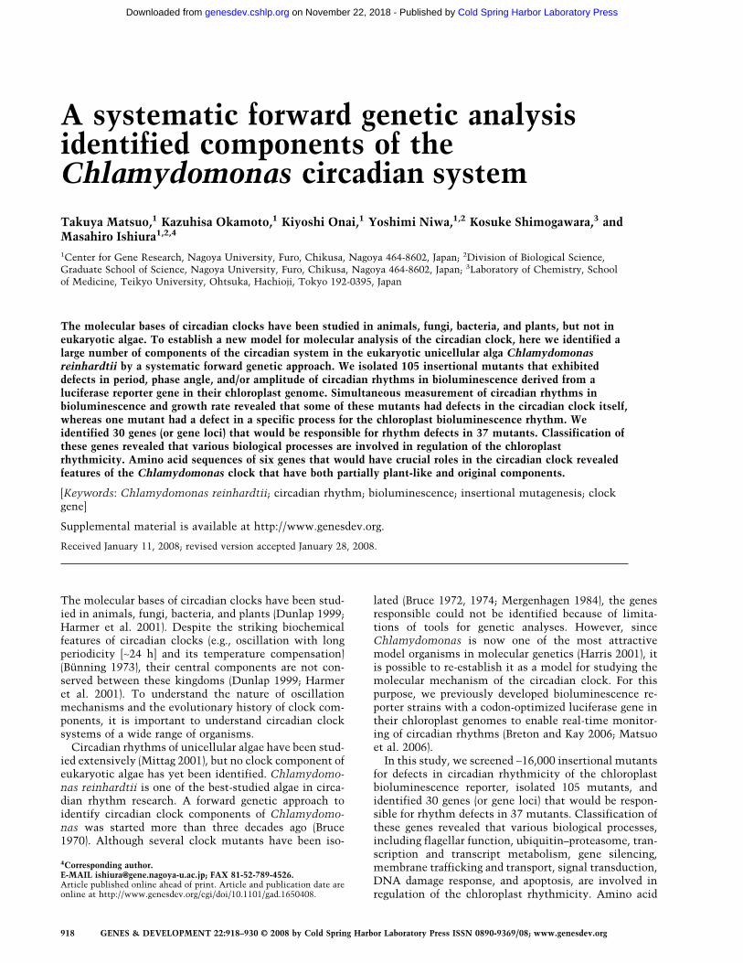
We measured bioluminescence rhythms of a reporterstrain (tufA-lucCP+; the plus symbol indicates plus mat-ing type [mt+]) that expresses the luciferase gene underthe control of the circadian-regulated chloroplast tufApromoter (Hwang et al. 1996) under high-throughput as-say conditions developed in this study (on agar media inconstant darkness [DD]) (see Materials and Methods).However, we failed to detect significant circadianrhythms in about half of the preparations because therhythms were not robust under the conditions (Fig. 1A).To overcome this problem, we screened for a geneticbackground suitable for the assay conditions. The tufA-lucCP+ strain was crossed with the minus mating typecells of genetically different wild-type strains (137c−,6145c−, and 11-32a−) (Harris 2001), and the progenieswere subjected to the high-throughput bioluminescence
assay. Surprisingly, the progenies exhibited various bio-luminescence rhythm patterns (Fig. 1B–D). Rhythm am-plitudes were quite variable, especially in the offspringfrom the cross with 11-32a− (Fig. 1B–D). Phase angles ofthe progenies from crosses with 137c− and 11-32a− weredivided into two groups (Fig. 1B,D). On the other hand,period lengths were relatively maintained in the circa-dian range (Fig. 1B,D). We chose a clone that exhibited arobust rhythm from the cross with 11-32a−. All 96 inde-pendent preparations from the clone exhibited stable androbust rhythms (Fig. 1E). We named the clone CBR34+
and used it as a wild-type strain in this study. To gener-ate a mating type minus equivalent of CBR34+, the 11-32a− strain was crossed four times with CBR34+ to ob-tain a CBR− strain. All progenies from a cross betweenCBR34+ and CBR− exhibited robust rhythms (Fig. 1F),whereas the progenies from a cross between the originaltufA-lucCP+ and CBR− exhibited various biolumines-cence rhythm patterns (Fig. 1G). These results indicatethat the genetic background of the reporter strainstrongly affects the circadian rhythmicity of chloroplastbioluminescence, and that the CBR34 genetic back-ground is suitable for high-throughput screening for cir-cadian rhythm mutants and for their genetic backcrossanalysis. Moreover, these results imply that multiplegenes are involved in maintaining the robustness of chlo-roplast bioluminescence rhythms.
Figure 1. Screening of a genetic background suitable for the high-throughput assay of bioluminescence rhythms. All samples wereprepared according to the high-throughput assay protocol (see Materials and Methods). Traces of bioluminescence from the 96 sampleson agar media are shown on the left, and numbers of samples (per total samples examined) that exhibited a significant circadianrhythmicity (error value [calculated by the RAP] � 0.1) are indicated on the right of the traces. Histograms of period length, peak phaseangle, bottom phase angle, and amplitude of the rhythmic samples are shown. Strains or pairs of the crosses are shown on the right.
The circadian system of Chlamydomonas
GENES & DEVELOPMENT 919
Cold Spring Harbor Laboratory Press on November 22, 2018 - Published by genesdev.cshlp.orgDownloaded from

Screening and genetic analysis of circadian rhythmmutants
To generate circadian rhythm insertional mutants, theCBR34+ strain was transformed with a DNA fragmentcontaining a hygromycin-resistant gene (Berthold et al.2002). We screened ∼16,000 transformants for defects inperiod length, phase angle, and amplitude of biolumines-cence rhythm, and isolated 115 candidate rhythm mu-tants. Then, 91% of the candidates (105 mutants) thatreproduced their mutant phenotypes were analyzed fur-ther as rhythm of chloroplast (roc) mutants (Supplemen-tal Fig. 1; Supplemental Table 1). The roc mutants weredivided into five types (Fig. 2A): short period (SP), longperiod (LP), advanced phase angle (APA), delayed phaseangle (DPA), and low amplitude (LA). The LA mutantscould be further divided into four types (Fig. 2A): exhib-iting arrhythmic or very weak oscillations for a fewcycles (LA I), sustained low-amplitude rhythm with arelatively low bioluminescence level (LA IIa), sustainedlow-amplitude rhythm with a relatively high biolumi-nescence level (LA IIb), and damped rapidly (LA III). In-terestingly, 78% of isolated mutants were LA (Fig. 2B).This result supports the idea that multiple genes are in-volved in maintaining the robustness of chloroplast bio-luminescence rhythms. Next, all of the mutants werebackcrossed to the CBR− strain, and progenies were sub-jected to bioluminescence rhythm measurements andhygromycin resistance testing (cosegregation analysis)(Supplemental Fig. 2; Supplemental Table 1). If the mu-tation was caused by a single integration of the markergene into the genome, the mutant phenotype shouldhave strictly cosegregated with the hygromycin resis-tance. roc63 is representative of a “strict cosegregation”mutant in which almost all hygromycin-resistant prog-eny exhibited a mutant phenotype (Fig. 2C). If the inte-gration occurred at multiple loci, only a proportion ofhygromycin-resistant progeny (∼50% if integration oc-curred at two loci) should have exhibited the mutantphenotype. Thus, “partial cosegregation” was observed,as seen in roc4 (Fig. 2C). roc84 is representative of a“biased segregation” mutant in which all progenies werehygromycin-sensitive or -resistant and all exhibitedwild-type or mutant phenotypes, respectively (Fig. 2C). Ifthe mutation was independent from the integration ofthe marker gene, cosegregation should not have been ob-served. roc71 is representative of “no cosegregation”(Fig. 2C). Strict cosegregation was observed in 52 mu-tants (49%), whereas no cosegregation was observed inonly eight mutants (8%) (Fig. 2D). The mutant pheno-types of most of the strict-cosegregation mutants weresegregated into mt+ and mt− progenies (SupplementalTable 1), excluding the possibility that the strict-coseg-regation pattern is an artifact of the random progenyanalysis caused by surviving parental strains. We couldnot obtain progenies from crosses in 27 mutants (25%)(Fig. 2D). Most of these mutants exhibited a palmelloidphenotype or precipitated at the bottom in liquid cul-tures (Supplemental Table 1), suggesting an abnormalityin hatching processes or flagellar functions. Although we
used 300 ng and 30 ng of marker DNA to generate inser-tional mutants, there was no apparent difference in therate of appearance of strict-cosegregation mutants (300ng, 47.9% [23 of 48]; 30 ng, 50.9% [29 of 57]). Strict co-segregation does not necessarily mean the single integra-tion of a marker gene, because the cosegregation analysiswould not reveal additional integrations of the markergene into silent loci. To determine the number of inte-gration sites, we performed a Southern blot analysis ofgenomic DNA digested with NheI or PstI, which do not
Figure 2. Screening and genetic analysis of circadian rhythmmutants. (A) Representative bioluminescence traces of severaltypes of roc mutants. (SP) roc55; (LP) roc66; (APA) roc83; (DPA)roc25; (LA I) roc77; (LA IIa) roc39; (LA IIb) roc109; (LA III) roc76.The maximum values are expressed as 100. (B) Pie chart show-ing the distribution of all mutants by phenotype. (C) Represen-tative results of the cosegregation analysis. Histograms of hy-gromycin-resistant (red) and hygromycin-sensitive (blue) prog-enies are shown. (D) Pie chart showing the distribution of allmutants by segregation pattern. (E) Southern blot analysis. The3� region of the aph7� coding sequence was used as a probe. Thepositions of size markers (2 and 12 kb) are also shown on the left.
Matsuo et al.
920 GENES & DEVELOPMENT
Cold Spring Harbor Laboratory Press on November 22, 2018 - Published by genesdev.cshlp.orgDownloaded from

cut the marker gene itself. Except for the PstI-digestedroc20 genome, single bands were detected in 50 strictcosegregation mutants (Fig. 2E). These results suggestthat the responsible genes of the 50 mutants were taggedby a single integration of the marker gene.
Circadian rhythms in the growth rateof isolated mutants
To characterize isolated mutants further, we measuredcircadian rhythms in cell growth in liquid cultures undercontinuous light (LL) in several mutants. If these mu-tants had a defect in the circadian clock itself, the circa-dian rhythms in growth would also be affected. Thewild-type strain exhibited a robust growth rhythm (Fig.3A). The mutants roc55, roc75, and roc114 showed de-fects in growth rhythm similar to those in chloroplastbioluminescence rhythm on agar in DD (i.e., short periodfor roc55 and low amplitudes for roc75 and roc114) (Fig.3A; Supplemental Fig. 1). In contrast, growth rhythms ofthe other mutants exhibited different phenotypes tosome extent from their bioluminescence rhythms. Theshort period mutant roc15 and slightly short period mu-tant roc40 no longer exhibited robust growth rhythmic-ity (Fig. 3A; Supplemental Fig. 1). Interestingly, the in-terval between the first and second peaks of growthrhythm for roc40 was extremely long, although roc40was a short period mutant in terms of bioluminescencerhythm (Fig. 3A; Supplemental Fig. 1). roc66 exhibited aslightly long period (∼26.8 h) in growth rhythm, although
the period of its bioluminescence rhythm was very long(29.9 h) (Fig. 3A; Supplemental Fig. 1). The mutantsroc23 and roc81 exhibited robust growth rhythms de-spite their very low-amplitude rhythms in chloroplastbioluminescence (Fig. 3A; Supplemental Fig. 1). Toclarify whether these unexpected phenotypes are specificto growth rhythm, we measured chloroplast biolumines-cence and growth rhythms simultaneously in identicalliquid cultures. The wild-type strain exhibited robustrhythms in both growth and bioluminescence (Fig. 3B).As expected, roc55 and roc75 exhibited short periodand low-amplitude phenotypes, respectively, in bothrhythms (Fig. 3B). Phenotypes of the bioluminescencerhythms of roc40 and roc66 were similar to those of theirgrowth rhythms (Fig. 3B). This indicates that their un-expected phenotypes are not specific to the growthrhythm, but are thought to be due to a change in theoscillation of the circadian clock in these mutants de-pending on the culture conditions. The interval betweenthe first and second peaks of roc40 rhythms was ex-
Figure 3. Growth rhythms of isolated mutants. (A) Growthrhythms of mutants. Growth rhythms were monitored by a con-tinuous culture system (see Materials and Methods) in LL. Two-hour moving averages of growth rates (added fresh medium perculture volume per day) are shown. (B) Simultaneous measure-ment of growth and bioluminescence rhythms. Growth and bio-luminescence rhythms were monitored simultaneously from anidentical liquid culture by the continuous culture system.
Figure 4. Schematic representation of the integration sites ofroc15/roc74 (A), roc40 (B), roc11/roc55 (C), roc66 (D), roc75 (E),and roc108/roc114 (F). Boxes indicate exons. Black boxes indi-cate gene models of JGI Chlamydomonas version 3.0. Predicted(first) initiation codons and stop codons are shown. Red arrowsindicate integration sites of the marker in each mutant. Thephenotype of the mutants is indicated. Gray bars indicate thepredicted genome structures of the roc55 and roc114 mutants.Blue bars indicate the regions used for probes in the Northernblot analysis. The asterisk in B indicates an exon/intron bound-ary that does not follow the GT–AG rule. Scaffold numbers anddirections (arrows) are indicated on the left.
The circadian system of Chlamydomonas
GENES & DEVELOPMENT 921
Cold Spring Harbor Laboratory Press on November 22, 2018 - Published by genesdev.cshlp.orgDownloaded from
tremely long again (Fig. 3B). To determine the periodlength precisely, we prepared roc40 samples according tothe high-throughput assay protocol and measured biolu-minescence rhythm in LL. The periodicity of the rhythmwas barely detectable under the conditions, and we con-firmed that the period length was extremely long in LL(Supplemental Fig. 3). Interestingly, roc81 exhibited dif-ferent phenotypes of rhythm between growth and biolu-minescence. The bioluminescence rhythm was very lowamplitude, although the growth rhythm appeared to benormal (Fig. 3B). This indicates that the low-amplitudephenotype of roc81 is specific to chloroplast biolumines-cence rhythm, and suggests the possibility that roc81 hasa defect in a specific biological process to regulate circa-dian rhythmicity of chloroplast gene expression. Takentogether, these results suggest that our screening iso-lated not only circadian clock mutants, but also mutantsof specific processes for circadian rhythmicity of chloro-plast bioluminescence.
Systematic identification of the responsible genes
To identify integration sites of the marker gene, we de-termined flanking genomic sequences by the thermal
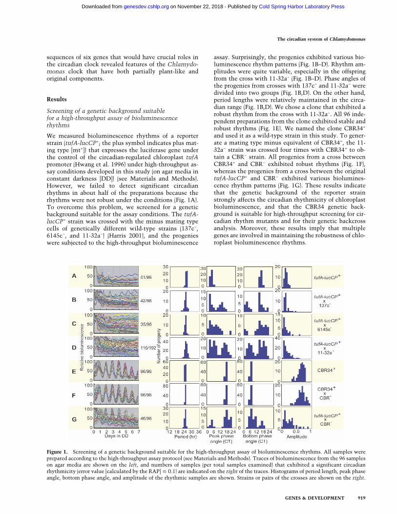
asymmetric interlaced (TAIL)–PCR method (Liu et al.1995). We analyzed 50 mutants tagged by a single inte-gration of the marker gene (Fig. 2E). We succeeded inidentifying flanking genomic sequences on both sides ofthe marker gene in 18 mutants and on one side in 21mutants. The opposite flanking sequences in the 21 mu-tants were analyzed by PCR using specific primer pairsdesigned for each mutant and were identified in 14 mu-tants. Overall, we identified both flanking sequences in32 mutants and a single flanking sequence in seven mu-tants. The rate of success in identification of both flank-ing sequences was apparently high in mutants generatedwith 30 ng of transforming DNA (300 ng, 42.9% [nine of21]; 30 ng, 79.3% [23 of 29]). A major problem in TAIL–PCR of the mutants generated with 300 ng of transform-ing DNA was the integration of concatameric markerDNA that led to amplification only of the marker se-quence.
Flanking sequences were compared with the Chlamy-domonas genome sequence (JGI Chlamydomonas rein-hardtii version 3.0: http://genome.jgi-psf.org/Chlre3/Chlre3.home.html; Merchant et al. 2007). All of theflanking sequences were found in the database. In mostmutants (27 of 32), the marker genes were integrated
Figure 5. Complementation of the mutant phenotypes. Transformants with the genomic DNA fragments were subjected to thehigh-throughput bioluminescence assay. (A) Representative traces of complemented (red) and control (blue) strains on agar. For roc55and roc114, traces of complemented strains with roc55HS and roc114KK genomic fragments, respectively, are shown. (B–G) Histo-grams of the complementation analysis of roc15 (B), roc40 (C), roc55 (D), roc66 (E), roc75 (F), and roc114 (G). For period length mutants(roc15, roc55, and roc66), transformants with period lengths in the range of 24.0–26.0 h were judged to be complemented. For roc40,transformants in the range of 24.0- to 27.0-h periods in LL were judged to be complemented. For low-amplitude mutants (roc75 androc114), transformants with amplitudes (calculated by the RAP) >0.4 and in the range of 0.2–0.4 were judged to be complemented andpartially complemented, respectively. Numbers of complemented (roc15, roc40, roc55, roc66, and roc75) and partially complemented(roc114) transformants among the transformants examined are shown in the graphs.
Matsuo et al.
922 GENES & DEVELOPMENT
Cold Spring Harbor Laboratory Press on November 22, 2018 - Published by genesdev.cshlp.orgDownloaded from
without any large changes in their original genome se-quences (Supplemental Table 2). On the other hand, theintegration site of roc97 was accompanied by a 65.6-kbdeletion of genomic DNA, and each flanking sequence ofroc9, roc55, roc88, and roc114 was found in differentscaffolds of the genome sequence (Supplemental Table2). These results suggest that a genomic deletion or re-arrangement occurred in these mutants accompanied byintegration of the marker gene. Two allelic mutants(roc11 and roc55, roc15 and roc74, roc23 and roc112,roc28 and roc59, roc30 and roc54, roc77 and roc105, androc108 and roc114) were found at seven gene loci(Supplemental Tables 2, 3). This result strongly suggeststhat the disruptions of these gene loci are responsible fortheir mutant phenotypes.
The candidate genes are listed in Supplemental Tables2 and 3. Based on the annotation of the genome sequence(JGI Chlamydomonas reinhardtii version 3.0), sequencesimilarity to known proteins, or conserved functional do-mains, we classified the candidate genes into several bio-logical processes: flagellar function (FAP256, ODA4, PF9,DLC7a, FAP131, and FLA14), ubiquitin–proteasome (SKP1and ROC114), transcription and transcript metabolism(ROC15, ROC40, ROC56, ROC59, ROC66, ROC75,ROC76, ROC93, and XRN1), gene silencing (MUT-9),membrane trafficking and transport (VPS11, ROC81,DRP2, and MFT10), signal transduction (MAPKKKK), DNAdamage response (ATR1), and apoptosis (ALIX). These re-sults suggest that various cellular processes are involved inmaintenance of the chloroplast circadian rhythmicity.
Structures of ROC genes that would have crucial rolesin the circadian clockwork
We further focused on six mutants (roc15, roc40, roc55,roc66, roc75, and roc114). Since they exhibited severedefects in circadian rhythmicity, the responsible genes ofthese mutants would be clock genes closely related tothe central oscillatory mechanism of the Chlamydomo-nas circadian clock. It should be noted, however, that thesevere phenotypes of roc40 and roc66 are conditional(Fig. 3). cDNA sequences of the candidate genes wereidentified by RT–PCR analyses. Comparison of thecDNA sequences to the genome sequence revealed thatall of these genes contained introns following the GT–AG rule, except for the splice donor site of an intron ofthe ROC40 gene (Fig. 4B). Alternative splicing was ob-served upstream of the predicted initiation codon of theROC75 gene (Fig. 4E). Since an (UG)�7 repeat in the 3�-untranslated region that is known to be bound by anRNA-binding protein, CHLAMY1, was found in theROC40 gene (Fig. 4B; Mittag 1996), it is possible thatCHLAMY1 participates in circadian clockwork, as sug-gested previously (Iliev et al. 2006), via the UG repeat ofROC40. Except for roc15 and roc74, it was evident thatthe integrations occurred within transcription units (Fig.4B–F). The integration sites of roc15 and roc74 were up-stream from the cDNA sequence identified by RT–PCR(Fig. 4A), and from the most 5�-terminal of availableESTs. Interestingly, roc15 and roc74 exhibited opposite
phenotypes, a short (21.3-h) and long (26.6-h) period, re-spectively, although their integration sites were veryclose (Fig. 4A; Supplemental Table 2). A comparativeanalysis of C. reinhardtii and Volvox carteri genome se-quences by the Berkeley Genome Pipeline (VISTA trackof JGI Chlamydomonas version 3.0) revealed that geno-mic sequences of the six gene loci are conserved in thesetwo species, suggesting the existence of homologousgenes in V. carteri. The gene model for the ROC114 lo-cus has an N-terminal extension compared with our RT–PCR result (Protein ID: 186976, initiation codon locates14.2 kb upstream); however, we could not amplify theN-terminal extension by an additional RT–PCR.
Complementation of the mutant phenotypesby ROC genes
We tested whether these six candidate genes comple-ment their mutant phenotypes or not. The mutants weretransformed with genomic DNA fragments (Supplemen-tal Fig. 4) linked to a selectable marker gene, aphVIII(Sizova et al. 2001). All mutants were complemented bythe genomic DNA fragments, including their candidategene, without any other predicted genes (Fig. 5). On theother hand, genomic DNA fragments corresponding tothe opposite flanking regions of the integrated marker ofroc55 and roc114 (roc55sc7 and roc114sc89, respectively;Supplemental Fig. 4C,F) did not complement their phe-notypes (Fig. 5D,G). These results indicate that theROC15, ROC40, ROC55, ROC66, ROC75, and ROC114genes are responsible for the circadian rhythm defects ofthese mutants. Restored rhythmicities of complementedstrains of roc114 were, however, at a somewhat low am-plitude compared with the wild-type strain, whereas rhyth-micities of other mutants were fully restored (Fig. 5A,G).
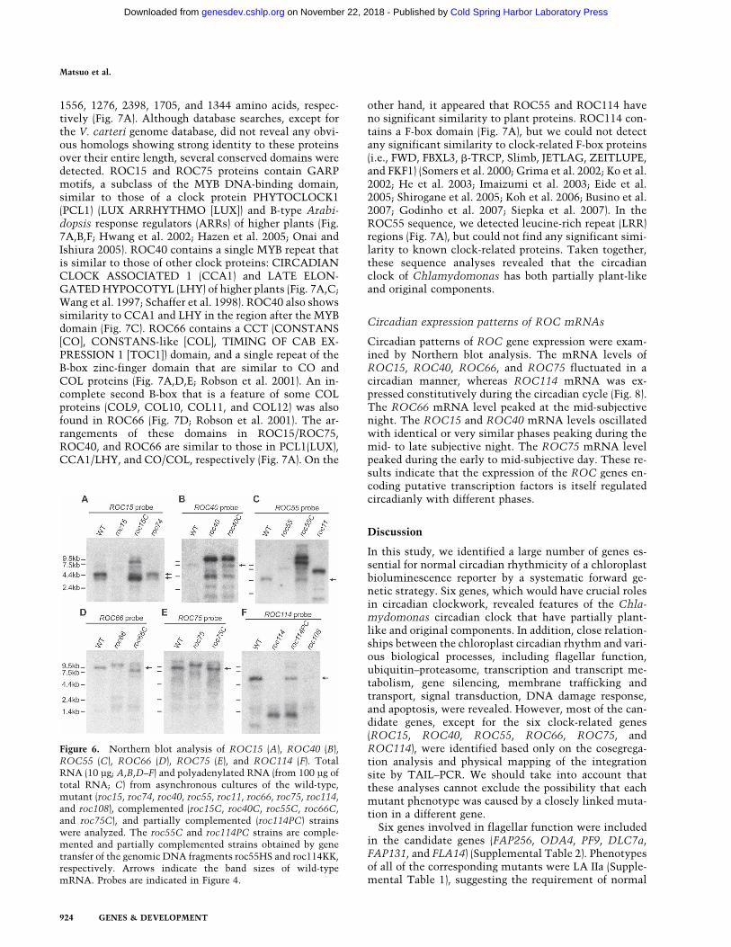
Confirmation of the disruption of ROC genesby Northern blot analysis
Northern blot analyses of the RNA from these mutantsconfirmed disruption of these genes. No transcript ortranscripts with abnormal sizes were observed in themutants, whereas transcripts of normal sizes were de-tected in complemented strains (Fig. 6). One exceptionwas a complemented strain of roc66 (roc66C) in which aslightly short transcript was detected (Fig. 6D). Addi-tional transcripts were detected in the roc15C androc55C strains (Fig. 6A,C). These transcripts with unex-pected sizes might be due to truncation of the transform-ing DNA fragments. The ROC114 transcript of the par-tially complemented strain (roc114PC) was comparablein size to that of the wild-type strain (Fig. 6F). This resultstrongly suggests that the roc114KK genomic DNA frag-ment contains an entire transcriptional unit of theROC114 gene (Supplemental Fig. 4F). In addition, thefailure of complete restoration in the roc114 mutant im-plies that roc114 is not a loss-of-function mutant.
Predicted amino acid sequences of ROC proteins
The predicted proteins encoded by ROC15, ROC40,ROC55, ROC66, ROC75, and ROC114 consist of 631,
The circadian system of Chlamydomonas
GENES & DEVELOPMENT 923
Cold Spring Harbor Laboratory Press on November 22, 2018 - Published by genesdev.cshlp.orgDownloaded from
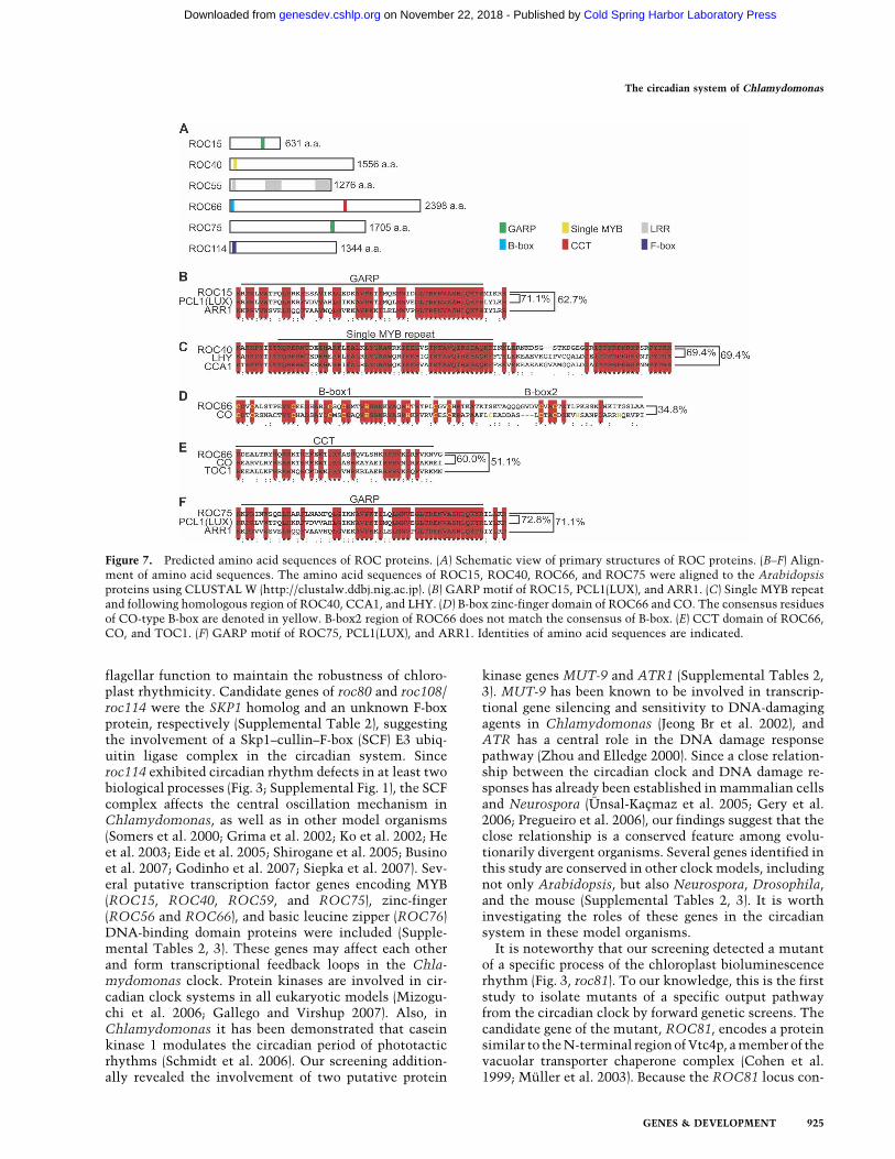
1556, 1276, 2398, 1705, and 1344 amino acids, respec-tively (Fig. 7A). Although database searches, except forthe V. carteri genome database, did not reveal any obvi-ous homologs showing strong identity to these proteinsover their entire length, several conserved domains weredetected. ROC15 and ROC75 proteins contain GARPmotifs, a subclass of the MYB DNA-binding domain,similar to those of a clock protein PHYTOCLOCK1(PCL1) (LUX ARRHYTHMO [LUX]) and B-type Arabi-dopsis response regulators (ARRs) of higher plants (Fig.7A,B,F; Hwang et al. 2002; Hazen et al. 2005; Onai andIshiura 2005). ROC40 contains a single MYB repeat thatis similar to those of other clock proteins: CIRCADIANCLOCK ASSOCIATED 1 (CCA1) and LATE ELON-GATED HYPOCOTYL (LHY) of higher plants (Fig. 7A,C;Wang et al. 1997; Schaffer et al. 1998). ROC40 also showssimilarity to CCA1 and LHY in the region after the MYBdomain (Fig. 7C). ROC66 contains a CCT (CONSTANS[CO], CONSTANS-like [COL], TIMING OF CAB EX-PRESSION 1 [TOC1]) domain, and a single repeat of theB-box zinc-finger domain that are similar to CO andCOL proteins (Fig. 7A,D,E; Robson et al. 2001). An in-complete second B-box that is a feature of some COLproteins (COL9, COL10, COL11, and COL12) was alsofound in ROC66 (Fig. 7D; Robson et al. 2001). The ar-rangements of these domains in ROC15/ROC75,ROC40, and ROC66 are similar to those in PCL1(LUX),CCA1/LHY, and CO/COL, respectively (Fig. 7A). On the
other hand, it appeared that ROC55 and ROC114 haveno significant similarity to plant proteins. ROC114 con-tains a F-box domain (Fig. 7A), but we could not detectany significant similarity to clock-related F-box proteins(i.e., FWD, FBXL3, �-TRCP, Slimb, JETLAG, ZEITLUPE,and FKF1) (Somers et al. 2000; Grima et al. 2002; Ko et al.2002; He et al. 2003; Imaizumi et al. 2003; Eide et al.2005; Shirogane et al. 2005; Koh et al. 2006; Busino et al.2007; Godinho et al. 2007; Siepka et al. 2007). In theROC55 sequence, we detected leucine-rich repeat (LRR)regions (Fig. 7A), but could not find any significant simi-larity to known clock-related proteins. Taken together,these sequence analyses revealed that the circadianclock of Chlamydomonas has both partially plant-likeand original components.
Circadian expression patterns of ROC mRNAs
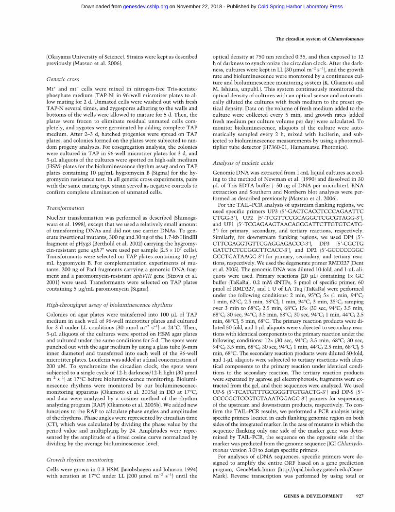
Circadian patterns of ROC gene expression were exam-ined by Northern blot analysis. The mRNA levels ofROC15, ROC40, ROC66, and ROC75 fluctuated in acircadian manner, whereas ROC114 mRNA was ex-pressed constitutively during the circadian cycle (Fig. 8).The ROC66 mRNA level peaked at the mid-subjectivenight. The ROC15 and ROC40 mRNA levels oscillatedwith identical or very similar phases peaking during themid- to late subjective night. The ROC75 mRNA levelpeaked during the early to mid-subjective day. These re-sults indicate that the expression of the ROC genes en-coding putative transcription factors is itself regulatedcircadianly with different phases.
Discussion
In this study, we identified a large number of genes es-sential for normal circadian rhythmicity of a chloroplastbioluminescence reporter by a systematic forward ge-netic strategy. Six genes, which would have crucial rolesin circadian clockwork, revealed features of the Chla-mydomonas circadian clock that have partially plant-like and original components. In addition, close relation-ships between the chloroplast circadian rhythm and vari-ous biological processes, including flagellar function,ubiquitin–proteasome, transcription and transcript me-tabolism, gene silencing, membrane trafficking andtransport, signal transduction, DNA damage response,and apoptosis, were revealed. However, most of the can-didate genes, except for the six clock-related genes(ROC15, ROC40, ROC55, ROC66, ROC75, andROC114), were identified based only on the cosegrega-tion analysis and physical mapping of the integrationsite by TAIL–PCR. We should take into account thatthese analyses cannot exclude the possibility that eachmutant phenotype was caused by a closely linked muta-tion in a different gene.
Six genes involved in flagellar function were includedin the candidate genes (FAP256, ODA4, PF9, DLC7a,FAP131, and FLA14) (Supplemental Table 2). Phenotypesof all of the corresponding mutants were LA IIa (Supple-mental Table 1), suggesting the requirement of normal
Figure 6. Northern blot analysis of ROC15 (A), ROC40 (B),ROC55 (C), ROC66 (D), ROC75 (E), and ROC114 (F). TotalRNA (10 µg; A,B,D–F) and polyadenylated RNA (from 100 µg oftotal RNA; C) from asynchronous cultures of the wild-type,mutant (roc15, roc74, roc40, roc55, roc11, roc66, roc75, roc114,and roc108), complemented (roc15C, roc40C, roc55C, roc66C,and roc75C), and partially complemented (roc114PC) strainswere analyzed. The roc55C and roc114PC strains are comple-mented and partially complemented strains obtained by genetransfer of the genomic DNA fragments roc55HS and roc114KK,respectively. Arrows indicate the band sizes of wild-typemRNA. Probes are indicated in Figure 4.
Matsuo et al.
924 GENES & DEVELOPMENT
Cold Spring Harbor Laboratory Press on November 22, 2018 - Published by genesdev.cshlp.orgDownloaded from
flagellar function to maintain the robustness of chloro-plast rhythmicity. Candidate genes of roc80 and roc108/roc114 were the SKP1 homolog and an unknown F-boxprotein, respectively (Supplemental Table 2), suggestingthe involvement of a Skp1–cullin–F-box (SCF) E3 ubiq-uitin ligase complex in the circadian system. Sinceroc114 exhibited circadian rhythm defects in at least twobiological processes (Fig. 3; Supplemental Fig. 1), the SCFcomplex affects the central oscillation mechanism inChlamydomonas, as well as in other model organisms(Somers et al. 2000; Grima et al. 2002; Ko et al. 2002; Heet al. 2003; Eide et al. 2005; Shirogane et al. 2005; Businoet al. 2007; Godinho et al. 2007; Siepka et al. 2007). Sev-eral putative transcription factor genes encoding MYB(ROC15, ROC40, ROC59, and ROC75), zinc-finger(ROC56 and ROC66), and basic leucine zipper (ROC76)DNA-binding domain proteins were included (Supple-mental Tables 2, 3). These genes may affect each otherand form transcriptional feedback loops in the Chla-mydomonas clock. Protein kinases are involved in cir-cadian clock systems in all eukaryotic models (Mizogu-chi et al. 2006; Gallego and Virshup 2007). Also, inChlamydomonas it has been demonstrated that caseinkinase 1 modulates the circadian period of phototacticrhythms (Schmidt et al. 2006). Our screening addition-ally revealed the involvement of two putative protein
kinase genes MUT-9 and ATR1 (Supplemental Tables 2,3). MUT-9 has been known to be involved in transcrip-tional gene silencing and sensitivity to DNA-damagingagents in Chlamydomonas (Jeong Br et al. 2002), andATR has a central role in the DNA damage responsepathway (Zhou and Elledge 2000). Since a close relation-ship between the circadian clock and DNA damage re-sponses has already been established in mammalian cellsand Neurospora (Ünsal-Kaçmaz et al. 2005; Gery et al.2006; Pregueiro et al. 2006), our findings suggest that theclose relationship is a conserved feature among evolu-tionarily divergent organisms. Several genes identified inthis study are conserved in other clock models, includingnot only Arabidopsis, but also Neurospora, Drosophila,and the mouse (Supplemental Tables 2, 3). It is worthinvestigating the roles of these genes in the circadiansystem in these model organisms.
It is noteworthy that our screening detected a mutantof a specific process of the chloroplast bioluminescencerhythm (Fig. 3, roc81). To our knowledge, this is the firststudy to isolate mutants of a specific output pathwayfrom the circadian clock by forward genetic screens. Thecandidate gene of the mutant, ROC81, encodes a proteinsimilar to the N-terminal region of Vtc4p, a member of thevacuolar transporter chaperone complex (Cohen et al.1999; Müller et al. 2003). Because the ROC81 locus con-
Figure 7. Predicted amino acid sequences of ROC proteins. (A) Schematic view of primary structures of ROC proteins. (B–F) Align-ment of amino acid sequences. The amino acid sequences of ROC15, ROC40, ROC66, and ROC75 were aligned to the Arabidopsisproteins using CLUSTAL W (http://clustalw.ddbj.nig.ac.jp). (B) GARP motif of ROC15, PCL1(LUX), and ARR1. (C) Single MYB repeatand following homologous region of ROC40, CCA1, and LHY. (D) B-box zinc-finger domain of ROC66 and CO. The consensus residuesof CO-type B-box are denoted in yellow. B-box2 region of ROC66 does not match the consensus of B-box. (E) CCT domain of ROC66,CO, and TOC1. (F) GARP motif of ROC75, PCL1(LUX), and ARR1. Identities of amino acid sequences are indicated.
The circadian system of Chlamydomonas
GENES & DEVELOPMENT 925
Cold Spring Harbor Laboratory Press on November 22, 2018 - Published by genesdev.cshlp.orgDownloaded from
tains extended regions of undetermined nucleotides, thesequence of the C-terminal transmembrane domaincould not be predicted. Since Vtc4p is known to localizeto the membrane of vacuolar and other cellular compart-ments to control the distribution of membrane proteinsin yeast (Cohen et al. 1999; Müller et al. 2003), our re-sults suggest that regulation of membrane proteins isinvolved in the circadian output pathway for the chloro-plast rhythmicity. An interesting question is whetherVtc4p localizes to the outer membrane of chloroplasts,but to date there are no studies assessing its localizationin plant cells. Analysis of ROC81 localization in Chla-mydomonas will shed light on the molecular mecha-nisms for synchronization of the circadian rhythmicitybetween the nucleus and chloroplast.
ROC15/ROC75 and ROC40 have MYB-related DNA-binding domains similar to those of the Arabidopsisclock proteins PCL1(LUX) and CCA1/LHY, respectively(Fig. 7A–C,F). In Arabidopsis, they are critical to the gen-eration of a normal circadian rhythmicity (Schaffer et al.1998; Wang and Tobin 1998; Hazen et al. 2005; Onai andIshiura 2005). CCA1/LHY homologs in Oryza sativa andLemna gibba also play critical roles in the circadianclocks of these species (Miwa et al. 2006; Murakami etal. 2007). The identification of ROC15/ROC75 andROC40 as components of the circadian clock in Chla-mydomonas suggests that the two types of MYB-relatedDNA-binding domains of ROC15/ROC75/PCL1(LUX)and ROC40/CCA1/LHY are common features of circa-dian clock proteins over a wide range of species in thegreen lineage. Are the roles of the Chlamydomonas MYB
proteins in the circadian clock the same as those of theArabidopsis MYB proteins? In contrast to the Arabidop-sis cca1/lhy mutants (Green and Tobin 1999; Alabadí etal. 2002; Mizoguchi et al. 2002), the severe phenotype ofthe roc40 mutant in LL was a long period (SupplementalFig. 3), and the phenotype was conditional (Supplemen-tal Figs. 1, 3). Furthermore, the roc15 mutant exhibited ashort period phenotype, but its rhythm remained robustin DD (Supplemental Fig. 1), whereas the Arabidopsispcl1(lux) mutant is completely arrhythmic (Hazen et al.2005; Onai and Ishiura 2005). Concerning the circa-dian expression profile, Arabidopsis CCA1/LHY andPCL1(LUX) interact genetically with each other, result-ing in an antiphase circadian expression peaking at sub-jective dawn and subjective dusk, respectively (Schafferet al. 1998; Wang and Tobin 1998; Hazen et al. 2005;Onai and Ishiura 2005). These expression profiles areconserved in the CCA1 and PCL1(LUX) homologs inOryza sativa (Murakami et al. 2007), and in the LHYhomologs in Lemna gibba and Lemna paucicoatata(Miwa et al. 2006). In Chlamydomonas, the expressionpattern of ROC40 was comparable with those of CCA1/LHY (Fig. 8). However, in contrast to PCL1(LUX),ROC15 and ROC75 expression did not peak at subjec-tive dusk. The mRNA level of ROC15 oscillated in thesame phase with that of ROC40, and the ROC75 mRNAlevel oscillated in a slightly delayed phase comparedwith the ROC40 mRNA level and peaked at the early- tomid-subjective day (Fig. 8). Taken together, these resultssuggest the possibility that the interaction and roles ofthe MYB proteins in the circadian clock of Chlamydo-monas differ, at least to some extent, from those of Ara-bidopsis.
In the past decade, the transcriptional/translationalnegative feedback loop model had been believed to be thecommon mechanism for generation of the circadian os-cillation (Dunlap 1999; Harmer et al. 2001). However,recent findings in cyanobacteria (Nakajima et al. 2005;Tomita et al. 2005) indicate that the circadian oscillationis generated by a set of clock proteins without transcrip-tion and translation. One of the most interesting ques-tions is whether the new concept found in a bacterium isapplicable to eukaryotes or not. In the eukaryotic greenalga Acetabularia, the chloroplast photosyntheticrhythm persists even in enucleated cells (Sweeney andHaxo 1961). This fact eliminates the necessity of nucleartranscription for generation of the circadian oscillation,at least in Acetabularia. Although there is no evidencefor a transcription/translation-less circadian oscillator inChlamydomonas, its simple cellular composition willmake Chlamydomonas a good system to assess the newconcept of circadian oscillation in the eukaryotic cell.
Materials and methods
Strains
Wild-type strains 137c− (CC-124) and 6145c− (CC-1691) wereobtained from the Chlamydomonas Center (Duke University,NC), and 11-32a− was kindly provided by Dr. Tatsuaki Saito
Figure 8. Circadian expression patterns of ROC mRNAs. (A)Northern blot analysis of ROC15, ROC40, ROC66, ROC75,and ROC114 mRNAs. Total RNA (5 µg) from a synchronousculture of the wild-type strain in LL (10 µmol m−2 s−1) at 17°Cwas analyzed. The ROC55 mRNA was undetectable under theexperimental conditions. Probes are the same as those used inFigure 6. Equal RNA loading was confirmed by reprobing theblot for rbcL mRNA. (B) Temporal expression profiles of theROC mRNAs. The maximum values are expressed as 100.
Matsuo et al.
926 GENES & DEVELOPMENT
Cold Spring Harbor Laboratory Press on November 22, 2018 - Published by genesdev.cshlp.orgDownloaded from
(Okayama University of Science). Strains were kept as describedpreviously (Matsuo et al. 2006).
Genetic cross
Mt+ and mt− cells were mixed in nitrogen-free Tris-acetate-phosphate medium (TAP-N) in 96-well microtiter plates to al-low mating for 2 d. Unmated cells were washed out with freshTAP-N several times, and zygospores adhering to the walls andbottoms of the wells were allowed to mature for 5 d. Then, theplates were frozen to eliminate residual unmated cells com-pletely, and zygotes were germinated by adding complete TAPmedium. After 2–3 d, hatched progenies were spread on TAPplates, and colonies formed on the plates were subjected to ran-dom progeny analyses. For cosegregation analysis, the colonieswere cultured in TAP in 96-well microtiter plates for 3 d, and5-µL aliquots of the cultures were spotted on high-salt medium(HSM) plates for the bioluminescence rhythm assay and on TAPplates containing 10 µg/mL hygromycin B (Sigma) for the hy-gromycin resistance test. In all genetic cross experiments, pairswith the same mating type strain served as negative controls toconfirm complete elimination of unmated cells.
Transformation
Nuclear transformation was performed as described (Shimoga-wara et al. 1998), except that we used a relatively small amountof transforming DNAs and did not use carrier DNAs. To gen-erate insertional mutants, 300 ng and 30 ng of the 1.7-kb HindIIIfragment of pHyg3 (Berthold et al. 2002) carrying the hygromy-cin-resistant gene aph7� were used per sample (2.5 × 10⁷ cells).Transformants were selected on TAP plates containing 10 µg/mL hygromycin B. For complementation experiments of mu-tants, 200 ng of PacI fragments carrying a genomic DNA frag-ment and a paromomycin-resistant aphVIII gene (Sizova et al.2001) were used. Transformants were selected on TAP platescontaining 5 µg/mL paromomycin (Sigma).
High-throughput assay of bioluminescence rhythms
Colonies on agar plates were transferred into 100 µL of TAPmedium in each well of 96-well microtiter plates and culturedfor 3 d under LL conditions (30 µmol m−2 s−1) at 24°C. Then,5-µL aliquots of the cultures were spotted on HSM agar platesand cultured under the same conditions for 5 d. The spots werepunched out with the agar medium by using a glass tube (6-mminner diameter) and transferred into each well of the 96-wellmicrotiter plates. Luciferin was added at a final concentration of200 µM. To synchronize the circadian clock, the spots weresubjected to a single cycle of 12-h darkness/12-h light (30 µmolm−2 s−1) at 17°C before bioluminescence monitoring. Biolumi-nescence rhythms were monitored by our bioluminescence-monitoring apparatus (Okamoto et al. 2005a) in DD at 17°C,and data were analyzed by a cosiner method of the rhythmanalyzing program (RAP) (Okamoto et al. 2005b). We added newfunctions to the RAP to calculate phase angles and amplitudesof the rhythms. Phase angles were represented by circadian time(CT), which was calculated by dividing the phase value by theperiod value and multiplying by 24. Amplitudes were repre-sented by the amplitude of a fitted cosine curve normalized bydividing by the average bioluminescence level.
Growth rhythm monitoring
Cells were grown in 0.3 HSM (Jacobshagen and Johnson 1994)with aeration at 17°C under LL (200 µmol m−2 s−1) until the
optical density at 750 nm reached 0.35, and then exposed to 12h of darkness to synchronize the circadian clock. After the dark-ness, cultures were kept in LL (30 µmol m−2 s−1), and the growthrate and bioluminescence were monitored by a continuous cul-ture and bioluminescence monitoring system (K. Okamoto andM. Ishiura, unpubl.). This system continuously monitored theoptical density of cultures with an optical sensor and automati-cally diluted the cultures with fresh medium to the preset op-tical density. Data on the volume of fresh medium added to theculture were collected every 5 min, and growth rates (addedfresh medium per culture volume per day) were calculated. Tomonitor bioluminescence, aliquots of the culture were auto-matically sampled every 2 h, mixed with luciferin, and sub-jected to bioluminescence measurements by using a photomul-tiplier tube detector (H7360-01, Hamamatsu Photonics).
Analysis of nucleic acids
Genomic DNA was extracted from 1-mL liquid cultures accord-ing to the method of Newman et al. (1990) and dissolved in 30µL of Tris-EDTA buffer (∼50 ng of DNA per microliter). RNAextraction and Southern and Northern blot analyses were per-formed as described previously (Matsuo et al. 2006).
For the TAIL–PCR analysis of upstream flanking regions, weused specific primers UP3 (5�-GACTCACCTCCCAGAATTCCTGG-3�), UP2 (5�-TCGTTCCGCAGGCTCGCGTAGG-3�),and UP1 (5�-TCGAGAAGTAACAGGGATTCTTGTGTCATG-3�) for primary, secondary, and tertiary reactions, respectively.Similarly, for downstream flanking regions, we used DP4 (5�-CTTCGAGGTGTTCGAGGAGACCC-3�), DP3 (5�-CGCTGGATCTCTCCGGCTTCACC-3�), and DP2 (5�-GCCCCCGGCGCCTGATAAGG-3�) for primary, secondary, and tertiary reac-tions, respectively. We used the degenerate primer RMD227 (Dentet al. 2005). The genomic DNA was diluted 10-fold, and 1-µL ali-quots were used. Primary reactions (20 µL) containing 1× GCbuffer (TaKaRa), 0.2 mM dNTPs, 5 pmol of specific primer, 60pmol of RMD227, and 1 U of LA Taq (TaKaRa) were performedunder the following conditions: 2 min, 95°C; 5× (1 min, 94°C;1 min, 62°C; 2.5 min, 68°C); 1 min, 94°C; 3 min, 25°C; rampingover 3 min to 68°C; 2.5 min, 68°C; 15× (30 sec, 94°C; 3.5 min,68°C; 30 sec, 94°C; 3.5 min, 68°C; 30 sec, 94°C; 1 min, 44°C; 2.5min, 68°C); 5 min, 68°C. The primary reaction products were di-luted 50-fold, and 1-µL aliquots were subjected to secondary reac-tions with identical components to the primary reaction under thefollowing conditions: 12× (30 sec, 94°C; 3.5 min, 68°C; 30 sec,94°C; 3.5 min, 68°C; 30 sec, 94°C; 1 min, 44°C; 2.5 min, 68°C); 5min, 68°C. The secondary reaction products were diluted 50-fold,and 1-µL aliquots were subjected to tertiary reactions with iden-tical components to the primary reaction under identical condi-tions to the secondary reaction. The tertiary reaction productswere separated by agarose gel electrophoresis, fragments were ex-tracted from the gel, and their sequences were analyzed. We usedUP-S (5�-TCATGTTTGCGGGTTGTGACTG-3�) and DP-S (5�-CCCCGCTCCGTGTAAATGGAGG-3�) primers for sequencingof the upstream and downstream products, respectively. To con-firm the TAIL–PCR results, we performed a PCR analysis usingspecific primers located in each flanking genomic region on bothsides of the integrated marker. In the case of mutants in which thesequence flanking only one side of the marker gene was deter-mined by TAIL–PCR, the sequence on the opposite side of themarker was predicted from the genome sequence (JGI Chlamydo-monas version 3.0) to design specific primers.
For analyses of cDNA sequences, specific primers were de-signed to amplify the entire ORF based on a gene predictionprogram, GeneMark.hmm (http://opal.biology.gatech.edu/Gene-Mark). Reverse transcription was performed by using total or
The circadian system of Chlamydomonas
GENES & DEVELOPMENT 927
Cold Spring Harbor Laboratory Press on November 22, 2018 - Published by genesdev.cshlp.orgDownloaded from

polyadenylated RNA of CBR34+, SuperScript II (Invitrogen), andan oligo-dT primer. PCR amplification was performed by usingLA Taq with GC buffer (TaKaRa) and specific primer pairs forROC15 (Forward, 5�-AAACTCATTCCAAGCGTCCCAAACCTTCG-3�; Reverse, 5�-CACAGAAGACGTAGGAGCTCAACGG-3�), ROC40 (Forward, 5�-CCCAGACTCGGCACAGTTACTTGCCAACG-3�; Reverse, 5�-CCCAGCACGCAAGAGCAAACCCCTC-3�), ROC55 (Forward, 5�-CCTTCACCTCGTTGCCCGCCTTCAC-3�; Reverse, 5�-ACACACTTTCACACCGCAGCCCCCC-3�), ROC66 (Forward, 5�-GTGCGCCCTGGCGGTGCTACCAACC-3�; Reverse, 5�-AGGCTTGCCCAAGGCTTGCCGCCAC-3�), ROC75 (Forward, 5�-GTTGTTTGGCGGAGGACACTCGTTGCTTG-3�; Reverse, 5�-TAATTTGGGCGACGATTGCTGAGGCATGG-3�), ROC114 (Forward, 5�-GAGAGTGCGAGGAGCGCCGTTGAGC-3�; Reverse, 5�-GCATACCCTGGAAGTTGCAAGGCAC-3�), and ROC114 N-terminal exten-sion (Forward 1, 5�-GTTTACAGATAGCTCTGTATCGTCTTGAC-3�; Forward 2, 5�-GCCCTGAGACAAAGTGTGCGAAAGGAATC-3�; Reverse 1, 5�-GCATACCCTGGAAGTTGCAAGGCAC-3�; Reverse 2, 5�-GCGCGCCACTCCGCAAACCAGC-3� [all four combinations of the forward and reverse primers]).
Plasmid construction
To generate plasmids for genetic complementation experi-ments, we modified pSI103�NotI (Sizova et al. 2001) as fol-lows: The HindIII site was destroyed by blunting/religa-tion, a PacI linker (5�-TTAATTAAGTAC-3�) was inserted intothe KpnI site, and a polylinker (top strand, 5�-GGCCTTAATTAACTAGTGGTACCGATATCAAGCTTGAATTCGC-3�;bottom strand, 5�-GGCCGCGAATTCAAGCTTGATATCGGTACCACTAGTTAATTAA-3�) was inserted into the NotI sitein the same direction as the aphVIII gene, yielding pSI103PL.BAC clones containing a gene of interest were searched for onthe Genome browser of JGI Chlamydomonas version 3.0 andobtained from the Clemson University Genomics Institute.BAC clones were digested by restriction enzymes, and a geno-mic DNA fragment containing the gene was cloned into thepolylinker region of pSI103PL. The plasmids were digested byPacI, and a DNA fragment containing the genomic DNA withthe aphVIII marker was used for the complementation experi-ments. Names and sizes of the restriction fragments of genomicDNA segments (Supplemental Fig. 4) and clone numbers ofBACs carrying the DNA segments used in this study were asfollows: roc15KK, 7.4-kb KpnI fragment, 38G22; roc40HH, 10.6-kb HindIII fragment, 3O10; roc55HH, 11.9-kb HindIII fragment,16E8; roc55HS, 8.9-kb HindIII–SnaBI fragment, 16E8; roc55HX,4.8-kb HindIII–XhoI fragment, 16E8; roc55sc7, 11.4-kb HindIII–DraI fragment, 25L12; roc66ES, 13.3-kb EcoRI–SpeI fragment,36M13; roc75BB, 11.8-kb BamHI fragment, 26N16; roc114KK,14.3-kb KpnI fragment, 21B13; roc114PP, 9.4-kb PvuI fragment,21B13; roc114sc89, 12.9-kb EcoRI fragment, 17D2.
Accession numbers
cDNA sequences of ROC15, ROC40, ROC55, ROC66, longersplice variant of ROC75, shorter splice variant of ROC75, andROC114 have been deposited in the GenBank/EMBL/DDBJ un-der accession numbers AB363964, AB363965, AB363966,AB363967, AB363968, AB363969, and AB363970, respectively.
Acknowledgments
We thank W. Mages (Regensburg University) for the pHyg3 plas-mid, I. Sizova (St. Petersburg State University) for the
pSI103�NotI plasmid, T. Saito (Okayama University of Science)for the 11-32a− strain, Y. Nakaoka (Nagoya University) for tech-nical assistance, and D. Mrozek (Medical English Service) forprofessional editing. This work was supported by grants fromthe Japanese Ministry of Education, Culture, Sports, Scienceand Technology (MEXT) to M.I., and grants from Research Fel-lowships of the Japan Society for the Promotion of Science forYoung Scientists to T.M. The Division of Biological Science,Graduate School of Science, Nagoya University, was supportedby a 21st century COE grant from MEXT.
References
Alabadí, D., Yanovsky, M.J., Mas, P., Harmer, S.L., and Kay, S.A.2002. Critical role for CCA1 and LHY in maintaining circa-dian rhythmicity in Arabidopsis. Curr. Biol. 12: 757–761.
Berthold, P., Schmitt, R., and Mages, W. 2002. An engineeredStreptomyces hygroscopicus aph 7� gene mediates dominantresistance against hygromycin B in Chlamydomonas rein-hardtii. Protist 153: 401–412.
Breton, G. and Kay, S.A. 2006. Circadian rhythms lit up inChlamydomonas. Genome Biol. 7: 215. doi: 10.1186/gb-2006-7-4-215.
Bruce, V.G. 1970. The biological clock in Chlamydomonasreinhardi. J. Protozool. 17: 328–334.
Bruce, V.G. 1972. Mutants of the biological clock in Chlamydo-monas reinhardi. Genetics 70: 537–548.
Bruce, V.G. 1974. Recombinants between clock mutants ofChlamydomonas reinhardi. Genetics 77: 221–230.
Bünning, E. 1973. The physiologycal clock: Circadian rhythmsand biologycal chronometry, 3rd ed., Springer-Verlag, NewYork.
Busino, L., Bassermann, F., Maiolica, A., Lee, C., Nolan, P.M.,Godinho, S.I., Draetta, G.F., and Pagano, M. 2007. SCFFbxl3
controls the oscillation of the circadian clock by directingthe degradation of cryptochrome proteins. Science 316: 900–904.
Cohen, A., Perzov, N., Nelson, H., and Nelson, N. 1999. A novelfamily of yeast chaperons involved in the distribution ofV-ATPase and other membrane proteins. J. Biol. Chem. 274:26885–26893.
Dent, R.M., Haglund, C.M., Chin, B.L., Kobayashi, M.C., andNiyogi, K.K. 2005. Functional genomics of eukaryotic pho-tosynthesis using insertional mutagenesis of Chlamydomo-nas reinhardtii. Plant Physiol. 137: 545–556.
Dunlap, J.C. 1999. Molecular bases for circadian clocks. Cell 96:271–290.
Eide, E.J., Woolf, M.F., Kang, H., Woolf, P., Hurst, W., Camacho,F., Vielhaber, E.L., Giovanni, A., and Virshup, D.M. 2005.Control of mammalian circadian rhythm by CKI�-regulatedproteasome-mediated PER2 degradation. Mol. Cell. Biol. 25:2795–2807.
Gallego, M. and Virshup, D.M. 2007. Post-translational modifi-cations regulate the ticking of the circadian clock. Nat. Rev.Mol. Cell Biol. 8: 139–148.
Gery, S., Komatsu, N., Baldjyan, L., Yu, A., Koo, D., and Koef-fler, H.P. 2006. The circadian gene per1 plays an importantrole in cell growth and DNA damage control in human can-cer cells. Mol. Cell 22: 375–382.
Godinho, S.I., Maywood, E.S., Shaw, L., Tucci, V., Barnard, A.R.,Busino, L., Pagano, M., Kendall, R., Quwailid, M.M., Ro-mero, M.R., et al. 2007. The after-hours mutant reveals arole for Fbxl3 in determining mammalian circadian period.Science 316: 897–900.
Green, R.M. and Tobin, E.M. 1999. Loss of the circadian clock-
Matsuo et al.
928 GENES & DEVELOPMENT
Cold Spring Harbor Laboratory Press on November 22, 2018 - Published by genesdev.cshlp.orgDownloaded from

associated protein 1 in Arabidopsis results in altered clock-regulated gene expression. Proc. Natl. Acad. Sci. 96: 4176–4179.
Grima, B., Lamouroux, A., Chelot, E., Papin, C., Limbourg-Bouchon, B., and Rouyer, F. 2002. The F-box protein slimbcontrols the levels of clock proteins period and timeless.Nature 420: 178–182.
Harmer, S.L., Panda, S., and Kay, S.A. 2001. Molecular bases ofcircadian rhythms. Annu. Rev. Cell Dev. Biol. 17: 215–253.
Harris, E.H. 2001. Chlamydomonas as a model organism. Annu.Rev. Plant Physiol. Plant Mol. Biol. 52: 363–406.
Hazen, S.P., Schultz, T.F., Pruneda-Paz, J.L., Borevitz, J.O.,Ecker, J.R., and Kay, S.A. 2005. LUX ARRHYTHMO encodesa Myb domain protein essential for circadian rhythms. Proc.Natl. Acad. Sci. 102: 10387–10392.
He, Q., Cheng, P., Yang, Y., Yu, H., and Liu, Y. 2003. FWD1-mediated degradation of FREQUENCY in Neurospora estab-lishes a conserved mechanism for circadian clock regulation.EMBO J. 22: 4421–4430.
Hwang, S., Kawazoe, R., and Herrin, D.L. 1996. Transcription oftufA and other chloroplast-encoded genes is controlled by acircadian clock in Chlamydomonas. Proc. Natl. Acad. Sci.93: 996–1000.
Hwang, I., Chen, H.C., and Sheen, J. 2002. Two-component sig-nal transduction pathways in Arabidopsis. Plant Physiol.129: 500–515.
Iliev, D., Voytsekh, O., Schmidt, E.M., Fiedler, M., Nykytenko,A., and Mittag, M. 2006. A heteromeric RNA-binding pro-tein is involved in maintaining acrophase and period of thecircadian clock. Plant Physiol. 142: 797–806.
Imaizumi, T., Tran, H.G., Swartz, T.E., Briggs, W.R., and Kay,S.A. 2003. FKF1 is essential for photoperiodic-specific lightsignalling in Arabidopsis. Nature 426: 302–306.
Jacobshagen, S. and Johnson, C.H. 1994. Circadian rhythms ofgene expression in Chlamydomonas reinhardtii: Circadiancycling of mRNA abundances of cab II, and possibly of �-tubulin and cytochrome c. Eur. J. Cell Biol. 64: 142–152.
Jeong Br, B.R., Wu-Scharf, D., Zhang, C., and Cerutti, H. 2002.Suppressors of transcriptional transgenic silencing inChlamydomonas are sensitive to DNA-damaging agents andreactivate transposable elements. Proc. Natl. Acad. Sci. 99:1076–1081.
Ko, H.W., Jiang, J., and Edery, I. 2002. Role for Slimb in thedegradation of Drosophila Period protein phosphorylated byDoubletime. Nature 420: 673–678.
Koh, K., Zheng, X., and Sehgal, A. 2006. JETLAG resets theDrosophila circadian clock by promoting light-induced deg-radation of TIMELESS. Science 312: 1809–1812.
Liu, Y.G., Mitsukawa, N., Oosumi, T., and Whittier, R.F. 1995.Efficient isolation and mapping of Arabidopsis thaliana T-DNA insert junctions by thermal asymmetric interlacedPCR. Plant J. 8: 457–463.
Matsuo, T., Onai, K., Okamoto, K., Minagawa, J., and Ishiura,M. 2006. Real-time monitoring of chloroplast gene expres-sion by a luciferase reporter: Evidence for nuclear regulationof chloroplast circadian period. Mol. Cell. Biol. 26: 863–870.
Merchant, S.S., Prochnik, S.E., Vallon, O., Harris, E.H., Karpow-icz, S.J., Witman, G.B., Terry, A., Salamov, A., Fritz-Laylin,L.K., Marechal-Drouard, L., et al. 2007. The Chlamydomo-nas genome reveals the evolution of key animal and plantfunctions. Science 318: 245–250.
Mergenhagen, D. 1984. Circadian clock: Genetic characteriza-tion of a short period mutant of Chlamydomonas reinhardii.Eur. J. Cell Biol. 33: 13–18.
Mittag, M. 1996. Conserved circadian elements in phylogeneti-cally diverse algae. Proc. Natl. Acad. Sci. 93: 14401–14404.
Mittag, M. 2001. Circadian rhythms in microalgae. Int. Rev.Cytol. 206: 213–247.
Miwa, K., Serikawa, M., Suzuki, S., Kondo, T., and Oyama, T.2006. Conserved expression profiles of circadian clock-re-lated genes in two Lemna species showing long-day andshort-day photoperiodic flowering responses. Plant CellPhysiol. 47: 601–612.
Mizoguchi, T., Wheatley, K., Hanzawa, Y., Wright, L., Mizogu-chi, M., Song, H.R., Carre, I.A., and Coupland, G. 2002. LHYand CCA1 are partially redundant genes required to main-tain circadian rhythms in Arabidopsis. Dev. Cell 2: 629–641.
Mizoguchi, T., Putterill, J., and Ohkoshi, Y. 2006. Kinase andphosphatase: The cog and spring of the circadian clock. Int.Rev. Cytol. 250: 47–72.
Müller, O., Neumann, H., Bayer, M.J., and Mayer, A. 2003. Roleof the Vtc proteins in V-ATPase stability and membranetrafficking. J. Cell Sci. 116: 1107–1115.
Murakami, M., Tago, Y., Yamashino, T., and Mizuno, T. 2007.Comparative overviews of clock-associated genes of Arabi-dopsis thaliana and Oryza sativa. Plant Cell Physiol. 48:110–121.
Nakajima, M., Imai, K., Ito, H., Nishiwaki, T., Murayama, Y.,Iwasaki, H., Oyama, T., and Kondo, T. 2005. Reconstitutionof circadian oscillation of cyanobacterial KaiC phosphoryla-tion in vitro. Science 308: 414–415.
Newman, S.M., Boynton, J.E., Gillham, N.W., Randolph-Ander-son, B.L., Johnson, A.M., and Harris, E.H. 1990. Transforma-tion of chloroplast ribosomal RNA genes in Chlamydomo-nas: Molecular and genetic characterization of integrationevents. Genetics 126: 875–888.
Okamoto, K., Onai, K., Furusawa, T., and Ishiura, M. 2005a. Aportable integrated automatic apparatus for the real-timemonitoring of bioluminescence in plants. Plant Cell Envi-ron. 28: 1305–1315.
Okamoto, K., Onai, K., and Ishiura, M. 2005b. RAP, an inte-grated program for monitoring bioluminescence and analyz-ing circadian rhythms in real time. Anal. Biochem. 340: 193–200.
Onai, K. and Ishiura, M. 2005. PHYTOCLOCK 1 encoding anovel GARP protein essential for the Arabidopsis circadianclock. Genes Cells 10: 963–972.
Pregueiro, A.M., Liu, Q., Baker, C.L., Dunlap, J.C., and Loros, J.J.2006. The Neurospora checkpoint kinase 2: A regulatorylink between the circadian and cell cycles. Science 313: 644–649.
Robson, F., Costa, M.M., Hepworth, S.R., Vizir, I., Pineiro, M.,Reeves, P.H., Putterill, J., and Coupland, G. 2001. Functionalimportance of conserved domains in the flowering-time geneCONSTANS demonstrated by analysis of mutant alleles andtransgenic plants. Plant J. 28: 619–631.
Schaffer, R., Ramsay, N., Samach, A., Corden, S., Putterill, J.,Carre, I.A., and Coupland, G. 1998. The late elongated hy-pocotyl mutation of Arabidopsis disrupts circadian rhythmsand the photoperiodic control of flowering. Cell 93: 1219–1229.
Schmidt, M., Gessner, G., Luff, M., Heiland, I., Wagner, V., Ka-minski, M., Geimer, S., Eitzinger, N., Reissenweber, T.,Voytsekh, O., et al. 2006. Proteomic analysis of the eyespotof Chlamydomonas reinhardtii provides novel insights intoits components and tactic movements. Plant Cell 18: 1908–1930.
Shimogawara, K., Fujiwara, S., Grossman, A., and Usuda, H.1998. High-efficiency transformation of Chlamydomonasreinhardtii by electroporation. Genetics 148: 1821–1828.
Shirogane, T., Jin, J., Ang, X.L., and Harper, J.W. 2005. SCF�-TRCP
controls clock-dependent transcription via casein ki-
The circadian system of Chlamydomonas
GENES & DEVELOPMENT 929
Cold Spring Harbor Laboratory Press on November 22, 2018 - Published by genesdev.cshlp.orgDownloaded from
nase 1-dependent degradation of the mammalian period-1(Per1) protein. J. Biol. Chem. 280: 26863–26872.
Siepka, S.M., Yoo, S.H., Park, J., Song, W., Kumar, V., Hu, Y.,Lee, C., and Takahashi, J.S. 2007. Circadian mutant Over-time reveals F-box protein FBXL3 regulation of crypto-chrome and period gene expression. Cell 129: 1011–1023.
Sizova, I., Fuhrmann, M., and Hegemann, P. 2001. A Strepto-myces rimosus aphVIII gene coding for a new type phos-photransferase provides stable antibiotic resistance toChlamydomonas reinhardtii. Gene 277: 221–229.
Somers, D.E., Schultz, T.F., Milnamow, M., and Kay, S.A. 2000.ZEITLUPE encodes a novel clock-associated PAS proteinfrom Arabidopsis. Cell 101: 319–329.
Sweeney, B.M. and Haxo, F.T. 1961. Persistence of a photosyn-thetic rhythm in enucleated Acetabularia. Science 134:1361–1363.
Tomita, J., Nakajima, M., Kondo, T., and Iwasaki, H. 2005. Notranscription–translation feedback in circadian rhythm ofKaiC phosphorylation. Science 307: 251–254.
Ünsal-Kaçmaz, K., Mullen, T.E., Kaufmann, W.K., and Sancar,A. 2005. Coupling of human circadian and cell cycles by thetimeless protein. Mol. Cell. Biol. 25: 3109–3116.
Wang, Z.Y. and Tobin, E.M. 1998. Constitutive expression ofthe CIRCADIAN CLOCK ASSOCIATED 1 (CCA1) gene dis-rupts circadian rhythms and suppresses its own expression.Cell 93: 1207–1217.
Wang, Z.Y., Kenigsbuch, D., Sun, L., Harel, E., Ong, M.S., andTobin, E.M. 1997. A Myb-related transcription factor is in-volved in the phytochrome regulation of an ArabidopsisLhcb gene. Plant Cell 9: 491–507.
Zhou, B.B. and Elledge, S.J. 2000. The DNA damage response:Putting checkpoints in perspective. Nature 408: 433–439.
Matsuo et al.
930 GENES & DEVELOPMENT
Cold Spring Harbor Laboratory Press on November 22, 2018 - Published by genesdev.cshlp.orgDownloaded from
10.1101/gad.1650408Access the most recent version at doi: originally published online March 11, 200822:2008, Genes Dev.
Takuya Matsuo, Kazuhisa Okamoto, Kiyoshi Onai, et al.
circadian system ChlamydomonasA systematic forward genetic analysis identified components of the
Material
Supplemental
http://genesdev.cshlp.org/content/suppl/2008/03/11/gad.1650408.DC1
Related Content
Genes Dev. April , 2008 22: 825-831
Michael Brunner and Martha MerrowThe green yeast uses its plant-like clock to regulate its animal-like tail
References
http://genesdev.cshlp.org/content/22/7/918.full.html#related-urls
Articles cited in:
http://genesdev.cshlp.org/content/22/7/918.full.html#ref-list-1This article cites 59 articles, 29 of which can be accessed free at:
License
ServiceEmail Alerting
click here.right corner of the article or
Receive free email alerts when new articles cite this article - sign up in the box at the top
Copyright © 2008, Cold Spring Harbor Laboratory Press
Cold Spring Harbor Laboratory Press on November 22, 2018 - Published by genesdev.cshlp.orgDownloaded from