a robotic human masticatory system: kinematics · pdf filedofs of motion and, ... bound by...
TRANSCRIPT

Int. J. Intelligent Systems Technologies and Applications, Vol. 1, Nos. 1/2, 2005 3
Copyright © 2005 Inderscience Enterprises Ltd.
A robotic human masticatory system: kinematics simulations
J-S. Pap, W.L. Xu* and J. Bronlund Institute of Technology and Engineering, College of Sciences, Massey University, New Zealand E-mail: [email protected] E-mail: [email protected] E-mail: [email protected] *Corresponding author
Abstract: A robotic device is proposed by means of which the mastication process could be reproduced in a mechanically controllable way whilst the masticatory efficiency and/or food dynamics are assessed quantitatively. A robotic model of the mastication system is developed according to biomechanical findings about the jaw structure and muscles of mastication, where each of the major muscles are, temporalis, masseter and pterygoid, responsible for the masticatory movements is represented in a linear actuator and muscle origin and insertion are modelled as a spherical joint placed between the mandible and the skull. This forms a robotic platform model with the mandible being a moving plate and the skull a ground plate. To validate the robotic model, extensive simulations of forward kinematics, i.e., producing the mandible movements with given muscular actuations, and inverse kinematics, i.e., generating the muscular actuations required for a given human chewing pattern have been conducted using the SolidWorks and COSMOS/Motion. Results are given and discussed in terms of trajectories of key reference points, such as incisor and time-history of actuations.
Keywords: mastication system; robotic jaw; masticatory performance; chewing behaviours.
Reference to this paper should be made as follows: Pap, J-S., Xu, W.L. and Bronlund, J. (2005) ‘A robotic human masticatory system: kinematics simulations’, Int. J. Intelligent Systems Technologies and Applications, Vol. 1, Nos. 1/2, pp.3–17.
Biographical notes: J-S. Pap was an exchange student at the Institute of Technology and Engineering, Massey University, New Zealand, between May and August 2003. He received a Diploma in Mechanical Engineering at the University of Applied Science, Zittau/Goerlitz, Germany, 2004. Currently he studies for a Master degree in Mechatronics at Massey University.
W.L. Xu received a BE in Manufacturing Engineering and a ME in Mechanical Engineering from Southeast University, China, 1982 and 1985, respectively, and a PhD in Mechatronics and Robotics from Beijing University of Aeronautics and Astronautics, China, in 1988. He is now an Associate Professor in Mechatronics at the Institute of Technology and Engineering, Massey University, Palmerston North, New Zealand. Prior to joining Massey in 1999, he worked at the City University of Hong Kong, the University of Stuttgart, Germany and Southeast University, China. His current research interests include intelligent mechatronics, advanced robotics and intelligent

4 J-S. Pap, W.L. Xu and J. Bronlund
control. He is a senior member of IEEE and serves as Associate Editor for IEEE Transactions on Industrial Electronics and Regional Editor for International Journal of Intelligent Systems Technologies and Applications.
J. Bronlund is a Senior Lecturer in Bioprocess Engineering at the Institute of Technology and Engineering at Massey University, Palmerston North, New Zealand. He completed his PhD at Massey University in 1997 on heat and mass transport in bulk powders. Since then his research focus is on the application of mathematical modelling techniques to industrial food processing systems for improved process understanding and optimisation of food quality.
1 Introduction
Masticatory performance is associated with the quantitative movement parameters of duration (rhythm), velocity and displacement of the mandible and the bite force, in relation to the chewing cycle. Trajectories of reference points such as the incisor, working condyle and/or balancing condyle in the frontal, sagittal and/or horizontal planes have been used for analysis of masticatory performance (Ow et al., 1998; Nakajima et al., 2001; Tsuruta et al., 2002). An improved masticatory performance is associated with a reduced duration or an increased rhythm of the chewing cycle, and an increased mandibular velocity (Ow et al., 1998). At the occlusal phase in the horizontal plane, the incisal movement exhibits distinct closure paths for foods of different textures, even though the overall paths and chewing patterns are individually different (Nakajima et al., 2001). The range of occluding phase at the lower incisal point during chewing movements, and how chewing cycle excursions and velocities change have been examined with varying food hardness (Hayasaki et al., 1999; Palla et al., 1997; Anderson et al., 2002; Peyron et al., 2002). Some of the interesting findings are: the amount of opening during chewing is apparently unaffected by food hardness; jaw velocities during chewing of hard foods is lower than when chewing soft foods and; the maximum bite force is most reproducible and the variation in the maximum bite force is very small when measured on different occasions (Tortopidis et al., 1998).
The masticatory system has been modelled mathematically according to biomechanical principles. A 3D model of the human masticatory system for static biting forces was firstly developed (Osborn and Baragar, 1985; Osborn, 1996). As the masticatory system is mechanically redundant and, different muscle activation patterns can be applied to produce a bite force, physiological constraints were considered in modelling the patterns of muscle activation (Koolstra, van Eijden, et al., 1988; Koolstra and van Eijden, 1992). A 3D dynamic model of jaw motion was then developed by applying Newton’s laws to the masticatory system, where a geometrically simplified joint model was used for TMJ and a linear displacement model for all masticatory muscles. However, the simplified muscle recruitment and material properties severely limits its reliability and the model is too complex to be computed even with high-performance desktop PCs. A six-degree-of-freedom model for jaw dynamics was later developed which could simulate the changes in lengths and contraction velocities of the sarcomeres of the human jaw-opening and jaw-closing muscles as well as the consequences for force production during jaw open-close movements (Koolstra and van Eijden, 1997; Koolstra and van Eijden, 2001).

A robotic human masticatory system: kinematics simulations 5
For the purpose of human mastication simulation, the JSN/2A and the WJ-series robots have been built at Niigata University, Japan (Hayashi et al., 1999, 2000) and Wasaeda University, Japan (Takanobu et al., 1997, 1998), respectively. They have three DOFs of motion and, are made up of a skeleton including condylar housing for TMJ, wire-tendon DC-servo actuators for dominant muscles, sensors for both actuated motion and bite force and controls for coordination of actuators. The problems with these two robots are firstly, that they lack sufficient DOFs for reproducing complete human chewing patterns in terms of both jaw motion and biting forces in 3D space and secondly, that the TMJ modelled as a fixed form of joint constrains the condyle moving along a fixed trajectory which violates the biomechanical findings with the concylar motion envelope (Nakajima et al., 2001; Palla et al., 1997; Scapino, 1997).
To analytically characterise masticatory efficiency, the measurements must be made continuously over the mastication cycle. These measurements may include: frequency, length of chewing, tracking of jaw movement, force distribution, application of compression and shear forces on the food and particle size and structure of the bolus just prior to swallowing. These variables vary between subjects (e.g., due to differences in jaw geometry, teeth shape, sensitivity to pain) and food texture (e.g., elasticity, hardness, adhesion especially to dentures, etc.). Because of the complex nature of the chewing process there is a real need for the development of quantitative methods for evaluating the capability of a person to effectively chew foods. It is proposed to fulfil this need through the development of a robotic jaw that simulates the chewing behaviour of a specific human subject. Reproduction of the mandibular motion of a human subject with this device will allow collection of detailed information on force application and the dynamics of food breakdown. The device would be used to objectively evaluate the differences between masticatory efficiency of edentulous and denture-wearing persons and how these differences are related to masticatory patterns, and it would also be used to quantitatively evaluate the dynamic changes to the texture of foods during chewing which is vital information required in the development of new foods.
A robotic model of the mastication system has been developed according to biomechanical findings about the jaw structure and muscles of mastication. This is a platform robotic model with the mandible being a moving plate and the skull a ground plate. To validate the robotic model, extensive simulations of forward kinematics, i.e., producing the mandible movements with given muscular actuations, and inverse kinematics, i.e., generating the muscular actuations required for a given human chewing pattern have been conducted using the SolidWorks and COSMOS/Motion. Results are given and discussed in terms of trajectories of key reference points, such as incisor and time-history of actuations.
2 Description of the chewing patterns
Masticatory movements of a healthy subject are rhythmically repeated, coordinated and smooth movements, although they vary greatly among individuals. In order for the robotic jaw to be used to quantitatively evaluate the masticatory performance, it is necessary to describe the chewing patterns of a sequence of chewing cycles. Many studies described the chewing behaviour of a subject by the incisor trajectory, which is bound by Posselt (1957) envelope, and interpreted in the frontal, sagittal or horizontal planes.

6 J-S. Pap, W.L. Xu and J. Bronlund
Figure 1 illustrates a representative cycle of incisor path while a healthy subject chews hard and soft foods. In the frontal plane the mandible begins its opening excursion toward the balancing side and reaches its maximum opening speed halfway to maximum opening at which the incisor is located about 2 mm lateral. During the closing the mandible moves towards the working side until about 4 mm inferior to its starting position and then moves towards the midline. The excursions between the opening of 1 mm are for occlusal phase. In the sagittal plane the closing path is normally posterior to the opening path due to the asynchronous retracted movement of both condyles during closing. It has also been found that, during the occlusal phase, the excursions are almost identical regardless of the hardness of the food; hard food is chewed more slowly and with wide lateral movements Anderson et al., 2002 and; more importantly, the working condyle and balancing condyle trace different trajectories which vary with the foods being chewed (Scapino, 1997). Thus, to fully characterise the complex chewing patterns, simultaneous analysis of the entire excursions of at least three reference points on the mandible (e.g., incisor, working condyle and balancing condyle) must be performed.
Figure 1 Trajectories of the incisal point in the frontal and sagittal planes, the dotted line for soft food, and the solid one for hard food: (a) the sagittal plane and (b) the frontal plane
Source: Adapted from Anderson et al. (2002).
In addition to the shape of chew cycles, other spatial and temporal parameters of chewing sequence, e.g., the cycle duration, excursion range, velocity can be recorded in the frontal, sagittal and horizontal planes. In general, opening and closing phases having nearly equal durations; the degree of opening decreases during the chewing sequence, as the jaw gap is related to the thickness of the bolus; at the end of the closing phase the mandible stops in maximum intercuspation for a very short period of about 0.2 seconds (Anderson et al., 2002).
3 A robotic model of the masticatory system
Major jaw muscles are temporalis, masseter and pterygoid. The temporalis, masseter and pterygoideus internus raise the mandible against the maxilla with great force. The pterygoideus externus assists in opening the mouth, but its main action is to draw

A robotic human masticatory system: kinematics simulations 7
forward the condyle and articular disk so that the mandible is protruded and the inferior incisors projected in front of the upper; in this action it is assisted by the pterygoideus internus (Hannam, 1997). The mandible can be regarded as a rigid body, suspended from the skull through the two TMJs and driven coordinately by the three muscles under the central nervous system. In the robotic model developed (Xu et al., 2004) a linear cylindrical actuator is used in place of a group of muscles. As an actuator can act bi-directionally and its two ends are attached onto the skull and the mandible via spherical joints, the actuating force is always in the direction of the resultant muscle forces. As shown in Figure 2, L1 and L2 stand for actuators for the right and left pterygoid externus, respectively; L3 and L4 the right and left temporalis, respectively; L5 and L6 the right and left masseter, respectively; and Gi and Mi (i = 1, 2, …, 6) denote the muscles’ origin and insertion locations on the skull and the mandible, respectively.
Figure 2 A robotic model of mastication system: (a) nomenclature and coordinate systems and (b) the robot covered by the skull in SolidWorks
(a)
(b)

8 J-S. Pap, W.L. Xu and J. Bronlund
The pterygoid externus actuators (L1 and L2) are placed posterior to the TMJs (or, M1 and M2) for ease of their placement, which is made possible thanks to the bi-directionally acting actuators. The pterygoid internus are not present in the model because they play only an assisting role in closing and opening the mouth. Its absence helps avoid any redundancy of the actuations, which may be introduced due to the use of the actuators in place of the muscles, and consequently eliminates any overconstraints that prevent the mandible of the robotic model from being movable. In the model the point M1 and M2 represent also the right and left condylar points, respectively, and each of them will trace a different trajectory when the mandible moves around. This matches both clinical and biomechanical findings that, during the jaw movements, the mandible does not rotate around a fixed condylar axis but around instantaneous axes that continuously change their position in space (Hayasaki et al., 1999; Palla et al., 1997; Hannam, 1997); and the working and balancing condylar points exhibit different trajectories that vary themselves with type of foods being chewed (Anderson et al., 2002).
4 Kinematics simulation
4.1 Kinematic parameters of the robotic model
As can be seem in Figure 2, the robotic model consists mainly of the skull (or the ground), the six cylindrical actuators and the mandible (or the end-effector). The mandible has an irregular shape and is approximated from a replica skull. The skull, as the immovable member of the model, does not need the physical parameters specified. The locations of both actuators’ insertions on the mandible and origins on the skull are approximated from the same replica skull, refer to Figure 2, and their coordinates are given in Tables 1 and 2. The actuators are regarded a cylindrical joint that allows both translation along and rotation about the axis MiGi(i = 1, 2, …, 6).
Table 1 Coordinates of the locations where the actuators are attached on the skull in OXYZS (unit: mm)
Attaching points Initials XS YS ZS Joint between skull and L1 G1 –158.5 –41.91 25.4 Joint between skull and L2 G2 –158.5 41.91 25.4 Joint between skull and L3 G3 –85 –48.75 85.5 Joint between skull and L4 G4 –85 48.75 85.5 Joint between skull and L5 G5 –53.8 –38.75 82.5 Joint between skull and L6 G6 –53.8 38.75 82.5
Table 2 Coordinates of the locations where the actuators are attached on the mandible in OXYZM (unit: mm)
Attaching points Initials XM YM ZM Joint between jaw and L1 M1 –101.344 –42 34.5 Joint between jaw and L2 M2 –101.344 42 34.5 Joint between jaw and L3 M3 –80.844 –44.75 –1 Joint between jaw and L4 M4 –80.844 44.75 –1 Joint between jaw and L5 M5 –62.044 –38.75 31.5 Joint between jaw and L6 M6 –62.044 38.75 31.5
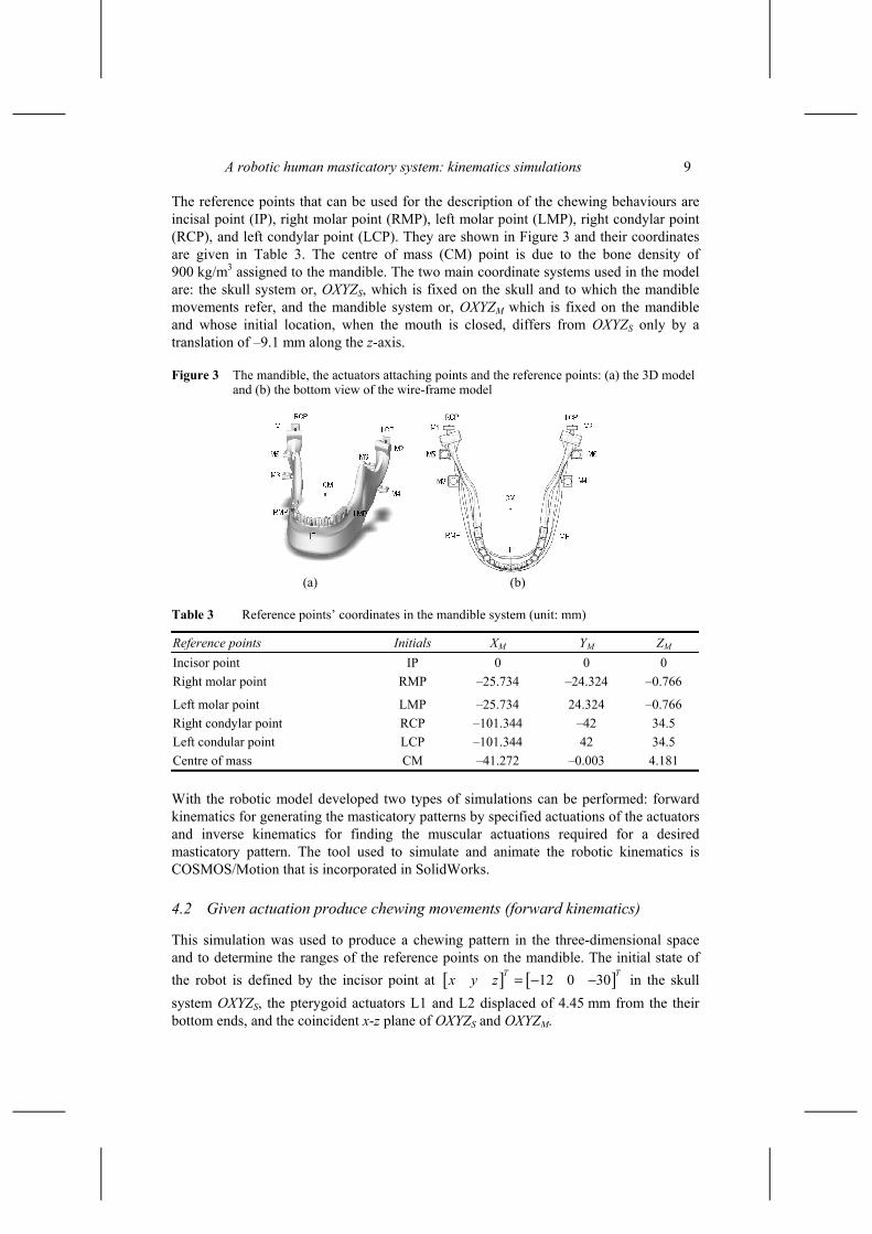
A robotic human masticatory system: kinematics simulations 9
The reference points that can be used for the description of the chewing behaviours are incisal point (IP), right molar point (RMP), left molar point (LMP), right condylar point (RCP), and left condylar point (LCP). They are shown in Figure 3 and their coordinates are given in Table 3. The centre of mass (CM) point is due to the bone density of 900 kg/m3 assigned to the mandible. The two main coordinate systems used in the model are: the skull system or, OXYZS, which is fixed on the skull and to which the mandible movements refer, and the mandible system or, OXYZM which is fixed on the mandible and whose initial location, when the mouth is closed, differs from OXYZS only by a translation of –9.1 mm along the z-axis.
Figure 3 The mandible, the actuators attaching points and the reference points: (a) the 3D model and (b) the bottom view of the wire-frame model
(a) (b)
Table 3 Reference points’ coordinates in the mandible system (unit: mm)
Reference points Initials XM YM ZM Incisor point IP 0 0 0 Right molar point RMP –25.734 –24.324 –0.766
Left molar point LMP –25.734 24.324 –0.766 Right condylar point RCP –101.344 –42 34.5 Left condular point LCP –101.344 42 34.5 Centre of mass CM –41.272 –0.003 4.181
With the robotic model developed two types of simulations can be performed: forward kinematics for generating the masticatory patterns by specified actuations of the actuators and inverse kinematics for finding the muscular actuations required for a desired masticatory pattern. The tool used to simulate and animate the robotic kinematics is COSMOS/Motion that is incorporated in SolidWorks.
4.2 Given actuation produce chewing movements (forward kinematics)
This simulation was used to produce a chewing pattern in the three-dimensional space and to determine the ranges of the reference points on the mandible. The initial state of the robot is defined by the incisor point at [ ] [ ]12 0 30T Tx y z = − − in the skull system OXYZS, the pterygoid actuators L1 and L2 displaced of 4.45 mm from the their bottom ends, and the coincident x-z plane of OXYZS and OXYZM.

10 J-S. Pap, W.L. Xu and J. Bronlund
The six actuators are driven by the following harmonic time function. z = 4sin(0.5 + π/2), for pterygoid externus actuators L1 and L2 z = sin(0.5 time), for temporalis actuators L3 and L4 z = 4sin(0.5 time – 0.083π), for right masseter actuator L5 z = 4.175sin(0.5time – 0.1π), for left masseter actuator L6
where z is the actuator’s displacement around its initial position. The spatial trajectories of IP, RMP, LMP, RCP and LCP in frontal, sagittal and
horizontal planes are shown in Figures 4 and 5. It can be found that the lateral, inferior-superior and anterior-posterior ranges of the incisor are 8, 25 and 20 mm, respectively, and the lateral, inferior-superior and anterior-posterior ranges of the RCP are 8, 2 and 8 mm, respectively. The incisal point moves within the Posselt envelope (Posselt, 1957) while the condylar movements match the clinical and biomechanical findings (Nakajima et al., 2001 and Scapino, 1997). The shape of the chewing path looks like one from a left-side chewing subject. However, it should be noted that the periodic actuations were arbitrarily chosen only for simulation purpose, and they do not reflect real human chewing processes, in particular, in terms of the temporal information of chewing. Figures 6 and 7 shows side and frontal views of the jaw with the trajectories of the reference points superimposed.
Figure 4 IP, RMP and LMP trajectories: (a) the frontal plane, (b) the sagittal plane and (c) the horizantal plane
(a)
(b)

A robotic human masticatory system: kinematics simulations 11
Figure 4 IP, RMP and LMP trajectories: (a) the frontal plane, (b) the sagittal plane and (c) the horizantal plane (continued)
(c)
Figure 5 RCP and LCP trajectories: (a) the frontal plane, (b) the sagittal plane and (c) the horizantal plane
(a)
(b)

12 J-S. Pap, W.L. Xu and J. Bronlund
Figure 5 RCP and LCP trajectories: (a) the frontal plane, (b) the sagittal plane and (c) the horizantal plane (continued)
(c)
Figure 6 Jaw with IP, RMP and LMP trajectories: (a) side view and (b) frontal view
Figure 7 TMJ with RCP and LCP trajectories: (a) side view and (b) frontal view

A robotic human masticatory system: kinematics simulations 13
4.3 Given chewing patterns generate actuations (inverse kinematics)
The data for this simulation were measured from the record of the masticatory sequence of a subject, provided by INRA, France. The subject chewed on the right side and the test food had hard elastic properties (Peyron et al., 2002). Only available data were the history of y and z coordinates of IP in OXYZS, as plotted in Figures 8 and 9. With these data, however, the mandible movements cannot be fully specified. To have a complete description, the mandible is further constrained as follows: the middle point of the two condyles M1 and M2 moves along a straight line in the x-z plane and 25.4 mm distant from the origin of OXYZS. Figures 10 and 11 show the trajectories of IP, RMP and LMP of the mandible, and RCP and LCP of the TMJ with respect to the specified motion.
Figure 8 Temporal chewing trajectories of IP from experiments: (a) lateral movement and (b) superior-inferior movement
(a)
(b)

14 J-S. Pap, W.L. Xu and J. Bronlund
Figure 9 Spatial chewing trajectories of IP in the frontal plane
Figure 10 IP, RMP and LMP trajectories for a person: (a) frontal view and (b) side view
Figure 11 RCP and LCP trajectories: (a) right side view and (b) top view
It can be found from the lateral movements (Figures 8 and 9) that the subject chewed at right side and performed three peaks of the lateral excursion movements. This can be explained as work of the tongue to collect or replace the food bolus in an accurate position. It can also be seen from the superior-inferior movements that for the first couple of chewing cycles the mouth openings are maximum of 30 mm, for the next few chewing cycles the maximum openings of about 25 mm and for the rest of chewing process the maximum opening of 20 mm.

A robotic human masticatory system: kinematics simulations 15
The time functions that six actuators must follow to reproduce the given real human chewing pattern were obtained. One of them (i.e., the right pterygoid actuator) is shown in Figure 12, with the maximum strokes of 19 mm, velocity of 86 mm/sec and acceleration of 5,834 mm/sec2, approximately. The maximum pterygoid actuations occur in the first few chewing cycles, and for the most of the chewing process their actuations are less than 9.3 mm and 7.7 mm, respectively. From these actuations an average chewing cycle of 1.9 seconds has been found, and other time related chewing parameters could also be found if needed.
Figure 12 Right pterygoid actuation required for the specified chewing patterns: (a) displacement, (b) velocity and (c) acceleration

16 J-S. Pap, W.L. Xu and J. Bronlund
5 Conclusion
For reproducing human chewing movements a robotic model was developed based on the biomechanical findings of the mastication system. The model allows performing two different kinds of simulations: specify actuators’ displacements to animate the mandible movements and use measurements from real chewing processes as input data to generate the actuations required to reproduce the human chewing movements. The robotic model was validated by extensive simulations in SolidWorks with COSMOS/Motion. While this work presented in the paper is preliminary, it does provide a viable robotic model to be worked on. The issues to be researched include such as simulations of dynamic forces using COSMOS/Works, kinematics, dynamics, force-motion control of the robot, and mechatronics design.
Acknowledgements
The work in this paper was supported by a MURF grant, Massey University, New Zealand. It is acknowledged that the real chewing data in the paper were provided by INRA, France.
References Anderson, K. and Throckmorton, G.S. et al. (2002) ‘The effects of bolus hardness on masticatory
kinematics’, J. Oral Rehabilitation, Vol. 29, pp.689–696. Hannam, A.C. (1997) ‘Jaw muscle structure and function’, in McNeill, C. (Ed.): Science and
Practice of Occlusion, Quintessence Publishing Co, Inc., pp.41–49. Hayasaki, H. and Nakata, S. et al. (1999) ‘A calculation method for the range of occluding phase at
the lower incisal point during chewing movements using the curved mesh diagram of mandibular excursion (CMDME)’, J. Oral Rehabilitation, Vol. 26, pp.236–242.
Hayashi, T. and Kato, S. et al. (1999) ‘Physiological control scheme of jaw simulator JSN/2A for improving reproducibility of open-close movement’, Proc. the First Joint BMES/EMBS Conference Serving Humanity, Advancing Technology, October 13–16, Atlanta, USA.
Hayashi, T. and Kato, S. et al. (2000) ‘A physiological control of chewing-like jaw movement for robotized jaw simulator JSN/2A’, Proc. of the 22nd Annual EMBS International Conference, July, Chicago, USA.
Koolstra, J.H. and van Eijden, T.M. (1992) ‘Application and validation of a three-dimensional mathematical model of the human masticatory system in vivo’, J. Biomech, Vol. 25, pp.175–187.
Koolstra, J.H. and van Eijden, T.M. (2001) ‘A method to predict muscle control in the kinematically and mechanically indeterminate human masticatory system’, J. Biomech., Vol. 34, pp.1179–1188.
Koolstra, J.H., van Eijden, T.M. et al. (1988) ‘A three-dimensinal mathematical model of the human masticatory system predicting maximum possible bite force’, J. Biomechanics, Vol. 21, pp.563–567.
Koolstra, J.H.and van Eijden, T.M., (1997) ‘Dynamics of the human masticatory muscles during a jaw open-close movement’, J Biomech, Vol. 30, No. 9, pp.883–889.
Nakajima, J. and Hidesshima, M. et al. (2001) ‘Masticatory mandibular movements for different food texture related to onomatopoetic words’, J. Med. Dent. Sci., Vol. 48, pp.121–129.

A robotic human masticatory system: kinematics simulations 17
Osborn, J.W. (1996) ‘Features of human jaw design which maximize the bite force’, J. Biomechanics, Vol. 29, No. 5, pp.589–595.
Osborn, J.W. and Baragar, F.A. (1985) ‘Predicted pattern of human muscle activity during clenching derived from a computer assisted model: symmetric vertical bite force’, J. Biomechanics, Vol. 29, No. 5, pp.589–595.
Ow, R.K.K., Carlsson, G.E. and Karlsson, S. (1998) ‘Relationship of masticatory mandibular movements to masticatory performance of dentate adults: a method study’, J. Oral Rehabilitation, Vol. 25, pp.821–829.
Palla, S., Krebs, M. and Gallo, L.M. (1997) ‘Jaw tracking and temporomandibular joint animation’, in McNeill, C. (Ed.): Science and Practice of Occlusion, Quintessence Publishing Co, Inc., pp.365–378.
Peyron, M.A., Lassauzay, C. and Woda, A. (2002) ‘Effects of increased hardness on jaw movement and muscle activity during chewing of visco-elastic model foods’, Exp. Brain Research, Vol. 142, pp.41–51.
Posselt, U. (1957) ‘Movement areas of the mandible’, J. Pros. Dent., Vol. 7, pp.375–385. Scapino, R.P. (1997) ‘Morphology and mechanism of the jaw joint’, in McNeill, C. (Ed.): Science
and Practice of Occlusion, Quintessence Publishing Co., pp.23–40. Takanobu, H. and Yajima, T. et al. (1997) ‘Development of a mastication robot using nonlinear
viscoelastic mechanism’, Proc. of the IEEE/RSJ International Conference on Intelligent Robots and Systems, pp.1527–1532.
Takanobu, H. and Yajima, T. et al. (1998) ‘Quantification of masticatory efficiency with a mastication robot’, Proc. of the IEEE Internal Conference on Robotics & Automation, pp.1635–1640.
Tortopidis, D. and Lyons, M.F. et al. (1998) ‘The variability of bite force measurement between sessions in different positions within the dental arch’, J. Oral Rehabilitation, Vol. 25, pp.681–686.
Tsuruta, J. and Mayanagi, A. et al. (2002) ‘An index for analysing the stability of lateral excursions’, J. Oral Rehabilitation, Vol. 29, pp. 274–281.
Xu, W.L., Bronlund, J. and Kieser, J. (2004) ‘A robotic model of human mastication system for reproducing chewing behaviours’, IEEE Robotics and Automation Magazine, in print.