a rapid, flexible alternative to elisa for protein … rapid, flexible alternative to elisa for...
TRANSCRIPT
A Rapid, Flexible Alternative to ELISA for Protein
Detection, Characterization and Quantitation
Broadcast Date: Tuesday, September 25, 2012
Time: 11:00 am EDT
Sponsored by
A Rapid, Flexible Alternative to ELISA for Protein
Detection, Characterization and Quantitation
A Rapid, Flexible Alternative to ELISA for Protein
Detection, Characterization and Quantitation
Your Moderator
Tamlyn Oliver Managing Editor
Genetic Engineering & Biotechnology News
A Rapid, Flexible Alternative to ELISA for Protein
Detection, Characterization and Quantitation
Francesco Lipari, Ph.D.
Senior Scientist
Biotherapeutic Applications
PerkinElmer Life Sciences and Technology
© 2009 PerkinElmer © 2009 PerkinElmer © 2009 PerkinElmer © 2009 PerkinElmer
Alpha Technology: Introduction Francesco Lipari, Ph.D. September 2012
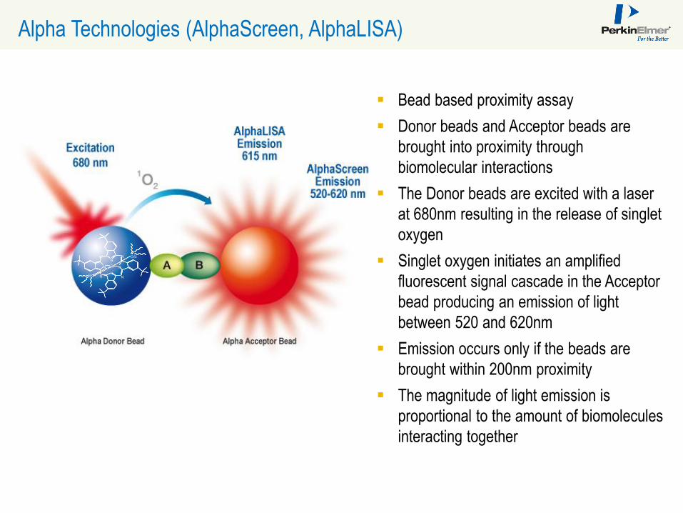
Alpha Technologies (AlphaScreen, AlphaLISA)
Bead based proximity assay
Donor beads and Acceptor beads are
brought into proximity through
biomolecular interactions
The Donor beads are excited with a laser
at 680nm resulting in the release of singlet
oxygen
Singlet oxygen initiates an amplified
fluorescent signal cascade in the Acceptor
bead producing an emission of light
between 520 and 620nm
Emission occurs only if the beads are
brought within 200nm proximity
The magnitude of light emission is
proportional to the amount of biomolecules
interacting together
Si N
N
N
N
N
N
N
N
OSi
SiO
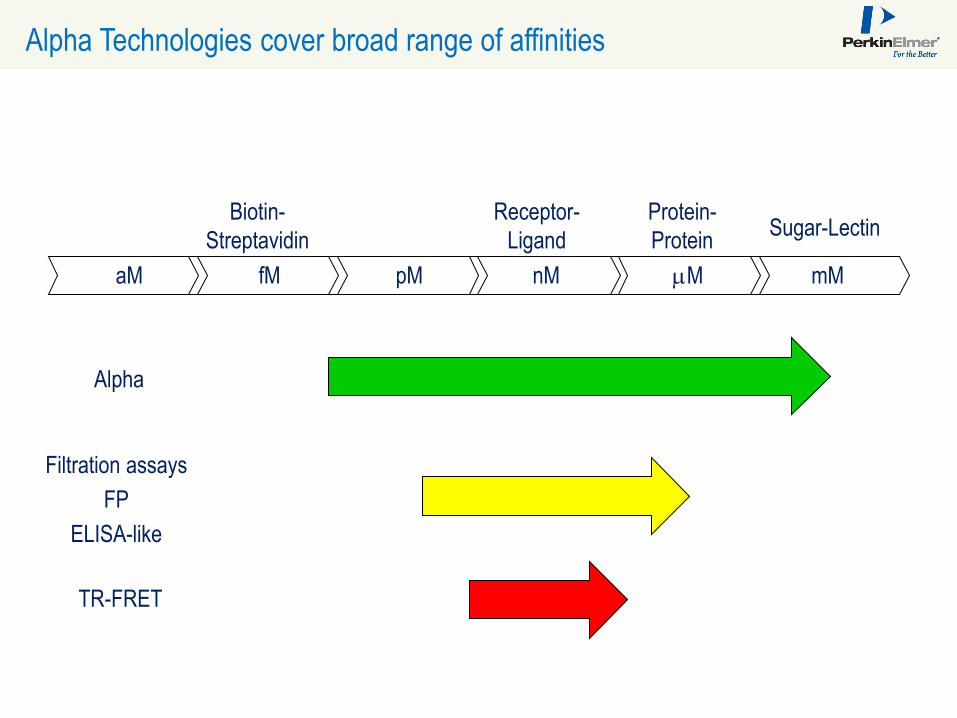
Alpha Technologies cover broad range of affinities
aM fM pM nM mM mM
Sugar-Lectin Protein-
Protein
Biotin-
Streptavidin
Receptor-
Ligand
Alpha
Filtration assays
FP
ELISA-like
TR-FRET
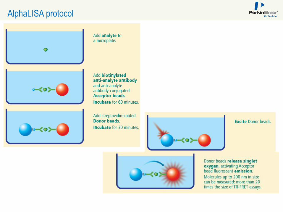
AlphaLISA protocol

A Rapid, Flexible Alternative to ELISA for Protein Detection, Characterization, and Quantitation
Michael Bembenek, Ph.D., Lead Discovery, Associate Scientific Fellow; Millennium:
The Takeda Oncology Company
“Determination of Complementary Antibody Pairs using Protein A Capture with the
AlphaScreen Assay Format”
Chamindie Punyadeera, Ph.D., Senior Research Fellow, Australian Institute for
Bioengineering and Nanotechnology, The University of Queensland
“Saliva Biomarker Detection Powered by AlphaLISA technology”
Francesco Lipari, Ph.D., Senior Scientist, Biotherapeutics Applications,
PerkinElmer Life Sciences and Technologies
“AlphaLISA: A Simple, Homogeneous Immunoassay Platform for Bioprocess Analytics”
A Rapid, Flexible Alternative to ELISA for Protein
Detection, Characterization and Quantitation
Michael E. Bembenek, Ph.D. Lead Discovery
Scientific Fellow
Millennium: The Takeda Oncology Company
2012/9/17 Millennium: The Takeda Oncology Company
Michael E. Bembenek, Ph.D.
Determination of Complementary Antibody Pairs using Protein A Capture with the AlphaScreen Assay Format
Lead Discovery
Scientific Fellow
|○○○○ | 09/2012 10

Millennium: The Takeda Oncology Company
GOAL = HOW TO FIND ANTIBODY PAIRS QUICKLY FOR USE IN BIOMARKER DISCOVERY
Benefits of complementary pairing antibodies include:
– use of sandwich format improves selectivity and specificity for biomarker.
– ability to move away from Western blot based assay methods because of limitations with quantitation & labor intensity.
No current high throughput method for identifying complementary antibody pairs.
|○○○○ | 09/2012 11
Millennium: The Takeda Oncology Company
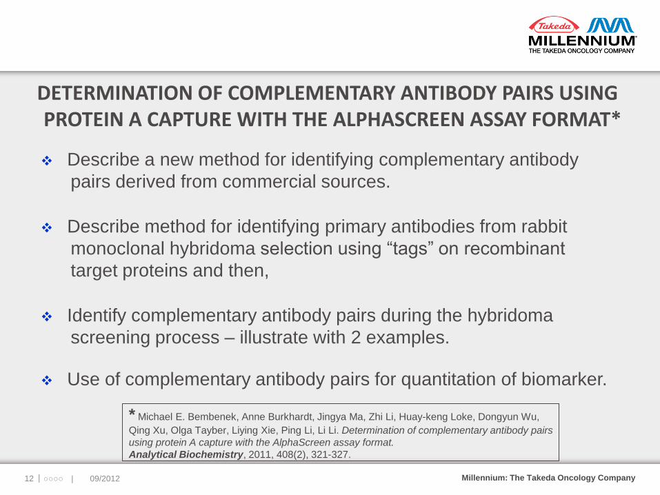
DETERMINATION OF COMPLEMENTARY ANTIBODY PAIRS USING PROTEIN A CAPTURE WITH THE ALPHASCREEN ASSAY FORMAT*
Describe a new method for identifying complementary antibody
pairs derived from commercial sources.
Describe method for identifying primary antibodies from rabbit
monoclonal hybridoma selection using “tags” on recombinant
target proteins and then,
Identify complementary antibody pairs during the hybridoma
screening process – illustrate with 2 examples.
Use of complementary antibody pairs for quantitation of biomarker.
|○○○○ | 09/2012 12
* Michael E. Bembenek, Anne Burkhardt, Jingya Ma, Zhi Li, Huay-keng Loke, Dongyun Wu,
Qing Xu, Olga Tayber, Liying Xie, Ping Li, Li Li. Determination of complementary antibody pairs
using protein A capture with the AlphaScreen assay format.
Analytical Biochemistry, 2011, 408(2), 321-327.
Millennium: The Takeda Oncology Company
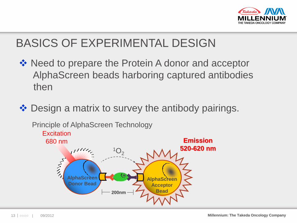
BASICS OF EXPERIMENTAL DESIGN
Need to prepare the Protein A donor and acceptor
AlphaScreen beads harboring captured antibodies
then
Design a matrix to survey the antibody pairings.
|○○○○ | 09/2012 13
Emission
520-620 nm
AlphaScreen
Acceptor
Bead
1O2
Excitation
680 nm
AlphaScreen
Donor Bead
200nm
Principle of AlphaScreen Technology
Millennium: The Takeda Oncology Company
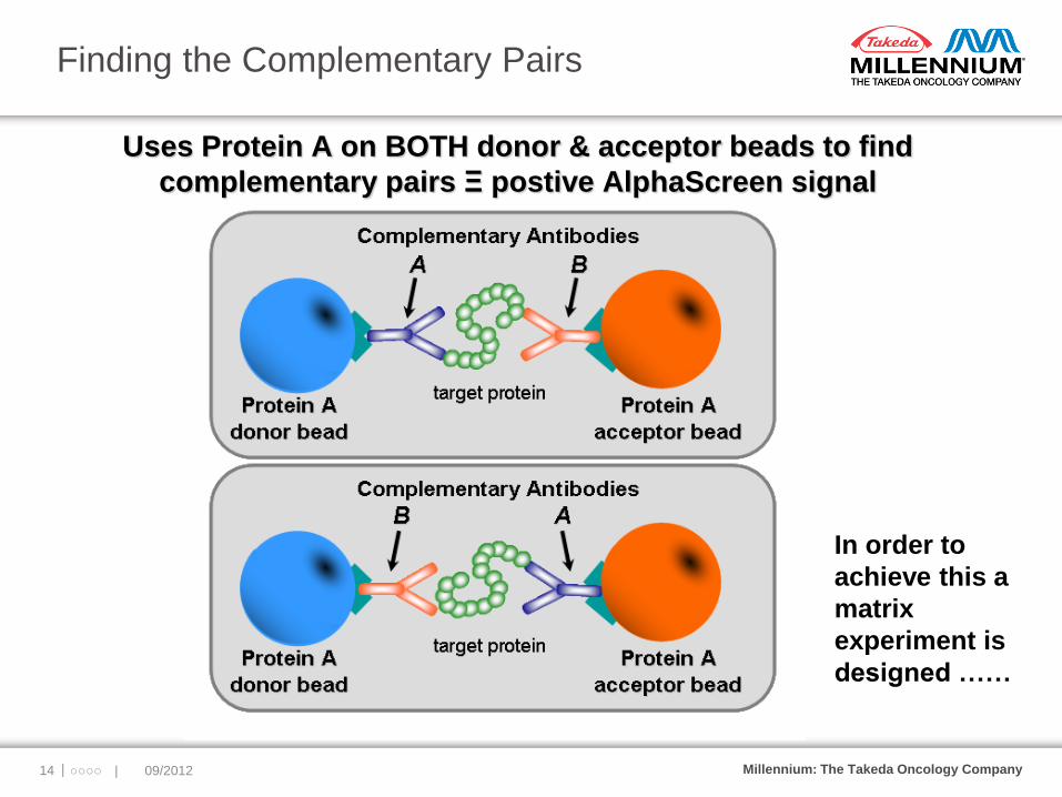
Finding the Complementary Pairs
|○○○○ | 09/2012 14
Uses Protein A on BOTH donor & acceptor beads to find
complementary pairs Ξ postive AlphaScreen signal
In order to
achieve this a
matrix
experiment is
designed ……
Millennium: The Takeda Oncology Company
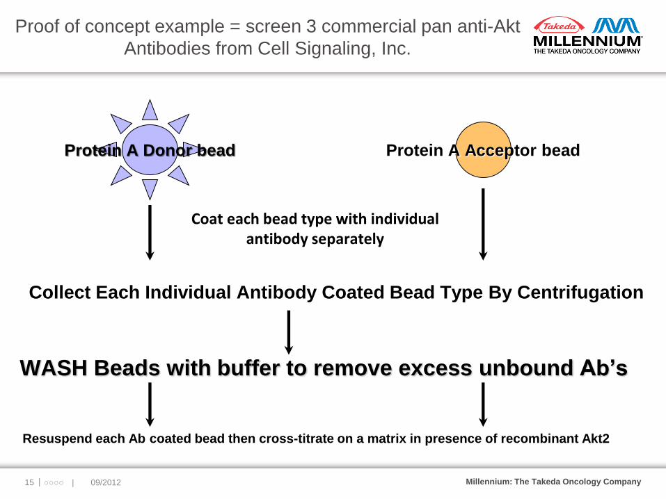
Proof of concept example = screen 3 commercial pan anti-Akt
Antibodies from Cell Signaling, Inc.
|○○○○ | 09/2012 15
Protein A Donor bead
Protein A Acceptor bead
Coat each bead type with individual antibody separately
WASH Beads with buffer to remove excess unbound Ab’s
Collect Each Individual Antibody Coated Bead Type By Centrifugation
Resuspend each Ab coated bead then cross-titrate on a matrix in presence of recombinant Akt2
Millennium: The Takeda Oncology Company
“AlphaMatrix” for Finding Complementary Pairs
|○○○○ | 09/2012 16
3 X 3 Matrix
pan-Akt antibody
(CST:#4685)
on
Protein A Donor
pan-Akt antibody
(CST:#4691)
on
Protein A donor
pan-Akt
polyclonal
antibody
(CST:#9272)
on
Protein A donor
pan-Akt antibody
(CST:#4685)
on
Protein A
acceptor
Same Ab
against itself
pan-Akt antibody
(CST:#4691)
on
Protein A
acceptor
Same Ab
against itself
pan-Akt polyclonal
antibody
(CST:#9272)
on
Protein A acceptor
Same Ab
against itself
Pairs are represented TWICE
on the matrix on different bead
types = off the diagonal
Each column
contains an equal
aliquot of
antibody coated
onto Protein A
donor beads
Each row contains an
equal aliquot of antibody
coated onto Protein A
acceptor beads

Millennium: The Takeda Oncology Company
“AlphaMatrix” for Finding Complementary
Total Akt Pairs: the Data
|○○○○ | 09/2012 17
17
pan-Akt antibody
(CST:#4685)
on
Protein A Donor
pan-Akt antibody
(CST:#4691)
on
Protein A donor
pan-Akt polyclonal
antibody
(CST:#9272)
on
Protein A donor
pan-Akt antibody
(CST:#4685)
on
Protein A acceptor
3439 3363 86906
pan-Akt antibody
(CST:#4691)
on
Protein A acceptor
5890 15124 459515
pan-Akt polyclonal
antibody
(CST:#9272)
on
Protein A acceptor
54815 306242 15409
3 X 3 Matrix
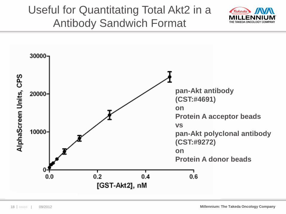
Millennium: The Takeda Oncology Company |○○○○ | 09/2012 18
Useful for Quantitating Total Akt2 in a
Antibody Sandwich Format
pan-Akt antibody
(CST:#4691)
on
Protein A acceptor beads
vs
pan-Akt polyclonal antibody
(CST:#9272)
on
Protein A donor beads
Millennium: The Takeda Oncology Company
HIGHER THROUGHPUT HYBRIDOMA ANTIBODY SCREENING
Use method for identifying positive rabbit hybridoma producing
antibodies to recombinant “tagged” protein antigens.
Select positive hybridoma clones with verification by Western blot &
immunoprecipitation.
Capture positive antibodies from hybridoma derived cell culture
supernatants onto both Protein A donor and acceptor beads –
requires no tagging of Ab’s like many other methods.
Use “AlphaMatrix” format to identify complementary pairs and
for final monoclonal selection.
|○○○○ | 09/2012 19
Millennium: The Takeda Oncology Company
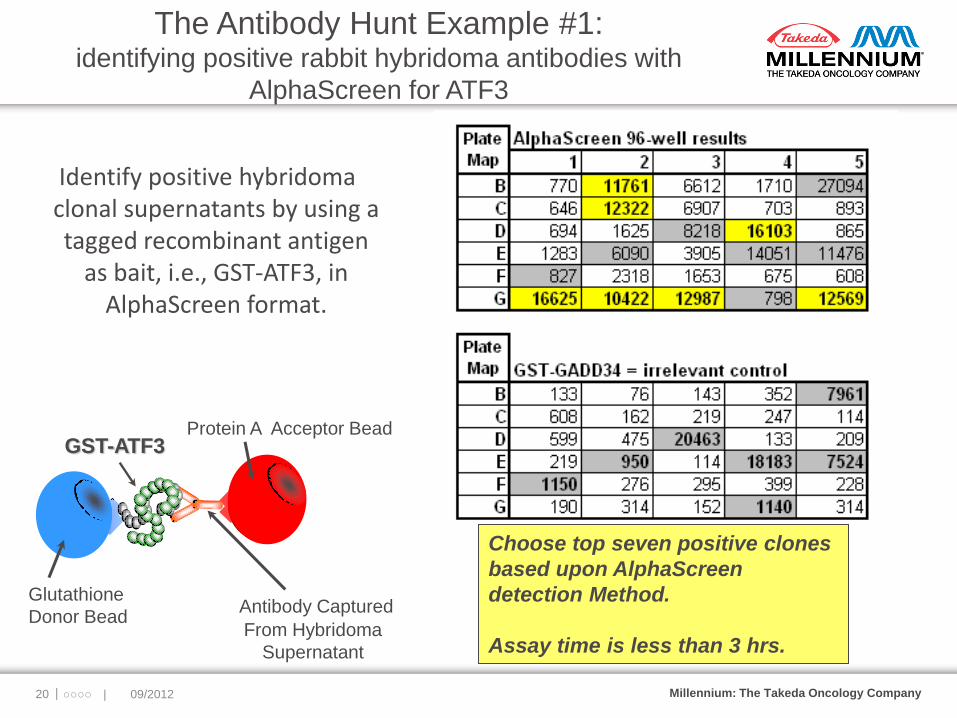
The Antibody Hunt Example #1: identifying positive rabbit hybridoma antibodies with
AlphaScreen for ATF3
|○○○○ | 09/2012 20
Identify positive hybridoma clonal supernatants by using a tagged recombinant antigen
as bait, i.e., GST-ATF3, in AlphaScreen format.
Glutathione
Donor Bead Antibody Captured
From Hybridoma
Supernatant
Protein A Acceptor Bead
Choose top seven positive clones
based upon AlphaScreen
detection Method.
Assay time is less than 3 hrs.
GST-ATF3

Millennium: The Takeda Oncology Company
Confirmed by Western Blot & Immunoprecipitation
|○○○○ | 09/2012 21
Western
blot of
transiently
transfected
293T cells
vs
hybridoma
clones(#’d).
Immunoppt. of
transiently
transfected
293T cell lysates vs
hybridoma
clones(#’d).
Millennium: The Takeda Oncology Company
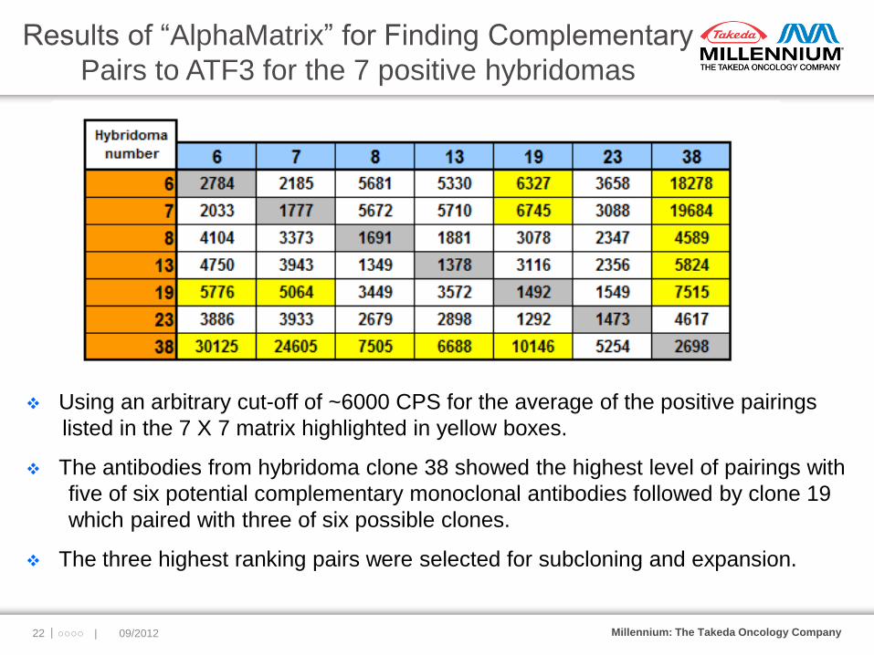
Results of “AlphaMatrix” for Finding Complementary
Pairs to ATF3 for the 7 positive hybridomas
|○○○○ | 09/2012 22
Using an arbitrary cut-off of ~6000 CPS for the average of the positive pairings
listed in the 7 X 7 matrix highlighted in yellow boxes.
The antibodies from hybridoma clone 38 showed the highest level of pairings with
five of six potential complementary monoclonal antibodies followed by clone 19
which paired with three of six possible clones.
The three highest ranking pairs were selected for subcloning and expansion.

Millennium: The Takeda Oncology Company
Example #2: Identifying positive hybridomas with AlphaScreen
for the b subunit of Neddylation activating enzyme(NAE)
|○○○○ | 09/2012 23
Top 20 hybridoma clones based
upon AlphaScreen detection
method are listed in yellow
highlighted boxes.
Western blot & IP positives
reduced to the number to18.
Antibody captured from
hybridoma supernatant
Anti-Flag
Acceptor bead
Protein A
donor
bead
FLAG-NAEβ
Positives were then screened in an 18X18 AlphaMatrix for identifying complementary pairs
Millennium: The Takeda Oncology Company
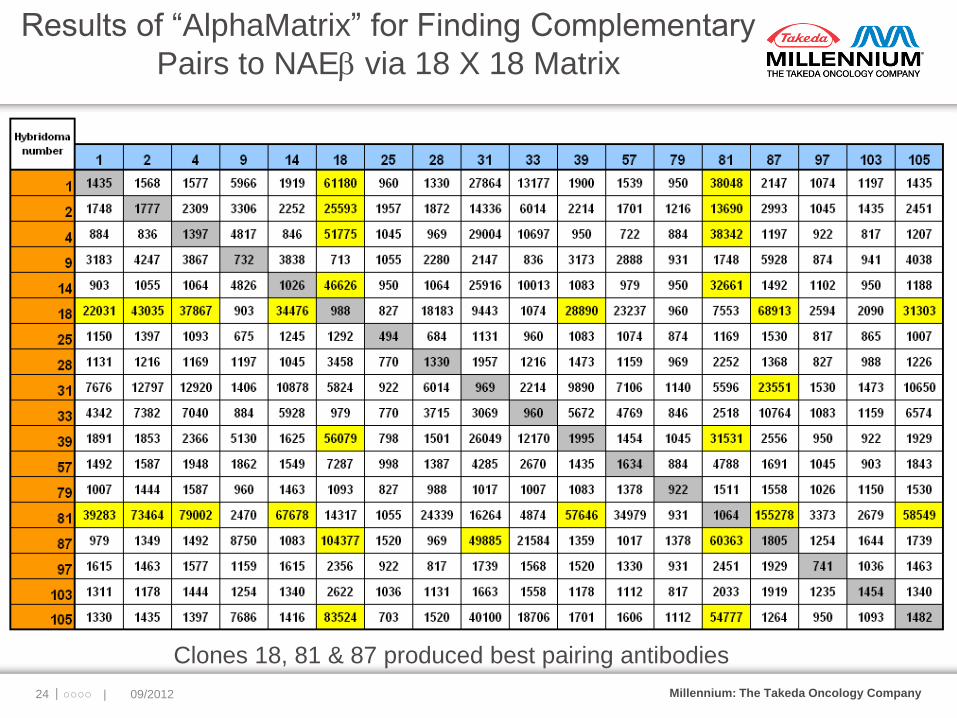
Results of “AlphaMatrix” for Finding Complementary
Pairs to NAEb via 18 X 18 Matrix
|○○○○ | 09/2012 24
Clones 18, 81 & 87 produced best pairing antibodies
Millennium: The Takeda Oncology Company
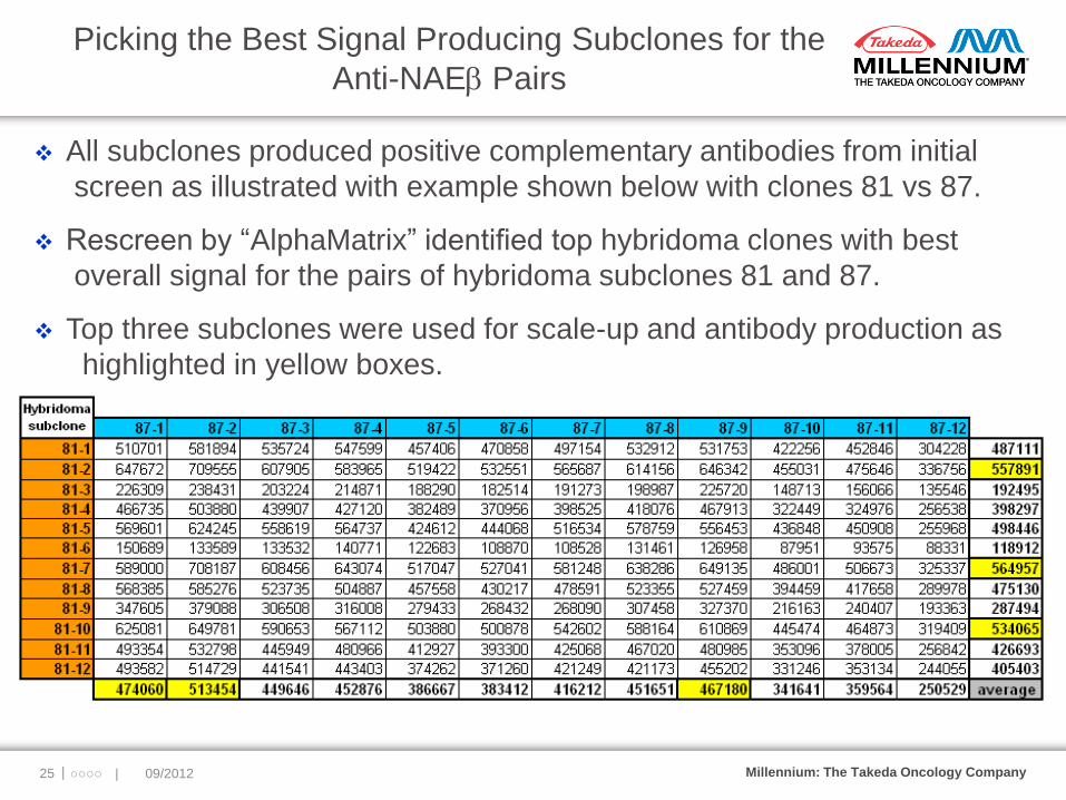
Picking the Best Signal Producing Subclones for the
Anti-NAEb Pairs
|○○○○ | 09/2012 25
All subclones produced positive complementary antibodies from initial
screen as illustrated with example shown below with clones 81 vs 87.
Rescreen by “AlphaMatrix” identified top hybridoma clones with best
overall signal for the pairs of hybridoma subclones 81 and 87.
Top three subclones were used for scale-up and antibody production as
highlighted in yellow boxes.
Millennium: The Takeda Oncology Company
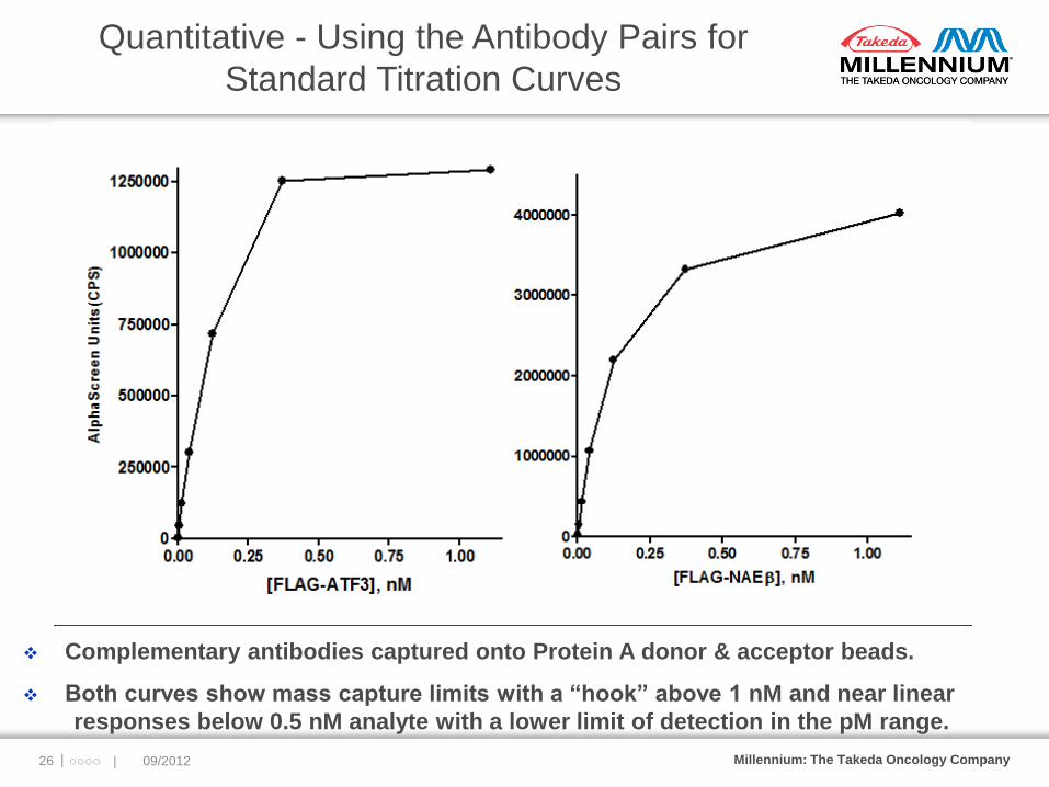
Quantitative - Using the Antibody Pairs for
Standard Titration Curves
|○○○○ | 09/2012 26
Complementary antibodies captured onto Protein A donor & acceptor beads.
Both curves show mass capture limits with a “hook” above 1 nM and near linear
responses below 0.5 nM analyte with a lower limit of detection in the pM range.
Millennium: The Takeda Oncology Company
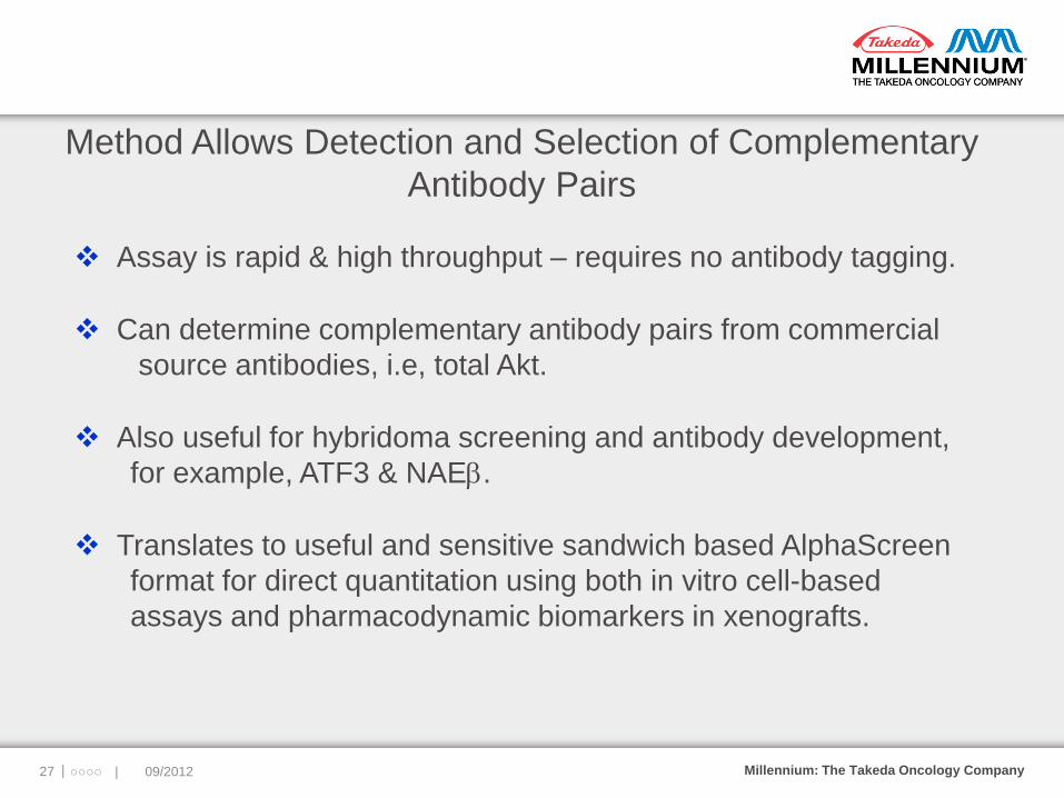
Method Allows Detection and Selection of Complementary
Antibody Pairs
Assay is rapid & high throughput – requires no antibody tagging.
Can determine complementary antibody pairs from commercial
source antibodies, i.e, total Akt.
Also useful for hybridoma screening and antibody development,
for example, ATF3 & NAEb.
Translates to useful and sensitive sandwich based AlphaScreen
format for direct quantitation using both in vitro cell-based
assays and pharmacodynamic biomarkers in xenografts.
|○○○○ | 09/2012 27
Millennium: The Takeda Oncology Company
Collaborators Reference:
Michael E. Bembenek, Anne Burkhardt,
Jingya Ma, Zhi Li, Huay-keng Loke,
Dongyun Wu, Qing Xu, Olga Tayber,
Liying Xie, Ping Li, Li Li. Determination
of complementary antibody pairs using
protein A capture with the AlphaScreen
assay format. Analytical Biochemistry,
2011, 408(2), 321-327.
MPI - Anne Burkhardt, Jingya
Ma, Zhi Li, Huay-keng Loke,
Dongyun Wu, Qing Xu, Olga
Tayber, Ping Li
Epitomics, Inc. - Liying Xie and
Li Li
Special thanks to Mike Kuranda &
William Mallender
for helpful scientific discussions.
|○○○○ | 09/2012 28

2012/9/17
We aspire to cure cancer

A Rapid, Flexible Alternative to ELISA for Protein
Detection, Characterization and Quantitation
Chamindie Punyadeera, Ph.D. Senior Research Fellow
The University of Queensland Diamantina Institute
The Translational Research Institute Pty Ltd.

Chamindie Punyadeera (PhD)
Email: [email protected]
Saliva Biomarker Detection Powered
by AlphaLISA® Technology
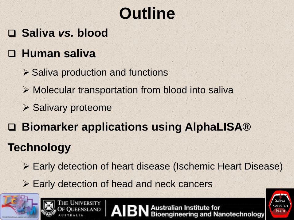
Outline Saliva vs. blood
Human saliva
Saliva production and functions
Molecular transportation from blood into saliva
Salivary proteome
Biomarker applications using AlphaLISA®
Technology
Early detection of heart disease (Ischemic Heart Disease)
Early detection of head and neck cancers
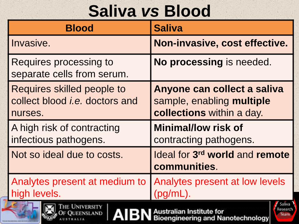
Saliva vs Blood Blood Saliva
Invasive. Non-invasive, cost effective.
Requires processing to
separate cells from serum.
No processing is needed.
Requires skilled people to
collect blood i.e. doctors and
nurses.
Anyone can collect a saliva
sample, enabling multiple
collections within a day.
A high risk of contracting
infectious pathogens.
Minimal/low risk of
contracting pathogens.
Not so ideal due to costs. Ideal for 3rd world and remote
communities.
Analytes present at medium to
high levels.
Analytes present at low levels
(pg/mL).
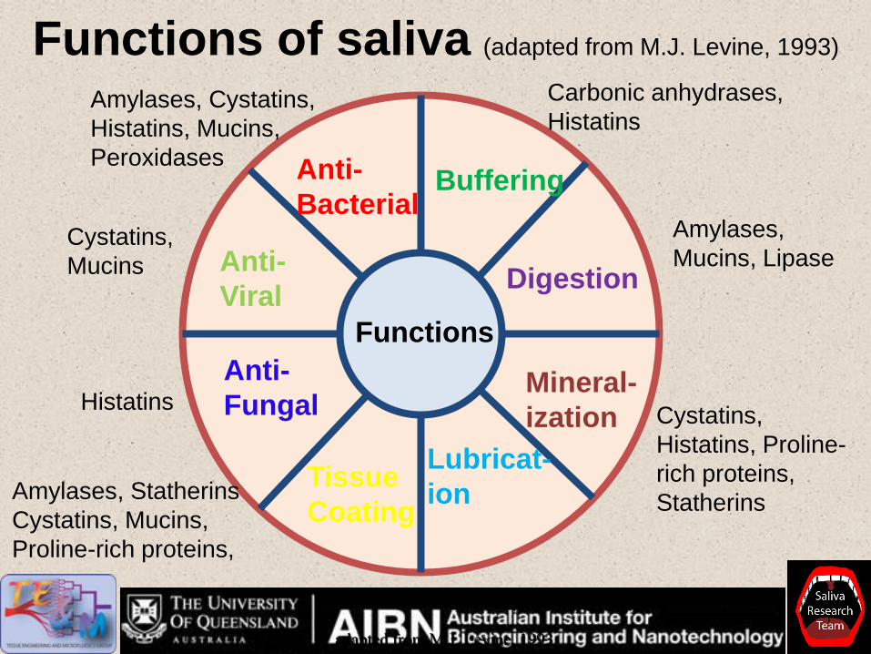
Functions of saliva (adapted from M.J. Levine, 1993)
Functions
Anti-
Bacterial Buffering
Digestion
Mineral-
ization
Lubricat-
ion Tissue
Coating
Anti-
Fungal
Anti-
Viral
Carbonic anhydrases,
Histatins
Amylases,
Mucins, Lipase
Cystatins,
Histatins, Proline-
rich proteins,
Statherins
Mucins, Statherins
Amylases, Statherins
Cystatins, Mucins,
Proline-rich proteins,
Histatins
Cystatins,
Mucins
Amylases, Cystatins,
Histatins, Mucins,
Peroxidases
adapted from M.J. Levine, 1993

Molecular transportation from blood into saliva
Pfaffe et al.,
Clin.Chem,
March 2011
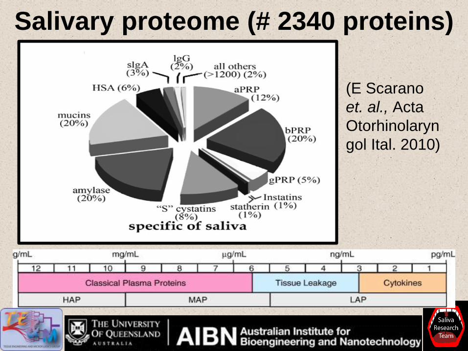
Salivary proteome (# 2340 proteins)
(E Scarano
et. al., Acta
Otorhinolaryn
gol Ital. 2010)
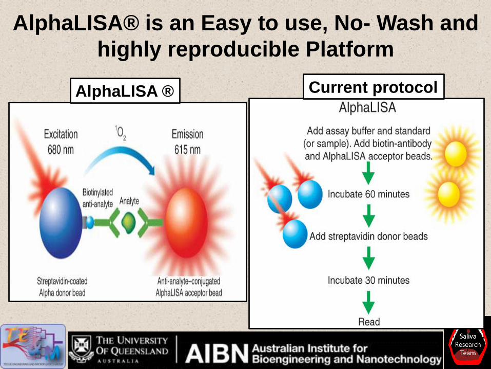
AlphaLISA® is an Easy to use, No- Wash and
highly reproducible Platform
Current protocol AlphaLISA ®
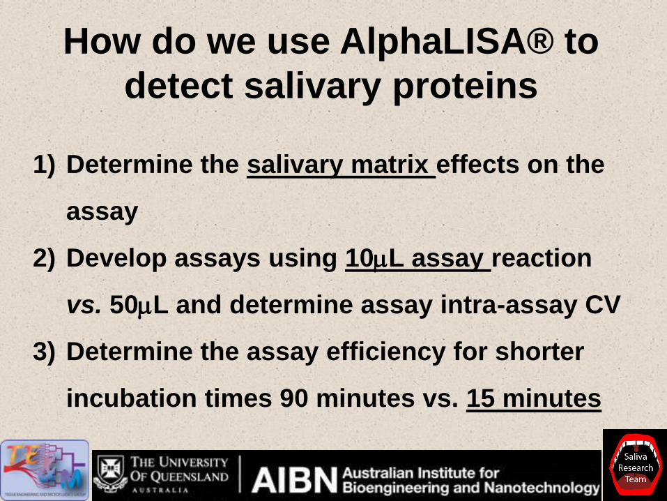
How do we use AlphaLISA® to
detect salivary proteins
1) Determine the salivary matrix effects on the
assay
2) Develop assays using 10mL assay reaction
vs. 50mL and determine assay intra-assay CV
3) Determine the assay efficiency for shorter
incubation times 90 minutes vs. 15 minutes
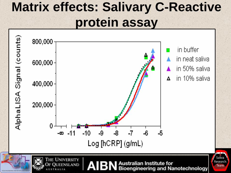
Matrix effects: Salivary C-Reactive
protein assay

Salivary C-Reactive Protein assay reproducibility
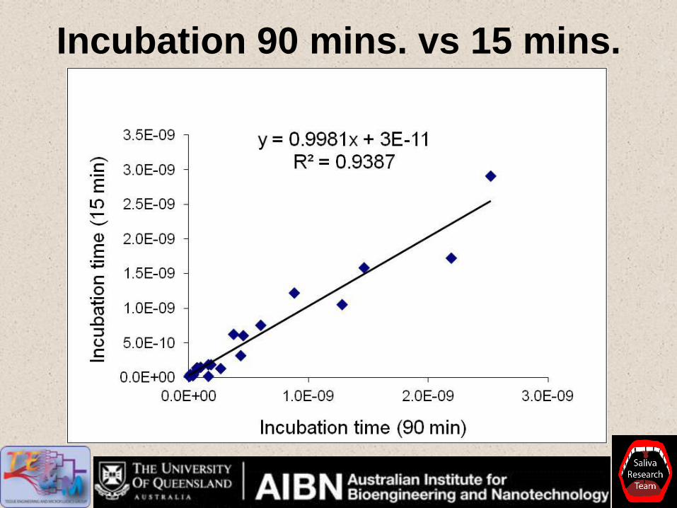
Incubation 90 mins. vs 15 mins.
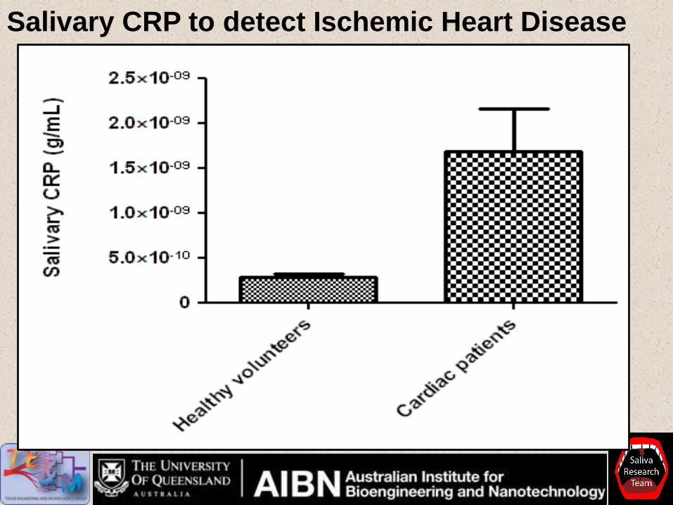
Salivary CRP to detect Ischemic Heart Disease
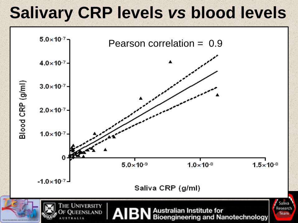
Salivary CRP levels vs blood levels
Pearson correlation = 0.9
A
Drool method (passive)
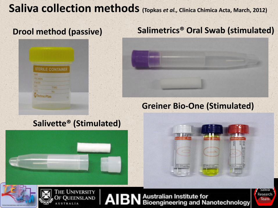
Saliva collection methods (Topkas et al., Clinica Chimica Acta, March, 2012)
Salimetrics® Oral Swab (stimulated)
Salivette® (Stimulated)
Greiner Bio-One (Stimulated)
Dro
ol
SOS
Sal
ivet
te® C
otton
Sal
ivet
te® S
ynth
etic
0
50
100
150
200
250
*
C-R
eacti
ve p
rote
in
(pg
/ml)
Dro
ol
SOS
Sal
ivet
te® C
otton
Sal
ivet
te® S
ynth
etic
0
200
400
600
800
*
Imm
un
og
lob
in E
(p
g/m
l)
Dro
ol
SOS
Sal
ivet
te® C
otton
Sal
ivet
te® S
ynth
etic
0
200
400
600
800
** *
Myo
glo
bin
(p
g/m
l)
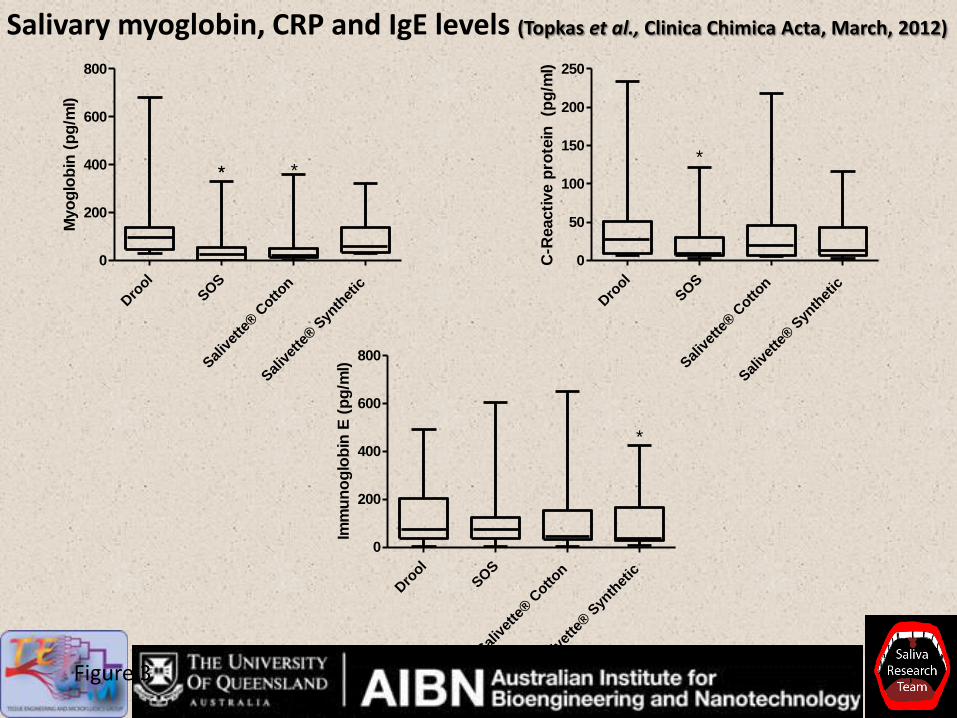
Figure 3
Salivary myoglobin, CRP and IgE levels (Topkas et al., Clinica Chimica Acta, March, 2012)
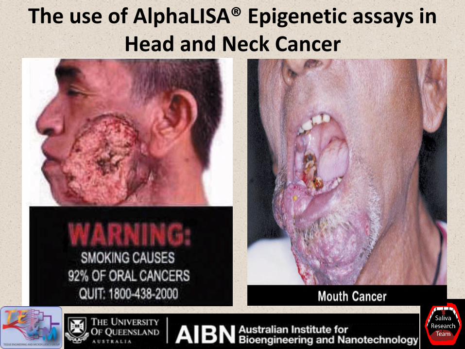
The use of AlphaLISA® Epigenetic assays in Head and Neck Cancer
Hard facts: head and neck cancers
Approximately 780,000 (p/a) new cases of head and neck
squamous cell carcinomas (HNSCC) diagnosed world wide and
300,000 deaths each year. (Boyle P. et al. World Cancer Report 2008).
Tobacco use is a major risk factor for HNSCC (other factors
diet, mouth wash use and HPV infections) (Spitz and Trizna, 1999).
Smoking kills over 1,000,000 people (p/a), causing 30% of all
cancer related deaths. Yet, 1 in 3 people, world-wide is addicted
to nicotine.
The direct impact of smoking can clearly be seen in the oral
cavity due to its proximity, thus, human saliva is ideal as a
detection medium.
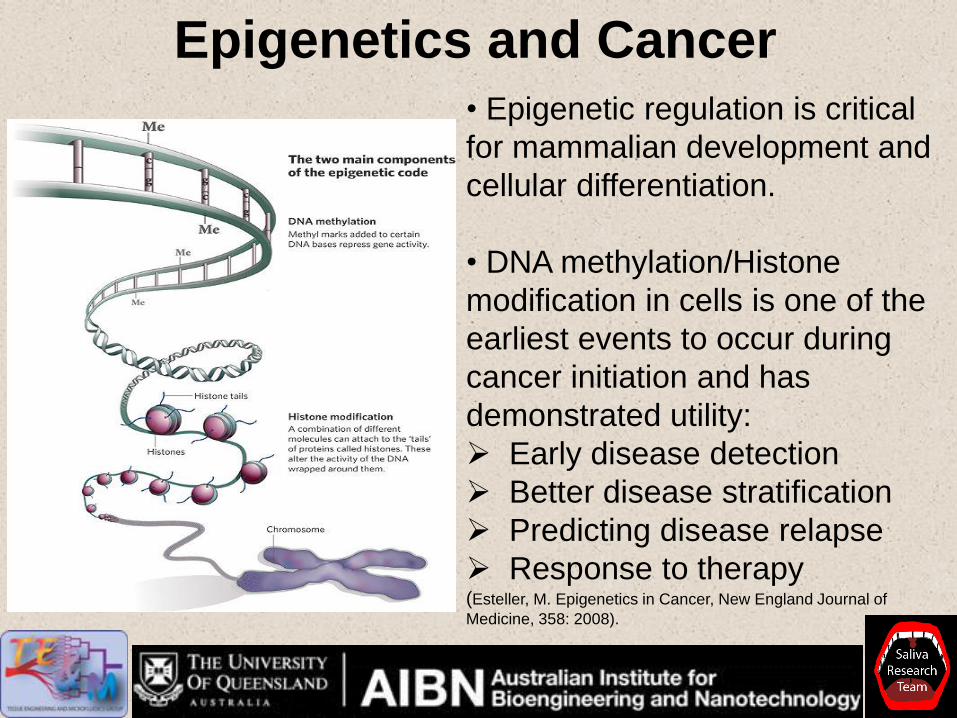
Epigenetics and Cancer • Epigenetic regulation is critical
for mammalian development and
cellular differentiation.
• DNA methylation/Histone
modification in cells is one of the
earliest events to occur during
cancer initiation and has
demonstrated utility:
Early disease detection
Better disease stratification
Predicting disease relapse
Response to therapy (Esteller, M. Epigenetics in Cancer, New England Journal of
Medicine, 358: 2008).

Future work using AlphaLISA
assays for cancer epigentics studies
The AlphaLISA Epigenetic Cellular Detection Kits in
cancer enable rapid and direct detection of endogenous
modification of epigenetic markers on histones H3 and
p53.
AlphaLISA cellular epigenetic assay advantages over
westerns/ELISA:
• Simple assay protocol which can be automated
• Suitable for endogenous and recombinant cell lines,
providing the flexibility to work with relevant cell models
Summary
• Advantages of AlphaLISA® technology over standard
immunoassays:
- AlphaLISA® is applicable with complex biological
fluids such as saliva with careful sample preparations
- Amenable to point of care assay platforms.
- Easy to use and optimize the assay to eventually
meet diagnostic assay quality for clinical research.
- Cost effective
• One of the disadvantages of AlphaLISA® is the
inability to multiplex.
Acknowledgments Saliva Team (present)
• Michael Caragata
• Yunxia Wan
• Prati Pandit
• Jared Foo
• Rahul Nagadia
• Alicia
• Pengxiang Ji
• Bob Xi Zhang
Past: •Tina Pfaffe
• James Dkhar
• Thea Cullen
• Eleni Topkas
• Patricia Keith
• Rosalinda Mohamed
• Jennifer
•Ling Li Long
SPIT Collaborators:
• Professor Justin Cooper white (UQ)
• Professor William Coman (UQ)
• Head and Neck Cancer clinic staff
• Dr Paul Slowey (Oasis Diagnostics, USA)
• Dr John Duley (UQ)
• Professor Ian Frazer (UQDI/TRI)
• Professor Matthew Cooper (UQ)
• Professor Karam Kostner (UQ/Mater)
• Professor John Atherton (RBWH/UQ)
• Dr Ben Schultz (UQ)
• Dr Goce Dimeski (PAH)
• Dr Dmitry Ovchinnikov (UQ)
• Professor David Wong (UCLA/USA) Research funding:
• Queensland Government Strategic Funds
• The University of Queensland Research Excellence Award
• University of Queensland Collaborative Industry Engagement

“The only true wisdom is not knowing nothing”
Socrates

A Rapid, Flexible Alternative to ELISA for Protein
Detection, Characterization and Quantitation
Francesco Lipari, Ph.D.
Senior Scientist
Biotherapeutic Applications
PerkinElmer Life Sciences and Technology
54 54 © 2009 PerkinElmer © 2009 PerkinElmer © 2009 PerkinElmer © 2009 PerkinElmer
AlphaLISA: A Simple, Homogeneous Immunoassay Platform for Bioprocess Analytics Francesco Lipari, Ph.D. September 2012
55 55
Advantages of AlphaLISA technology
Assays for the bioprocess lab
Outline

56 56
Trends in bioprocess analytics
More high throughput requirements due to the implementation of high throughput cell
culture and purification technologies
Homogeneous AlphaLISA assay allows:
Consistent, reproducible results - %CVs less than 10% (intra-assay)
Very simple protocol and easy automation- just 4 steps to results
Low affinity interactions are not washed away
Complete assay in < 3 hours
Sensitivity is similar to classical ELISA technology, but without wash steps
Low sample volumes (≤ 10 mL)
AlphaLISA Technology Advantages
57 57
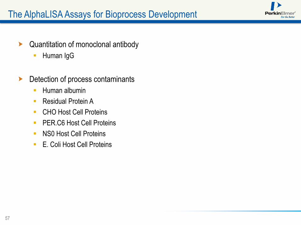
The AlphaLISA Assays for Bioprocess Development
Quantitation of monoclonal antibody
Human IgG
Detection of process contaminants
Human albumin
Residual Protein A
CHO Host Cell Proteins
PER.C6 Host Cell Proteins
NS0 Host Cell Proteins
E. Coli Host Cell Proteins
58 58
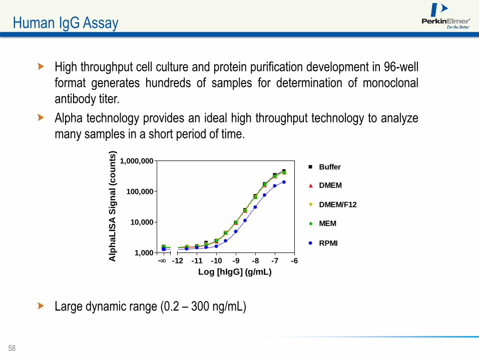
High throughput cell culture and protein purification development in 96-well
format generates hundreds of samples for determination of monoclonal
antibody titer.
Alpha technology provides an ideal high throughput technology to analyze
many samples in a short period of time.
Large dynamic range (0.2 – 300 ng/mL)
Human IgG Assay
1,000
10,000
100,000
1,000,000Buffer
DMEM
DMEM/F12
MEM
RPMI
-12 -11 -10 -9 -8 -7 -6-
Log [hIgG] (g/mL)
Alp
ha
LIS
A S
ign
al (c
ou
nts
)
59 59
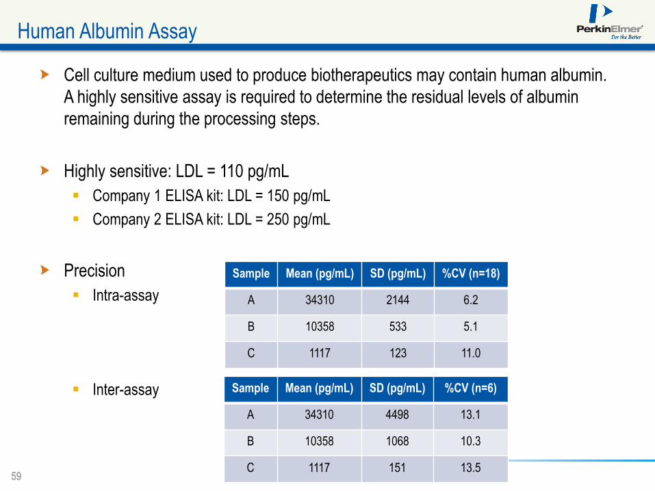
Cell culture medium used to produce biotherapeutics may contain human albumin.
A highly sensitive assay is required to determine the residual levels of albumin
remaining during the processing steps.
Highly sensitive: LDL = 110 pg/mL
Company 1 ELISA kit: LDL = 150 pg/mL
Company 2 ELISA kit: LDL = 250 pg/mL
Precision
Intra-assay
Inter-assay
Human Albumin Assay
Sample Mean (pg/mL) SD (pg/mL) %CV (n=18)
A 34310 2144 6.2
B 10358 533 5.1
C 1117 123 11.0
Sample Mean (pg/mL) SD (pg/mL) %CV (n=6)
A 34310 4498 13.1
B 10358 1068 10.3
C 1117 151 13.5
60 60
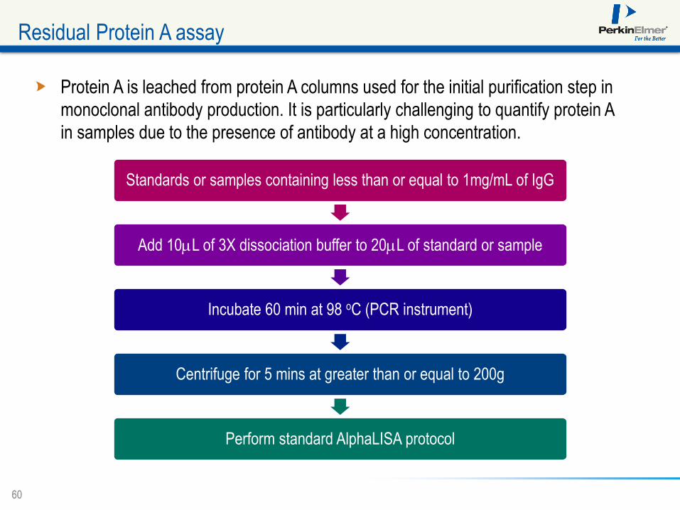
Residual Protein A assay
Protein A is leached from protein A columns used for the initial purification step in
monoclonal antibody production. It is particularly challenging to quantify protein A
in samples due to the presence of antibody at a high concentration.
Standards or samples containing less than or equal to 1mg/mL of IgG
Add 10mL of 3X dissociation buffer to 20mL of standard or sample
Incubate 60 min at 98 oC (PCR instrument)
Centrifuge for 5 mins at greater than or equal to 200g
Perform standard AlphaLISA protocol
61 61
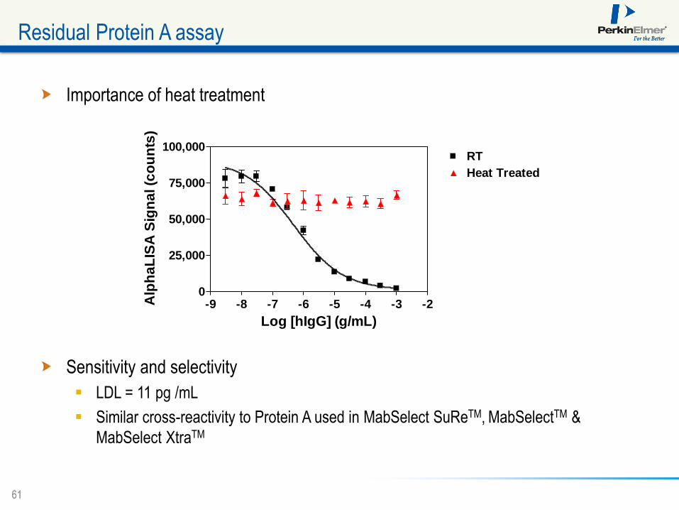
Residual Protein A assay
-9 -8 -7 -6 -5 -4 -3 -20
25,000
50,000
75,000
100,000RT
Heat Treated
Log [hIgG] (g/mL)
Alp
ha
LIS
A S
ign
al (c
ou
nts
)
Importance of heat treatment
Sensitivity and selectivity
LDL = 11 pg /mL
Similar cross-reactivity to Protein A used in MabSelect SuReTM, MabSelectTM &
MabSelect XtraTM
62 62
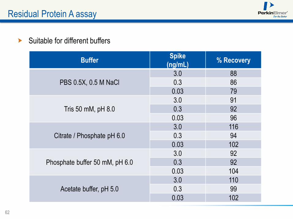
Residual Protein A assay
Buffer Spike
(ng/mL) % Recovery
PBS 0.5X, 0.5 M NaCl
3.0 88
0.3 86
0.03 79
Tris 50 mM, pH 8.0
3.0 91
0.3 92
0.03 96
Citrate / Phosphate pH 6.0
3.0 116
0.3 94
0.03 102
Phosphate buffer 50 mM, pH 6.0
3.0 92
0.3 92
0.03 104
Acetate buffer, pH 5.0
3.0 110
0.3 99
0.03 102
Suitable for different buffers
63 63
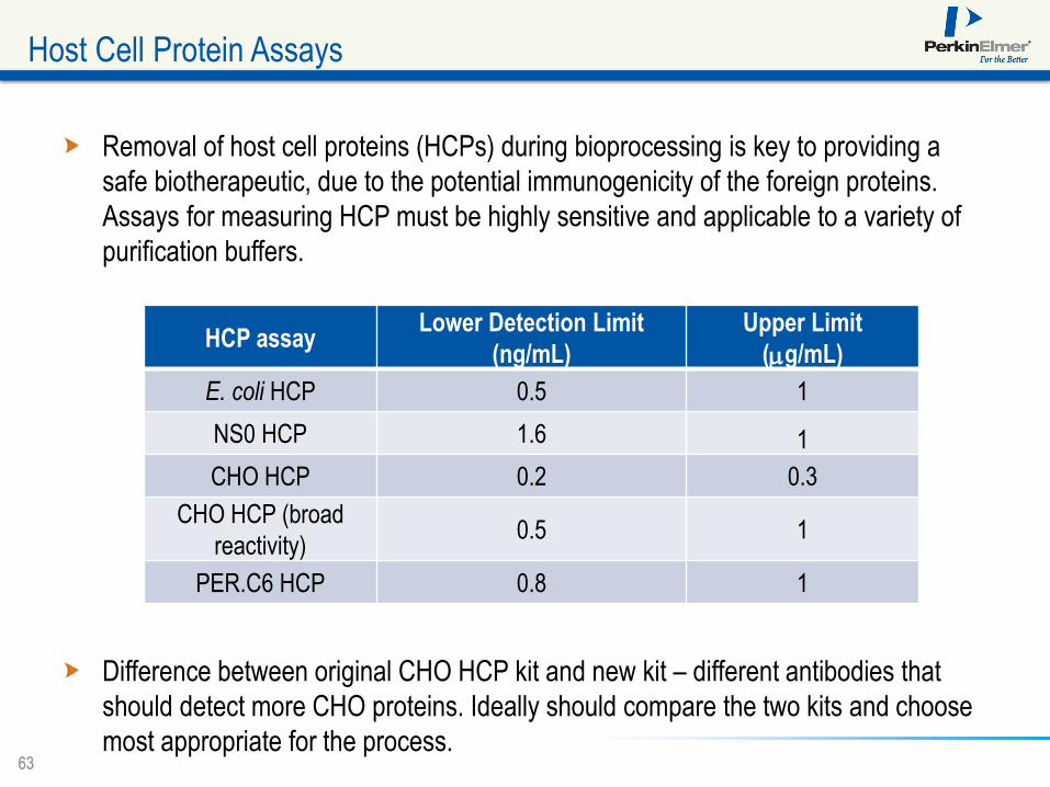
Removal of host cell proteins (HCPs) during bioprocessing is key to providing a
safe biotherapeutic, due to the potential immunogenicity of the foreign proteins.
Assays for measuring HCP must be highly sensitive and applicable to a variety of
purification buffers.
Difference between original CHO HCP kit and new kit – different antibodies that
should detect more CHO proteins. Ideally should compare the two kits and choose
most appropriate for the process.
Host Cell Protein Assays
HCP assay Lower Detection Limit
(ng/mL)
Upper Limit
(mg/mL)
E. coli HCP 0.5 1
NS0 HCP 1.6 1
CHO HCP 0.2 0.3
CHO HCP (broad
reactivity) 0.5 1
PER.C6 HCP 0.8 1
64 64
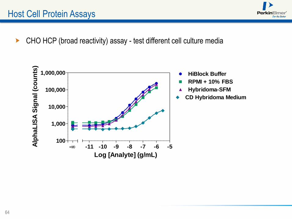
CHO HCP (broad reactivity) assay - test different cell culture media
Host Cell Protein Assays
100
1,000
10,000
100,000
1,000,000
-11 -10 -9 -8 -7 -6 -5
HiBlock Buffer
-
RPMI + 10% FBS
Hybridoma-SFM
CD Hybridoma Medium
Log [Analyte] (g/mL)
Alp
ha
LIS
A S
ign
al (c
ou
nts
)
65 65
Host Cell Protein Assays
0.0
0.5
1.0
1.5
2.0
2.5
3.0
0
20
40
60
80
100
0 50 100 150 200 250 300
Alpha HCP Kit: 2012.06.25
[Ho
st C
ell
Pro
tein
] (
mg/
mL)
Ce
ll Viab
ility (%)
Cultivation Time (h)
0.0
0.5
1.0
1.5
2.0
2.5
3.0
0
20
40
60
80
100
120
140
0 50 100 150 200 250 300
Alpha HCP Kit: 2012.06.25
[Ho
st C
ell
Pro
tein
] (
mg/
mL)
Viab
le ce
lls (x10
6 cells/mL)
Cultivation Time (h)
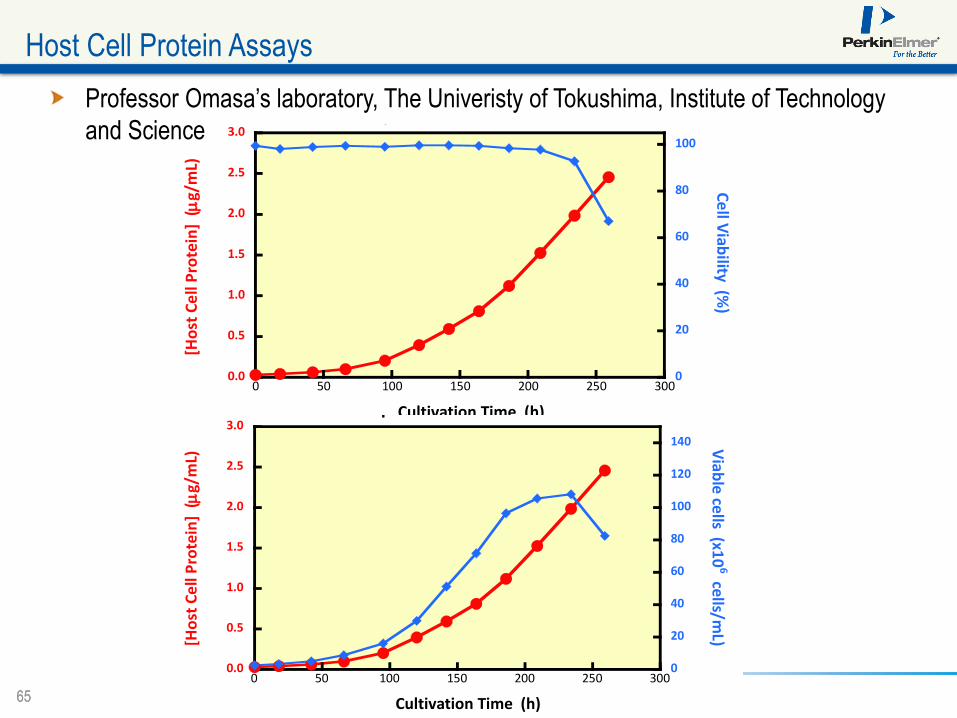
Professor Omasa’s laboratory, The Univeristy of Tokushima, Institute of Technology
and Science
66 66
Host Cell Protein Assays
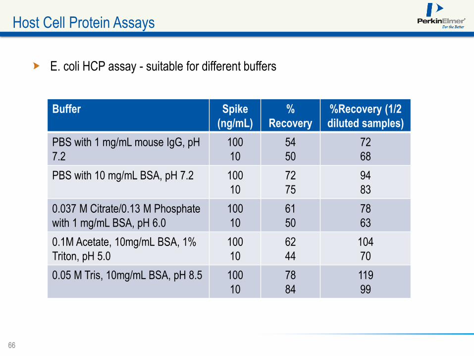
E. coli HCP assay - suitable for different buffers
Buffer Spike
(ng/mL)
%
Recovery
%Recovery (1/2
diluted samples)
PBS with 1 mg/mL mouse IgG, pH
7.2
100
10
54
50
72
68
PBS with 10 mg/mL BSA, pH 7.2 100
10
72
75
94
83
0.037 M Citrate/0.13 M Phosphate
with 1 mg/mL BSA, pH 6.0
100
10
61
50
78
63
0.1M Acetate, 10mg/mL BSA, 1%
Triton, pH 5.0
100
10
62
44
104
70
0.05 M Tris, 10mg/mL BSA, pH 8.5 100
10
78
84
119
99

67 67
Alpha technology offers:
Consistent, reproducible results - %CVs less than 10%
Simple protocol - just 4 steps to results in < 3 hrs
Equal or better sensitivity than other immunoassay technologies without tedious wash
steps
Suitable for a variety of bioprocess sample matrices
Wide dynamic range - up to 4 logs
Use anywhere you are currently using ELISA or electrochemiluminescent (ECL)
techniques
Conclusions
A Rapid, Flexible Alternative to ELISA for Protein
Detection, Characterization and Quantitation
A Rapid, Flexible Alternative to ELISA for Protein Detection,
Characterization and Quantitation
Q&A

A Rapid, Flexible Alternative to ELISA for Protein
Detection, Characterization and Quantitation
Your Moderator
Tamlyn Oliver Managing Editor
Genetic Engineering & Biotechnology News

A Rapid, Flexible Alternative to ELISA for Protein
Detection, Characterization and Quantitation
Francesco Lipari, Ph.D.
Senior Scientist
Biotherapeutic Applications
PerkinElmer Life Sciences and Technology

A Rapid, Flexible Alternative to ELISA for Protein
Detection, Characterization and Quantitation
Michael E. Bembenek, Ph.D. Lead Discovery
Scientific Fellow
Millennium: The Takeda Oncology Company
A Rapid, Flexible Alternative to ELISA for Protein
Detection, Characterization and Quantitation
Chamindie Punyadeera, Ph.D. Senior Research Fellow
The University of Queensland Diamantina Institute
The Translational Research Institute Pty Ltd.
A Rapid, Flexible Alternative to ELISA for Protein
Detection, Characterization and Quantitation
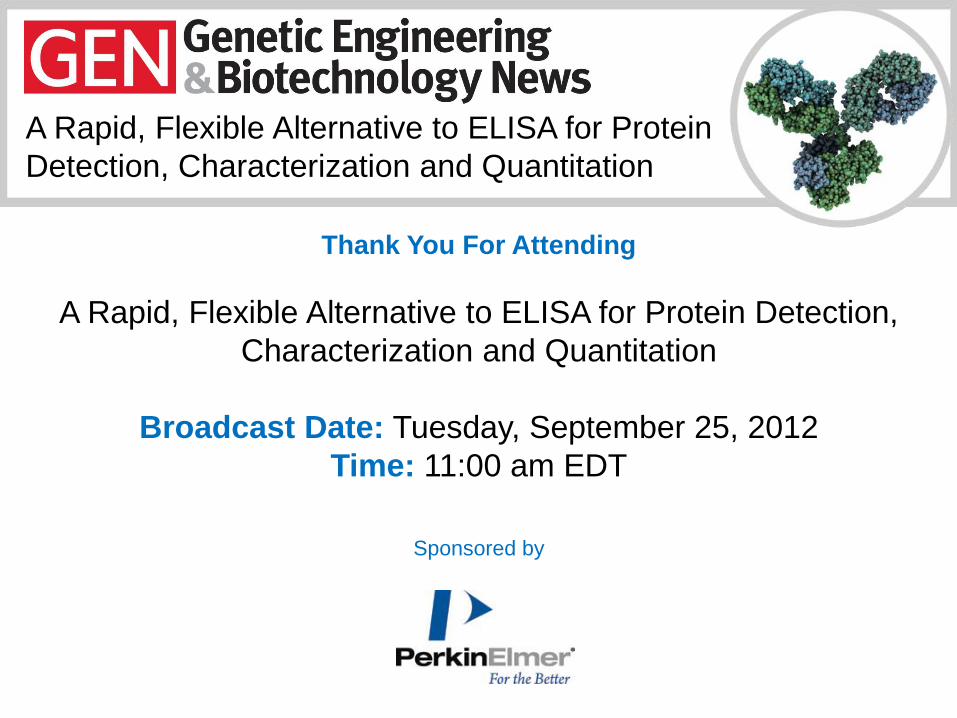
Thank You For Attending
A Rapid, Flexible Alternative to ELISA for Protein Detection,
Characterization and Quantitation
Broadcast Date: Tuesday, September 25, 2012
Time: 11:00 am EDT
Sponsored by