a new species of anatoma (vetigastropoda: anatomidae… · a new species of anatoma...
TRANSCRIPT

This reprint is protected by copyright and is provided to the purchaser or recipient for personal, non-commercial use.To obtain permission for any other use, please contact David R. Lindberg at University of California,
Department of Integrative Biology, 3060 VLSB MC# 3140, Berkeley, CA 94720-3140 or [email protected] Malacozoological Society, The Veliger, 2009
A New Species of Anatoma (Vetigastropoda: Anatomidae) from a
Hydrothermal Vent Field in Myojin Knoll Caldera, Izu-Ogasawara
Arc, Japan
TAKENORI SASAKI
The University Museum, The University of Tokyo, 7-3-1 Hongo, Bunkyo-ku, Tokyo, Japan 113-003
DANIEL L. GEIGER
Santa Barbara Museum of Natural History, 2559 Puesta del Sol Road, Santa Barbara, CA 93105, USA
(e-mail: [email protected])
AND
TAKASHI OKUTANI
Japan Agency for Marine-Earth Science and Technology, 2-15 Natsushima, Yokosuka, Kanagawa Prefecture,
Japan 237-0061
Abstract. Anatoma fujikurai sp. nov. is described from the hydrothermal-vent environment at Myojin Knoll,
southern Japan. The shell of the species is characterized by the predominant axial sculpture on the shoulder and base
and by the undulating selenizone. The animal lacks eyes and shows a radular structure not seen in any other
anatomid species examined to date. Hypotheses about its radular structure related to habitat depth, chemosynthetic
environment, and geography (Tethys Sea) are rejected; the most plausible explanation is simple interspecific
variation. The species is compared with all Japanese anatomid species, as well as to conchologically similar ones, i.e.,
those with stepped shell profiles from throughout the Indo-Pacific region. Comments on several misidentifications of
anatomid species in the literature are provided along with SEM images of the type material of Thieleella sagamiana
(Okutani, 1964) and Anatoma soyae (Habe, 1951) for comparison.
INTRODUCTION
The hydrothermal vent environment (see Van Dover,
2000, for a general review) has yielded many new
species, most of which are restricted geographically, as
well as in terms of habitat preference, to this unique
setting. Many species continue to be described from
chemosynthetic environments (e.g., Waren & Bouchet,
2001; Sasaki et al., 2005). The species of small size are
less well known in general (for a review of Japanese
species, see Sasaki, 2008), which is also the case for
deep-sea species. The family Anatomidae contains
many undescribed species worldwide (Geiger, 2008).
Here, we introduce a new taxon from the Myojin Knoll
submarine volcano off southern Japan. The examina-
tion of the anatomy of this species, and particularly the
radula, has yielded surprising results.
MATERIALS AND METHODS
Two specimens of the new species were collected from
the Myojin Knoll, south off Izu Islands, Japan,
32u06.209N, 139u52.179E, at 1224 m (see Sasaki et al.,
2003: fig. 1 for map) on June 24, 2003, on dive HD#185
of ROV Hyper-dolphin during cruise NT03-06 of R/V
Natsushima. The anterior parts of the animals were
removed from the shells by pulling the head-foot
complexes, and the visceral masses remained inside
the shells. The isolated animals were photographed
under a binocular microscope and dissected to remove
the radulae. The shells, opercula, radulae, and animals
were examined with a scanning electron microscope
(SEM) after being mounted on metal stubs and coated
with platinum-palladium. The shells and radulae were
cleaned in diluted commercial bleach, and the animals
were freeze-dried for SEM examination. Uncoated type
specimens of Thieleella sagamiana were examined using
variable-pressure SEM. The holotype and paratype of
the new species were deposited in the Department of
Historical Geology and Paleontology, The University
Museum, The University of Tokyo (UMUT).
Abbreviations for respositories of specimens and
descriptions are as follows: BMNH (The Natural
History Museum, London, UK), NSMT (National
Museum of Nature and Science, Tokyo, Japan
The Veliger 51(1):63–75 (March 31, 2010)
THE VELIGER# CMS, Inc., 2008
[formerly National Science Museum, Tokyo]);
SBMNH (Santa Barbara Museum of Natural History,
California, USA); SH (shell height); SW (shell width);
UMUT (The University Museum, The University of
Tokyo, Japan).
SYSTEMATICS
Class GASTROPODA
Clade VETIGASTROPODA
Family ANATOMIDAE McLean, 1989
Remarks: See Geiger (2003) for the differentiation of
the family from Scissurellidae and Zelaya & Geiger
(2007:395) for a diagnosis of the family. The elevation
to family rank is based on the molecular phylogeny of
Geiger & Thacker (2005, 2006).
Genus Anatoma Woodward, 1859
Type species: Scissurella crispata Fleming, 1828 (orig-
inal designation).
Anatoma s.l. fujikurai Sasaki, Geiger & Okutani,sp. nov.
(Figures 1–4)
Type material: Holotype: 3.2 (SW) 3 3.1 (SH) mm
(UMUT RM29549), paratype: 3.1 (SW) 3 2.7 (SH)
mm (UMUT RM29550).
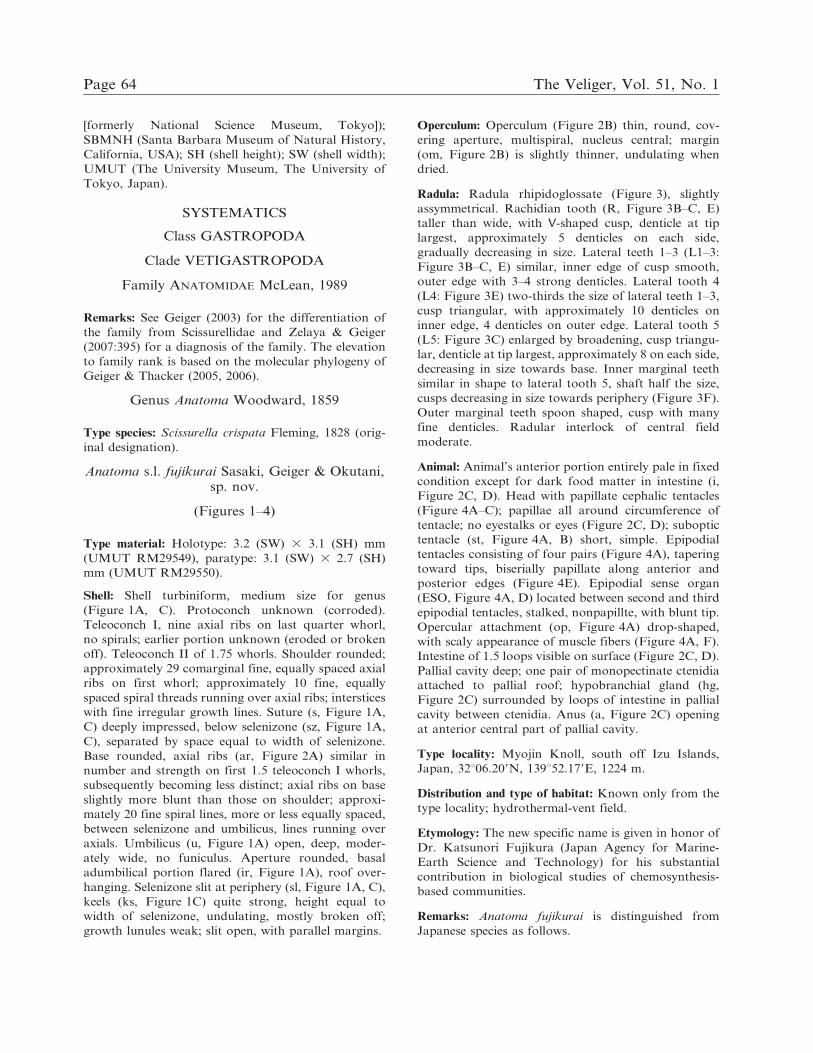
Shell: Shell turbiniform, medium size for genus
(Figure 1A, C). Protoconch unknown (corroded).
Teleoconch I, nine axial ribs on last quarter whorl,
no spirals; earlier portion unknown (eroded or broken
off). Teleoconch II of 1.75 whorls. Shoulder rounded;
approximately 29 comarginal fine, equally spaced axial
ribs on first whorl; approximately 10 fine, equally
spaced spiral threads running over axial ribs; interstices
with fine irregular growth lines. Suture (s, Figure 1A,
C) deeply impressed, below selenizone (sz, Figure 1A,
C), separated by space equal to width of selenizone.
Base rounded, axial ribs (ar, Figure 2A) similar in
number and strength on first 1.5 teleoconch I whorls,
subsequently becoming less distinct; axial ribs on base
slightly more blunt than those on shoulder; approxi-
mately 20 fine spiral lines, more or less equally spaced,
between selenizone and umbilicus, lines running over
axials. Umbilicus (u, Figure 1A) open, deep, moder-
ately wide, no funiculus. Aperture rounded, basal
adumbilical portion flared (ir, Figure 1A), roof over-
hanging. Selenizone slit at periphery (sl, Figure 1A, C),
keels (ks, Figure 1C) quite strong, height equal to
width of selenizone, undulating, mostly broken off;
growth lunules weak; slit open, with parallel margins.
Operculum: Operculum (Figure 2B) thin, round, cov-
ering aperture, multispiral, nucleus central; margin
(om, Figure 2B) is slightly thinner, undulating when
dried.
Radula: Radula rhipidoglossate (Figure 3), slightly
assymmetrical. Rachidian tooth (R, Figure 3B–C, E)
taller than wide, with V-shaped cusp, denticle at tip
largest, approximately 5 denticles on each side,
gradually decreasing in size. Lateral teeth 1–3 (L1–3:
Figure 3B–C, E) similar, inner edge of cusp smooth,
outer edge with 3–4 strong denticles. Lateral tooth 4
(L4: Figure 3E) two-thirds the size of lateral teeth 1–3,
cusp triangular, with approximately 10 denticles on
inner edge, 4 denticles on outer edge. Lateral tooth 5
(L5: Figure 3C) enlarged by broadening, cusp triangu-
lar, denticle at tip largest, approximately 8 on each side,
decreasing in size towards base. Inner marginal teeth
similar in shape to lateral tooth 5, shaft half the size,
cusps decreasing in size towards periphery (Figure 3F).
Outer marginal teeth spoon shaped, cusp with many
fine denticles. Radular interlock of central field
moderate.
Animal: Animal’s anterior portion entirely pale in fixed
condition except for dark food matter in intestine (i,
Figure 2C, D). Head with papillate cephalic tentacles
(Figure 4A–C); papillae all around circumference of
tentacle; no eyestalks or eyes (Figure 2C, D); suboptic
tentacle (st, Figure 4A, B) short, simple. Epipodial
tentacles consisting of four pairs (Figure 4A), tapering
toward tips, biserially papillate along anterior and
posterior edges (Figure 4E). Epipodial sense organ
(ESO, Figure 4A, D) located between second and third
epipodial tentacles, stalked, nonpapillte, with blunt tip.
Opercular attachment (op, Figure 4A) drop-shaped,
with scaly appearance of muscle fibers (Figure 4A, F).
Intestine of 1.5 loops visible on surface (Figure 2C, D).
Pallial cavity deep; one pair of monopectinate ctenidia
attached to pallial roof; hypobranchial gland (hg,
Figure 2C) surrounded by loops of intestine in pallial
cavity between ctenidia. Anus (a, Figure 2C) opening
at anterior central part of pallial cavity.
Type locality: Myojin Knoll, south off Izu Islands,
Japan, 32u06.209N, 139u52.179E, 1224 m.
Distribution and type of habitat: Known only from the
type locality; hydrothermal-vent field.
Etymology: The new specific name is given in honor of
Dr. Katsunori Fujikura (Japan Agency for Marine-
Earth Science and Technology) for his substantial
contribution in biological studies of chemosynthesis-
based communities.
Remarks: Anatoma fujikurai is distinguished from
Japanese species as follows.
Page 64 The Veliger, Vol. 51, No. 1
Anatoma lamellata (A. Adams, 1862) from Japan has
a similar overall shape and similar density of axial
lamellae. However, on teleoconch I, it has a distinct
spiral cord in the position of the selenizone (absent in
A. fujikurai), teleoconch I is of less then 0.5 whorls
(.0.75 in A. fujikurai), the spiral cords are approxi-
mately one-third the strength of the axial lamellae
(, one-fifth in A. fujikurai), the suture is 1.5 times the
width of the selenizone below the selenizone (space
equal to width of selenizone in A. fujikurai), and a
distinct funiculus is present in the umbilical cavity
(absent in A. fujikurai). The differentiation is based on
Figure 1. Anatoma fujikurai n. sp. Shell. A, Frontal view. Arrowhead indicates end of slit. B, Apical view. C, Back view. D, Basalview. Abbreviations: a 5 apex; ir 5 reflected part of inner lip; ks 5 keels along selenizone; s 5 suture; sl 5 slit; sz 5 selenizone; u 5
umbilicus. A–B, Holotype, UMUT RM29549. C–D, Paratype, UMUT RM29550.
T. Sasaki et al., 2008 Page 65

Figure 2. Anatoma fujikurai n. sp. A, Enlargement of sulpture of body whorl. B, Exterior view of operculum. C, Dorsal view ofanterior part of the animal. D, Lateral view of same part. Abbreviations: a 5 anus; ar 5 axial ribs; c 5 ctenidium; ct 5 cephalictentacle; ept 5 epipodial tentacle; f 5 foot; hg 5 hypobranchial gland; i 5 intestine; mm 5 mantle margin; om 5 thin margin ofoperculum; s 5 suture; sn 5 snout; ss 5 spiral streaks; sz 5 selenizone. A, Holotype, UMUT RM29549. B–D, Paratype,UMUT RM29550.
Page 66 The Veliger, Vol. 51, No. 1

the examination of type material in the BMNH by
SEM (Geiger, pers. obs.). The species has usually been
misidentified, e.g., Habe (1951) and Izawa & Matsuoka
(1999) illustrated an anomphalous species with a
distinct absence of spiral cords just below the
selenizone, characters not found in A. lamellata, but
quite typical for T. reticulata; McLean’s (1967: pl. 56,
fig. 8) illustrated specimen is an A. lyra (Berry, 1947);
Kuroda et al. (1971) showed an unidentified anatomid
species lacking the lamellae typical for A. lamellata;
Figure 3. Anatoma fujikurai n. sp. Radula. A, D, Six rows of whole radular teeth. B, E, Rachidian (R) and lateral teeth (L1–L4).C, Entire view of rachidian and part of lateral teeth, showing bases of teeth, F, Enlargement of left rows of marginal teeth. A–C,Holotype, UMUT RM29549. D–F, Paratype, UMUT RM29550.
T. Sasaki et al., 2008 Page 67

Numanami & Okutani (1990) showed an unidentified
species not conspecific with A. lamellata; Okutani &
Hasegawa (2000: pl. 18, figs. 1a, 1b) figured Thieleela
reticulata Bandel, 1998. Other published illustrations
(e.g., Habe, 1961) are so small as to make positive
identification impossible. Only Tsuchida et al. (1991)
and Tsuchida & Hori (1996) illustrated the true A.
lamellata, and Thiele’s (1912) line drawing agrees
exceptionally well with the SEM images of the type
material.
Anatoma japonica (A. Adams, 1862) from Japan has
an overall biconical shape (stepped in A. fujikurai); the
density of the axials is at least twice as high as in A.
fujikurai, while the spirals are almost as strong as the
axials (, one-fifth in A. fujikurai), and the keels of the
open slit converge towards the apertural margin (they
Figure 4. Anatoma fujikurai n. sp. Head-foot of animal. Paratype UMUT RM29550. A, Left lateral view of head-foot with buccalmass removed. B, Enlargement of head. C, Papillate tip of left cephalic tentacle. D, First to second left epipodial tentacles withepipodial sense organ. E, Fourth left epipodial tentacle. F, Surface of opercular attachment. Abbreviations: ct 5 cephalic tentacles;ESO 5 epipodial sense organ; L1–L4 5 first to fourth left epipodial tentacles; ol 5 outer lip of mouth; op 5 opercular lobe; R3–R45 third and fourth right epipodial tentacles; sn 5 snout; st 5 suboptic tentacle.
Page 68 The Veliger, Vol. 51, No. 1

maintain same width in A. fujikurai). The differentia-
tion is based on the examination of type material in the
BMNH by SEM (D. L. Geiger, personal observation).
Anatoma soyae (Habe, 1951) from Japan has an
overall biconical shape (stepped in A. fujikurai), has
denser axial sculpture (17 vs. 9 on the last quarter of
teleoconch I), and the suture inserts immediately below
the selenizone on early whorls (space equal to width of
selenizone in A. fujikurai), and the keels of the selenizone
are low [eroded?] (as high as selenizone width in A.
fujikurai). The differentiation is based on SEM images
of the holotype (NSMT Mo-38615: Figure 5).
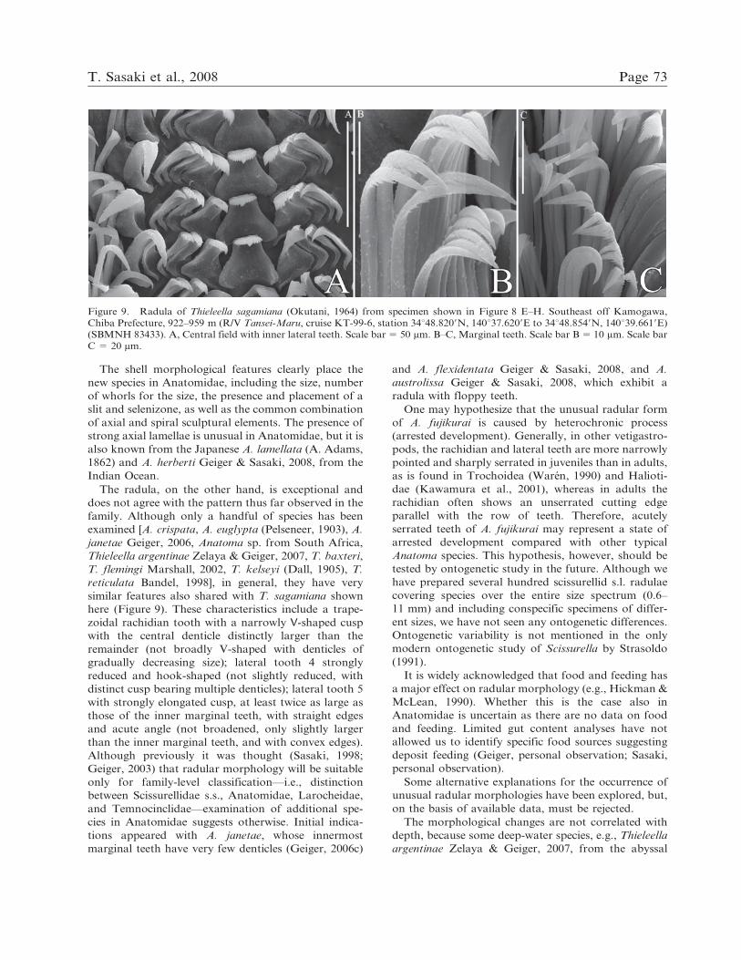
Thieleella sagamiana (Okutani, 1964) has a proto-
conch with reticulate sculpture, slightly shorter tele-
oconch I (0.66 vs. .0.75 whorls) with slightly more
axial cords (11 vs. 9 on last quarter whorl). On
teleoconch II, the axials are not elevated to low
lamellae as in A. fujikurai, and T. sagamiana is
anomphalous, while A. fujikurai shows a distinct
umbilicus. The comparison is based on SEM examina-
tion of the holotype (UMUT RM8808: Figure 6,
unfigured in the original description), the paratype
(NSMT Mo-69582: Figure 7, Okutani, 1964: pl. 5, fig.
2), and conspecific material collected from southeast off
Kamogawa, Chiba Prefecture, 922–959 m (R/V Tansei-
Maru, cruise KT-99-6, station 14, 34u48.8209N,
140u37.6209E to 34u48.8549N, 140u39.6619E), Hyuga
Basin, 779–803 m (cruise KT-93-9, station HY-4,
31u539220N, 131u529160E to 31u549160N, 131u539230E),
and Kumano Basin, 2029–2045 m (cruise KT-86-6,
Figure 5. Holotype of Anatoma soyae (Habe, 1951), NSMT Mo-38615. A, Frontal view. B, Basal view. C, Apical view.D, Protoconch.
T. Sasaki et al., 2008 Page 69

station KN6, 33u46960N 136u40900E to 33u46960N,
136u37960E) (Figure 8).
Among Indo-Pacific species with overall stepped
shell shape, A. fujikurai is distinguished as follows.
Anatoma indonesica Bandel, 1998, has on teleoconch
I a spiral cord in the position of the selenizone (absent
in A. fujikurai), has more strongly elevated spiral
lamellae on the shoulder, crossed by approximately 4–8
fine spiral lines concentrated in the middle of the
shoulder (equally distributed in A. fujikurai), and has
fine cancellate sculpture on the base (strong axials and
fine spirals in A. fujikurai).
Anatoma boucheti Geiger & Sasaki, 2008, from
Reunion Island, Indian Ocean, has a teleoconch of
1.125 whorls, shows fine reticulate sculpture of axials
and spiral lines of approximately equal strength, and
has the keels of the selenizone distinctly elevated.
Anatoma herberti Geiger & Sasaki, 2008, from
Reunion Island, Indian Ocean, has more strongly
elevated axial lamellae and a distinct constriction of
the base below the selenizone (absent in A. fujikurai),
which bears strongly elevated keels (low/eroded in A.
fujikurai) bearing three to four fine axial striae between
each axial cord (none in A. fujikurai).
Anatoma lamellata nanshaensis Feng, 1996, has on
teleoconch I a spiral cord in the position of the
selenizone (absent in A. fujikurai), the shoulder is at an
angle of approximately 10u (45u in A. fujikurai), the
selenizone is in the upper third of the whorl (below the
midline in A. fujikurai), and has fine reticulate sculpture
Figure 6. Holotype of Thieleella sagamiana (Okutani, 1964), UMUT RM-8808. A, Frontal view. B, Basal view. C, Apical view.
Page 70 The Veliger, Vol. 51, No. 1

on the base (more prominent axials in A. fujikurai). The
differentiation is based on the original figures of Feng
(1996).
Anatoma obtusata (Golikov & Gublin, 1978) is
overall more depressed (height-to-width ratio 5 0.86:
1.03 in A. fujikurai), seems to have reticulate sculpture
with axials and spiral of similar strength (axials
predominant in A. fujikurai) and has less distinct
(eroded?) keels of the selenizone. The differentiation is
based on the original line drawing and photographs of
the holotype in Kantor & Sysoev (2006).
DISCUSSION
The generic placement of Anatoma s.l. fujikurai is
uncertain, because the diagnostic protoconch sculpture
is not preserved in either of the two known specimens.
Anatoma s.s. has either smooth or flocculent sculpture,
while Thieleella has reticulate sculpture. Whether this
character is sufficient to justify generic distinction has
been discussed (Marshall, 2002; Geiger, 2003; Geiger &
Jansen, 2004; Zelaya & Geiger, 2007) and is provision-
ally accepted (see also Geiger, 2006a, b, c).
Figure 7. Paratype of Thieleella sagamiana (Okutani, 1964), NSMT Mo-69582. A, Frontal view. B, Basal view. C, Apical view.D, Protoconch.
T. Sasaki et al., 2008 Page 71

Figure 8. Thieleella sagamiana (Okutani, 1964). A–D, Kumano Basin, 2029–2045 m (R/V Tansei-Maru, cruise KT-86-6, stationKN6, 33u46960N, 136u40900E to 33u46960N, 136u37960E) (SBMNH 83432). E–H, Southeast off Kamogawa, Chiba Prefecture, 922–959 m (R/V Tansei-Maru, cruise KT-99-6, station 34u48.8209N, 140u37.6209E to 34u48.8549N, 140u39.6619E) (SBMNH 83433). A,E, Frontal view; B, F, Basal view; C, G, Apical view; D, H, Protoconch.
Page 72 The Veliger, Vol. 51, No. 1
The shell morphological features clearly place the
new species in Anatomidae, including the size, number
of whorls for the size, the presence and placement of a
slit and selenizone, as well as the common combination
of axial and spiral sculptural elements. The presence of
strong axial lamellae is unusual in Anatomidae, but it is
also known from the Japanese A. lamellata (A. Adams,
1862) and A. herberti Geiger & Sasaki, 2008, from the
Indian Ocean.
The radula, on the other hand, is exceptional and
does not agree with the pattern thus far observed in the
family. Although only a handful of species has been
examined [A. crispata, A. euglypta (Pelseneer, 1903), A.
janetae Geiger, 2006, Anatoma sp. from South Africa,
Thieleella argentinae Zelaya & Geiger, 2007, T. baxteri,
T. flemingi Marshall, 2002, T. kelseyi (Dall, 1905), T.
reticulata Bandel, 1998], in general, they have very
similar features also shared with T. sagamiana shown
here (Figure 9). These characteristics include a trape-
zoidal rachidian tooth with a narrowly V-shaped cusp
with the central denticle distinctly larger than the
remainder (not broadly V-shaped with denticles of
gradually decreasing size); lateral tooth 4 strongly
reduced and hook-shaped (not slightly reduced, with
distinct cusp bearing multiple denticles); lateral tooth 5
with strongly elongated cusp, at least twice as large as
those of the inner marginal teeth, with straight edges
and acute angle (not broadened, only slightly larger
than the inner marginal teeth, and with convex edges).
Although previously it was thought (Sasaki, 1998;
Geiger, 2003) that radular morphology will be suitable
only for family-level classification—i.e., distinction
between Scissurellidae s.s., Anatomidae, Larocheidae,
and Temnocinclidae—examination of additional spe-
cies in Anatomidae suggests otherwise. Initial indica-
tions appeared with A. janetae, whose innermost
marginal teeth have very few denticles (Geiger, 2006c)
and A. flexidentata Geiger & Sasaki, 2008, and A.
austrolissa Geiger & Sasaki, 2008, which exhibit a
radula with floppy teeth.
One may hypothesize that the unusual radular form
of A. fujikurai is caused by heterochronic process
(arrested development). Generally, in other vetigastro-
pods, the rachidian and lateral teeth are more narrowly
pointed and sharply serrated in juveniles than in adults,
as is found in Trochoidea (Waren, 1990) and Halioti-
dae (Kawamura et al., 2001), whereas in adults the
rachidian often shows an unserrated cutting edge
parallel with the row of teeth. Therefore, acutely
serrated teeth of A. fujikurai may represent a state of
arrested development compared with other typical
Anatoma species. This hypothesis, however, should be
tested by ontogenetic study in the future. Although we
have prepared several hundred scissurellid s.l. radulae
covering species over the entire size spectrum (0.6–
11 mm) and including conspecific specimens of differ-
ent sizes, we have not seen any ontogenetic differences.
Ontogenetic variability is not mentioned in the only
modern ontogenetic study of Scissurella by Strasoldo
(1991).
It is widely acknowledged that food and feeding has
a major effect on radular morphology (e.g., Hickman &
McLean, 1990). Whether this is the case also in
Anatomidae is uncertain as there are no data on food
and feeding. Limited gut content analyses have not
allowed us to identify specific food sources suggesting
deposit feeding (Geiger, personal observation; Sasaki,
personal observation).
Some alternative explanations for the occurrence of
unusual radular morphologies have been explored, but,
on the basis of available data, must be rejected.
The morphological changes are not correlated with
depth, because some deep-water species, e.g., Thieleella
argentinae Zelaya & Geiger, 2007, from the abyssal
Figure 9. Radula of Thieleella sagamiana (Okutani, 1964) from specimen shown in Figure 8 E–H. Southeast off Kamogawa,Chiba Prefecture, 922–959 m (R/V Tansei-Maru, cruise KT-99-6, station 34u48.8209N, 140u37.6209E to 34u48.8549N, 140u39.6619E)(SBMNH 83433). A, Central field with inner lateral teeth. Scale bar 5 50 mm. B–C, Marginal teeth. Scale bar B 5 10 mm. Scale barC 5 20 mm.
T. Sasaki et al., 2008 Page 73

plain of the South Atlantic, has the typical anatomid
radular pattern. It is tantalizing that two deep-water
species from Reunion Island (A. flexidentata, A.
austrolissa: Geiger & Sasaki, 2008) show a very similar
yet untypical radular pattern of floppy teeth, but the
broader significance is difficult to assess, given the
sparse data available.
The hydrothermal vent environment is also unlikely
as an explanatory factor, as shown by the only
moderately modified radula of A. janetae, also collected
from sites in the vicinity of hydrothermal vents.
The three species with the most aberrant radulae are
from the Tethys area: Japan (A. fujikurai) and the
Indian Ocean (A. flexidentata, A. austrolissa: Geiger &
Sasaki, 2008). The significance of this observation is
uncertain because of generally poor sampling of the
deep sea owing to a lack of suitable material and the
relatively few Atlantic species that have having been
examined (Geiger, 2008).
The current contribution highlights the wealth of
interesting new information that is obtained from small
molluscan species. The examination of such understud-
ied groups of micromollusks as Anatomidae shows
significant promise for the discovery of novel charac-
ters and adaptive pathways.
Acknowledgments. We thank Dr. Bruce Marshall (Museum ofNew Zealand Te Papa Tongarewa) and Prof. Carole S.Hickman (University of California, Berkeley) for criticallyreading the manuscript and helping to improve it. Thematerials of new species were collected with the kind assistanceof the operation team of ROV Hyper-dolphin and the crew ofR/V Natsushima (Japan Agency for Marine-Earth Science andTechnology: JAMSTEC). Thieleella sagamiana (Figures 8–9)was provided from the late Mr. Eiji Tuschida (formerly of theOcean Research Institute, The University of Tokyo: ORI) andalso collected with support by Prof. Suguru Ohta (formerly ofthe ORI) and the crew of R/V Tansei-Maru (JAMSTEC,formerly of the ORI). Dr. Kathie Way accommodated DLGduring visits to the BMNH. Drs. Tsunemi Kubodera, HiroshiSaito, and Kazunori Hasegawa assisted us during visits to theNational Museum of Nature and Science. This study wassupported by a grant-in-aid for Scientific Research from theJapan Society for the Promotion of Science (no. 18340165,20540455) and by US National Science Foundation grantMRI 0402726.
LITERATURE CITED
FENG, W. 1996. Microgastropods from Nansha Sea area,China. Pp. 85–205 in Studies on Marine Fauna and Floraand Biogeography of the Nansha Islands and Neighbour-ing Waters II. Ocean Press: Beijing.
GEIGER, D. L. 2003. Phylogenetic assessment of charactersproposed for the generic classification of Recent Scissur-ellidae (Gastropoda: Vetigastropoda) with description ofone new genus and six new species from Easter Island andAustralia. Molluscan Research 23:21–83.
GEIGER, D. L. 2006a. Eight new species of Scissurellidae andAnatomidae (Mollusca: Gastropoda: Vetigastropoda)
from around the world, with discussion of two new seniorsynonyms. Zootaxa 1128:1–33.
GEIGER, D. L. 2006b. Sasakiconcha elegantissima new genusand species (Vetigastropoda: Anatomidae?) with dis-jointly coiled base. The Nautilus 120:45–51.
GEIGER, D. L. 2006c. A new blind Anatoma species from thebathyal of the northeastern Pacific (Vetigastropoda:Anatomidae). Molluscan Research 28:108–112.
GEIGER, D. L. 2008. Monographing micromollusks: a casestudy on Scissurellidae s.l. (Vetigastropoda). In D. L.Geiger & B. Ruthensteiner (eds.), Micromolluscs: Meth-odological Challenges—Exciting Results. Zoosymposia 1:133–145.
GEIGER, D. L. & P. JANSEN. 2004. Revision of the Australianspecies of Anatomidae (Mollusca: Gastropoda: Vetigas-tropoda). Zootaxa 414:1–35.
GEIGER, D. L. & C. E. THACKER. 2005. Molecular phylogenyof Vetigastropoda reveals non-monophyletic Scissurelli-dae, Trochoidea, and Fissurelloidea. Molluscan Research25:47–55.
GEIGER, D. L. & C. E. THACKER. 2006. Molecular phylogenyof basal gastropods (Vetigastropoda) shows stochasticvent colonization at least from the mid Triassic. Cahiersde Biologie Marine 46:343–346.
GEIGER, D. L. & T. SASAKI. 2008. Four new species ofAnatomidae (Mollusca: Vetigastropoda) from the IndianOcean (Reunion, Mayotte) and Australia, with notes on anovel radular type for the family. In D. L. Geiger & B.Ruthensteiner (eds.), Micromolluscs: MethodologicalChallenges—Exciting Results). Zoosymposia 1:247–264.
HABE, T. 1951. Scissurellidae in Japan. Illustrated Catalogueof Japanese Shells 11:65–69.
HABE, T. 1961. Colored Illustrations of Shells of Japan, vol. 2,first ed. Hoikusha: Osaka. 183 pp. 1–42 Appendix.
HICKMAN, C. S. & J. H. MCLEAN. 1990. Systematic revisionand suprageneric classification of trochacean gastropods.Science Series Natural History Museum of Los AngelesCounty 35:1–169.
IZAWA, N. & K. MATSUOKA. 1999. Catalogue of shellcollection by Mr. Hiroshi Takakuwa presented toToyohashi Museum of Natural History. Miscellaneousreport of the Toyohashi Museum of Natural History 7:1–25.
KANTOR, I. YU & A. V. SYSOEV. 2006. Marine and BrackishWater Gastropoda of Russia and Adjacent Countries: AnIllustrated Catalogue. KMK Scientific Press: Moscow.371 pp., 140 pls.
KAWAMURA, T., H. TAKAMI, R. D. ROBERTS & Y.YAMASHITA. 2001. Radula development in abaloneHaliotis discus hannai from larva to adult in relation tofeeding transitions. Fisheries Science 67:596–605.
KURODA, T., T. HABE & K. OYAMA. 1971. The Sea Shells ofSagami Bay. Maruzen: Tokyo. 489 + 51 pp., 121 pls.
MARSHALL, B. A. 2002. Some Recent scissurellids from theNew Zealand region, and remarks on some scissurellidgenus group names (Mollusca: Gastropoda). MolluscanResearch 22:165–181.
MCLEAN, J. H. 1967. West American Scissurellidae. TheVeliger 9:404–410.
NUMANAMI, H. & T. OKUTANI. 1990. A new and two knownspecies of the genus Anatoma collected by the IcebreakerShirase from Breid Bay and Gunnerus Bank, Antarctica(Gastropoda: Scissurellidae). Venus 49:93–106.
OKUTANI, T. 1964. Report on the archibenthal and abyssalgastropod Mollusca mainly collected from Sagami Bay
Page 74 The Veliger, Vol. 51, No. 1

and adjacent waters by the R. V. Soyo-Maru during theyears 1955–1963. Journal of the Faculty of Science,University of Tokyo, Section II, 15:371–447, pls. 1–7.
OKUTANI, T. & K. HASEGAWA. 2000. Family Scissurellidae.Pp. 36–37 in T. Okutani (ed.), Marine Mollusks in Japan.Tokai University Press: Tokyo.
SASAKI, T. 1998. Comparative anatomy and phylogeny of theRecent Archaeogastropoda (Mollusca: Gastropoda). TheUniversity Museum, University of Tokyo, Bulletin 38:1–224.
SASAKI, T. 2008. Micromolluscs in Japan: taxonomiccomposition, habitats, and future topics. In D. L. Geiger& B. Ruthensteiner (eds.), Micromolluscs: Methodo-logical Challenges—Exciting Results. Zoosymposia 1:147–232.
SASAKI, T., T. OKUTANI & K. FUJIKURA. 2003. New taxa andnew records of patelliform gastropods associated withchemoautosynthesis-based communities in Japanese wa-ters (Mollusca: Gastropoda). The Veliger 46:189–210.
SASAKI, T., T. OKUTANI & K. FUJIKURA. 2005. Molluscsfrom hydrothermal vents and cold seeps in Japan: areview of taxa recorded in twenty recent years (1984–2004). Venus 64:87–133.
STRASOLDO, M. 1991. Anatomie und Ontogenie von Scissur-ella jucunda (Smith, 1890) und Anatomie von Anatoma sp.Dissertation, University of Vienna, Austria. 145 pp., 37pls.
THIELE, J. 1912. Scissurelliden und Fissurelliden. Pp. 1–34,pls. 1–4 in H. C. Kobelt & W. Kuster (eds.), System-atisches Conchylien-Cabinet von Martini und Chemnitz.Bauer & Raspe: Nurnberg.
TSUCHIDA, E., Y. SHIKANO, S. HORI & T. MITOKI. 1991.Study on the Mollusca of Yamaguchi Pref. - 3 Revision ofthe remarkable molluscan shells from Yamaguchi Pref. inMr. T. Kawamoto’s Collection. (2) Gastropoda andPelecypoda. Bulletin of the Yamaguchi Museum 17:1–40. (in Japanese).
TSUCHIDA, E. & S. HORI. 1996. Marine molluscs aroundMishima and Tsunoshima Islands, Japan Sea collected bythe R/V Tansei-Maru. Bulletin of the National ScienceMuseum Series A (Zoology) 22:219–261.
VAN DOVER, C. L. 2000. The Ecology of Deep-seaHydrothermal Vents. Princeton University Press: Prince-ton. 424 pp.
WAREN, A. 1990. Ontogenetic changes in the trochoidean(Archaeogastropoda) radula, with some phylogeneticinterpretations. Zoologica Scripta 19:179–187.
WAREN, A. & P. BOUCHET. 2001. Gastropoda and Mono-placophora from hydrothermal vents and seeps; new taxaand records. The Veliger 44:116–231.
ZELAYA, D. G. & D. L. GEIGER. 2007. Species ofScissurellidae and Anatomidae from Sub-Antarctic andAntarctic waters (Gastropoda: Vetigastropoda). Malaco-logia 49:393–443.
T. Sasaki et al., 2008 Page 75