a contemporary view of the human placenta
TRANSCRIPT

Midwifery (1991) 7, 31-39 © Longman Group UK Ltd 1991 Midwifery CLINICAL REVIEW
A contemporary view of the human placenta
H Fox
O u r c u r r e n t knowledge o f the h u m a n p lacenta is briefly rev iewed. Par t icu lar stress is p laced u p o n the cons ide rab le func t iona l reserve capacity o f the placenta , the u n i m p o r t a n c e o f mos t visible abnormal i t ies o f the placenta , the lack o f any ev idence that the p lacenta ages d u r i n g gestat ion an d the lack o f significance o f placental weight . T h e effects on the p lacenta o f in fec t ion a n d o f m a t e rn a l c igaret te smoking are c o n s i d e r e d and the concep t o f placental insuff ic iency critically discussed. I t is c o n c l u d e d tha t mos t cases o f 'p lacental insuff ic iency ' are, in reality, examples o f ma t e rna l vascular insuff ic iency resu l t ing f r o m i n a d e q u a t e p lacen ta t ion d u r i n g the ear ly stages o f p r egnancy .
The placenta is, in functional terms, a highly complex organ though, without question, its most important task is to transfer oxygen and nutrients from the mother to the fetus. In a short review it is impossible to consider all aspects of the anatomy, physiology, endocrinology, immu- nology and pathology of the placenta and it is intended to discuss here only selected topics. Selectivity of this type can be invidious but the topics chosen are those which are considered to be of greatest practical importance to the midwife.
PLACENTAL STRUCTURE
Despite its many functional roles the placenta has a relatively simple structure. It is a villous haemochorial organ i.e. it has a villous structure
H Fox MD, FRC Path, FRCOG, Professor of Reproductive Pathology, Department of Pathological Sciences, Stopford Building, University of Manchester, Manchester M13 9PT. Requests for offprints to HF
and the chorion, or trophoblast, is in direct contact with maternal blood in the intervillous space. The organ consists of a number of sub- units, or lobules, each of which resembles an inverted tree (Fig. 1). These lobules, it should be noted, do not correspond with the 'lobes' seen on the maternal surface whilst the term 'cotyledon' should be avoided when describing the non- cotyledonary human placenta.
Each lobule consists of a framework of stem villi which branch to form intermediate villi from which 'bud o f f the terminal villi. The villi have a central mesenchymal core and a covering mantle of trophoblast (Fig. 2). The villous core contains mesenchymal tissue, macrophages (Hofbauer cells) and fetal capillaries which are initially small but dilate progressively throughout pregnancy until, at term, they occupy most of the cross sectional area of the villus.
In early pregnancy the villous throphoblast is of uniform thickness and consists of two layers of cells, an inner stratum of cytotrophoblastic cells with well defined limiting membranes and an outer layer of syncytiotrophoblast. The latter is a true syncytium for no cell boundaries are seen
31

32 MIDWIFERY
Fig. 1 Diagrammatic representation of two placental fetal Iobules and of the maternal blood f low through these Iobules
between the nuclei whilst microinjection studies show that substances flow freely through this layer and pass from villus to villus. This indicates that there is a continuous .common cytoplasm over the entire surface of the placental villi (Gaunt & Ockleford, 1986). The syncytiotropho- blast is derived from the cytotrophoblastic cells, the syncytiotrophoblast being a terminally differentiated tissue and incapable of mitotic activity. The cytotrophoblastic cells are there- fore the stem cells of the trophoblast but become less numerous and prominent as pregnancy progresses.
In the later stages of gestation the syncytiotro- phoblast is focally thinned and anuclear. These attenuated areas usually overlie a dilated fetal capillary and appear almost to fuse with the vessel wall. These 'vasculo-syncytial membranes' represent the points of closest approximation of maternal and fetal blood and are specialised zones of the trophoblast for the facilitation of materno-fetal oxygen transfer.
The placental villi are bathed in maternal blood which fills the intervillous space. The blood enters the space from the uteroplacental vessels, flows slowly around the villi and then drains into basal venous outlets. It is important to note that the placenta depends solely on the maternal blood for its oxygen and nutrient supply.
DEVELOPMENTAL ABNORMALITIES
The only common developmental abnormality of the placenta is extrachorial placentation, in which the chorionic plate of the placenta, f rom which the villi arise, is smaller than the basal plate. The transition from villous tissue to non- villous membranes takes place therefore not at the placental margin but at some distance within the circumference of the fetal surface of the placenta (Fig. 3). If this transition is marked by a flattened ring of membranes the placenta is classed as 'circummarginate' whilst if this ring has a raised, rolled edge the placenta is 'circum- vallate'. The clinical significance of extrachorial placentation has been much disputed but it is now clear that the circummarginate form is
Q
O Fig. 2 Diagrammatic representation of the placental villi at various stages of pregnancy. (a) A placental villus from a first trimester placenta. (b) A placental villus from a second trimester placenta. (c) A placental villus from a term placenta. FV = fetal vessel, S = Syncytiotrophoblast

Extrachorial placenta
Fetal vessels
Double layer of membranes
Fig. 3 Diagrammatic representation of an extra chorial placenta. A rim of placental tissue extends beyond the boundaries of the chorionic plate.
devoid of clinical significance and the circum- vallate placenta is frequently associated with a rather small baby; and possibly with a slight excess of congenital malformations (Laderma- cher et al, 1981). It is not accompanied by an increased perinatal mortality (Fox & Sen, 1972). Other aberrant forms of placentation are either of no functional significance, e.g. bilobate placenta, accessory lobes, or so rare that they can in practical terms be ignored, e.g. placenta membranacea, girdle placenta.
The site of cord insertion, (central, marginal or velamentous) is of no functional importance (Uyanwah-Akpom & Fox, 1977: Wood & Malan, 1978), though velamentous insertion does present a small risk because of the danger of trauma to the unprotected fetal vessels as they run through the membranes.
PLACENTAL WEIGHT
In routine practice it is virtually impossible to obtain a true estimate of placental mass after delivery, largely because of the variable quanti- ties of maternal and fetal blood t rapped within the placenta. Even if placental weight could be assessed accurately this information would, in itself, be of little value. Placental/fetal weight ratios are more meaningful but it would not be unfair to say that the only conclusion which can be drawn from the many studies of this ratio is that small babies usually have a small placenta
MIDWIFERY 33
and big babies have a big placenta. It then becomes almost an article of faith as to whether one believes that the baby is small because the placenta is small or that the placenta is small because the baby is small. I f however fetal growth was strictly limited by placental mass then it would, of necessity, have to be assumed that during a normal pregnancy the placenta is at the full stretch of its physiological capacity with little or no functional reserve. Many obser- vations suggest, however, that the placenta does have a considerable functional reserve. Thus, histopathological studies have shown that up to a third of the placental villi can be excluded from playing any role in materno-fetal transfer mechanisms, because they are entrapped in fibrin which fills in the intervillous space, without any effect on fetal growth or oxygenation (Fox,
1978). Further, experimental studies in sheep, involving either surgical reduction of placental mass (Robinson et al, 1979) or artificially increased fetal oxygen consumption (Lorijn & Longo, 1980), have confirmed the striking physiological reserve capacity of the placenta. It is therefore extremely unlikely that placental mass limits fetal weight and it thus appears that the placenta is small because the baby is small, the placenta sharing in the generally reduced growth of the fetal organs and its small size not acting as a contributory factor to fetal growth retardation.
PLACENTAL AGEING
There is a widely and tenaciously held view that the placenta ages as pregnancy progresses and that the term organ is on the verge of a decline into morphological and functional senescence. The villi of the term placenta are often described as showing all the morphological hallmarks of an aged tissue. However, this view is based almost entirely upon a misinterpretation of the structu- ral changes which mark villous maturation and trophoblastic differentiation. These changes, unlike true ageing changes, increase the func- tional efficiency of the placenta. There are no morphological features of the term placenta that

34 MIDWIFERY
can be considered as a manifestation of ageing (Fox, 1979).
Often adduced as evidence of placental ageing is the claim that placental growth and DNA synthesis cease at the 36th week of gestation (Winick et al, 1967). More recent studies have, however, shown that total placental DNA levels continue to rise in a linear fashion up to, and beyond, the 40th week of pregnancy (Sands & Dobbing, 1985). This finding is in accord with histological evidence of fresh villous growth in the term placenta (Fox, 1978) and with autora- diographic and cytophotometric studies which have shown continuing DNA synthesis in the villi o f the placenta at term (Geier et al, 1975: Hustin et al, 1984: Iversen & Farsund, 1985). The continuing growth of the placenta has also been confirmed by morphometr ic techniques showing a continuing expansion of the villous surface area and progressive branching of the villous tree up to and past term (Boyd, 1984).
GROSS LESIONS OF THE PLACENTA
expected to occur in a healthy vascular tree and it is therefore not surprising that infarction of a significant extent is virtually confined to placen- tae f rom women with pregnancy-induced hyper- tension. An acute atherosis is found in the uteroplacental vessels of women with preg- nancy-induced hypertension (Fox, 1988), and the women are predisposed to develop thrombo- sis. Far more importantly, however, in women with pregnancy-induced hypertension, whether thrombosis occurs or not, there is a severely restricted maternal blood flow to the placenta (the reason for this is discussed later). Extensive infarction only occurs, therefore, in the setting of a severely compromised uteroplacental circu- lation and it is this which is the true cause of the apparent effects of infarction on fetal oxy- genation and nutrition. Hence the true signifi- cance of extensive infarction is that it is the visible hallmark of an abnormal maternal vascu- lature and a severely compromised maternal blood flow to the placenta. The infarction is not the primary cause of the fetal complications and would be of no importance if it occurred in a placenta with a normal blood supply.
Few, if any, of the various plaques, thrombi and cysts seen on cutting the placenta are of any importance (Fox, 1978, 1986). Macroscopically visible calcification, however marked, is also a banal finding, devoid of any clinical significance.
It has already been remarked that lesions functionally inactivating a high proport ion of the villous population are not accompanied by any ill-effects on the fetus and hence it appears paradoxical that infarction of more than 10 per cent of the villous parenchyma is, by general concensus, associated with a high incidence of fetal hypoxia, growth retardat ion and death (Fox, 1978). A fresh infarct is moderately firm and dark red; as it ages it becomes progressively harder and its colour changes progressively to brown, yellow and white. An old infarct thus appears as an amorphous, hard, white plaque. An infarct is due to thrombosis of a maternal utero-placental vessel and infarction of more than 10 per cent of the organ implies that there must be multiple thrombi in the maternal vascu- lature. This is a process which would not be
PLACENTAL INFECTION
Infections reaching the placental tissue f rom the maternal blood or f rom the endometr ium, result in an inflammation of the villi - - a villitis. A predominant ly villous inflammation can be due to placental involvement in maternal infections such as rubella, toxoplasmosis, cytomegalovirus disease, syphilis or listeriosis. However such conditions account for only a very small propor- tion of cases of villitis, the vast majority of which are of unknown, or undetected, aetiology (Russell, 1987). Histological evidence of viUitis is found in between 8% and 10% of all placentae in Western countries (Russell, 1980; Knox & Fox, 1984) and there is a clear correlation between villitis and intrauterine fetal growth retardation. Not uncommonly it has been assumed that the deficit in fetal growth is due to damage inflicted on the placenta by the inflammatory process. Most cases of villitis are, however, of a focal

MIDWIFERY 35
nature with only a small proportion of the villous population showing evidence of either an active or a healed inflammatory process. This degree of villitis is unlikely to impair, let alone dissipate, the functional reserve of the placenta. It seems unlikely, therefore, that the association between villitis and intrauterine growth retardation can be explained simply in terms of placental damage and the most probable basis for this relationship is that a villitis is usually an indi- cation of fetal infection. The re is a widespread belief that the placenta acts as a barrier to fetal infection but, in reality, virtually all organisms can breach the placental defences and infect the fetus. The placenta acts largely as a physical barrier which delays, rather than prevents, the passage of organisms to the fetus. The effects of fetal infection vary with the stage of gestation but infection during the later stages of pregnancy inhibits fetal DNA synthesis and thus impairs fetal growth.
The only obvious exception to this general rule is malaria. In endemic areas, malarial infec- tion of the placenta is very common and is frequently associated with fetal growth retard- ation, transplacental passage of the parasite is nevertheless rare. In malaria there is, however, a massive accumulation of monocytic cells in the intervillous space and this is of a degree as to almost certainly impair maternal blood flow through the placenta (Galbraith et al, 1980).
Ascent of organisms from the birth canal, whether bacterial, viral or fungal, can lead to infection of the placental membranes - - a chorioamnionitis. Infection of the membranes results in a loss of their normal glistening trans- lucency and in severe cases the membranes may be opaque and foul smelling. Choriaomnionitis is commonly found in cases of prolonged mem- brane rupture, i.e. over 24h, and is also fre- quently present in placentae from babies delivered prematurely, particularly those weigh- ing less than 2.5 kg (Chellman & Rushton, 1985). There is a growing, but still not totally sub- stantiated, belief that chorioamnionitis is an important aetiological factor in pre-term delivery (Drife, 1989), being capable of causing premature onset of labour, because of prosta- glandins released from the inflamed membranes
(Lopaz-Bernal et al, 1989), and premature rupture of the membranes.
TOXIC DAMAGE TO THE PLACENTA
Depressingly little is known about the possible injurious effects on the placenta of toxins, drugs or environmental pollutants. There is, for ex- ample, virtually nothing known about the effects on the placenta of excessive intake of alcohol or the use of addictive drugs. Indeed, the only example of toxic damage which has been ade- quately studied is that of the effects of maternal cigarette smoking. Interest in this topic has been stimulated by the well known association between smoking and low birth weight.
Placentae from women who smoke show changes suggestive of a reduced maternal blood flow. This is probably due to the vasoconstrictive effects of nicotine on the uterine musculature, for it is known that smoking a single cigarette causes an acute reduction in blood flow through the intervillous space (Lehtovirta & Forss, 1978). Electron microscopy of placentae from women smoking during pregnancy shows focal degener- ative changes within the trophoblast which do not appear to be ischaemic in origin (van der Veen & Fox, 1982) and it has been suggested that this damage is due to cadmium. This substance is present in cigarette smoke and is specifically toxic to placental tissue (Di Sant' Agnese et al, 1983). Recently, Burton et al (1989) have described subtle changes in the villous fetal vessels of placentae from cigarette smokers which they thought could also be due to cadmium.
There is thus no doubt that the placenta is damaged by cigarette smoking. The degree of damage is, however, relatively slight and unlikely to dissipate the functional reserve of the organ. It is thus probable that the fetal growth deficit produced by cigarette smoking is due partly to nicotine-induced vasoconstriction of the uterine vessels, with a consequent inad- equate maternal supply of nutrients, and partly to the direct effects of nicotine and carbon monoxide on the fetus.

36 MIDWIFERY
PLACENTAL INSUFFICIENCY AND ABNORMAL PLACENTATION
It is still widely believed that a high proport ion of cases of otherwise unexplained fetal death, hypoxia or growth retardation are due to a failure of the placenta to fialfil its physiological role of transferring oxygen and nutrients from the mother to the fetus. It has to be stressed, however, that this concept of 'placental insuffi- ciency' is a purely clinical one for which a pathological basis has not, and cannot, be defined. It is becoming increasingly difficult to accept that placental failure, due to intrinsic placental damage, actually exists and it is increas- ingly clear that the common thread running through cases of presumed 'placental insuffi- ciency' is a reduced maternal blood flow to the feto-placental unit. Only in recent years has the pathological basis for this circulatory inadequacy been clearly defined and in order to understand this vascular abnormality it is necessary to con- sider the process of placentation during the early stages of pregnancy.
During the early stages of gestation tropho- blastic cells spread out from the placenta to colonise the decidua and adjacent myometr ium of the placental bed, these cells being known as the 'extravillous trophoblast'. In addition these
cells also stream into the lumens of the intradeci- dual portion of the spiral arteries of the placental bed. The intravascular extravillous trophoblastic cells destroy and replace the endothelium of these maternal vessels and then invade the media with resulting destruction of the medial muscular and elastic tissue (Brosens et al, 1967). The arterial wall becomes replaced by fibrinoid material which appears to be derived partly from fibrin in the maternal blood and partly from proteins secreted by the invading trophoblastic ceils (de Wolf et al, 1973). This process is complete by the end of the first trimester, by which time these 'physiological' changes within the spiral arteries of the placental bed extend to the myometrio-decidnal junction.
After this there is a 'rest phase' in this process but between the 14th and 16th weeks of ges- tation there is a resurgence of intravascular
trophoblastic migration with a second wave of cells moving down into the intramyometrial segments of the sprial arteries, the cells extend- ing as far as the origin of the spiral arteries from the radial vessels. Within the intramyometrial portion of the spiral arteries of the placental bed the same process as occurs in their intradecidual segment is repeated i.e. replacement of the endothelium, invasion and destruction of the medial musculoelastic tissue and fibrinoid change in the vessel wall.
The end result of this trophoblastic invasion of, and attack upon, the vessels of the placental bed is that the thick walled, muscular spiral arteries are convereted into flaccid sac-like uteroplacental vessels (Fig. 4). These can pass- ively dilate in order to accommodate the greatly augmented blood flow through this vascular system which is required as pregnancy advances.
The extravillous trophoblastic cells therefore play a key role in placentation and through the activity of these cells the placenta establishes the adequacy of its own blood supply and ensures an ample supply of oxygen and nutrients to the fetus. The crucial importance of this process is shown by the finding that in women destined to develop pregnancy-induced hypertension in the later stages of their pregnancy there is a partial failure of placentation. This results in a mark- edly restricted maternal blood flow to the utero- placental unit. This failure has two components. Firstly, in contrast to a normal pregnancy in which all the spiral arteries of the placental bed are invaded by extravillous trophoblast, this process occurs in only a proport ion of these vessels in women who later develop pregnancy- induced hypertension, with a significant fraction of the vessels of the placental bed showing a complete absence of physiological change (Khong et al, 1986). Secondly, although the first phase of the arterial invasion by trophoblast occurs quite normally, with these cells evoking a physiological change in the intradecidual portion of the spiral arteries, there is a complete failure of the second stage; endovascular tro- phoblast failing to advance into the intramyome- trial portion of these vessels (Robertson et al, 1967, 1975). As a result there is incomplete transformation of the spiral arteries into utero-
MIDWIFERY 37
At 21st day to implantallon 12th week 12th to 1Bthweek
Bosal I~ote
MyOITlet .... ~ ~ ~ ~
J
Fig. 4 Diagrammatic representation of conversion of spiral arteries in the placental bed into uteroplacental vessels. This takes place in two stages which results firstly in dilatation of the intradecidual portion of these vessels and, secondly, in the dilatation of the intramyometrial portion of these vessels
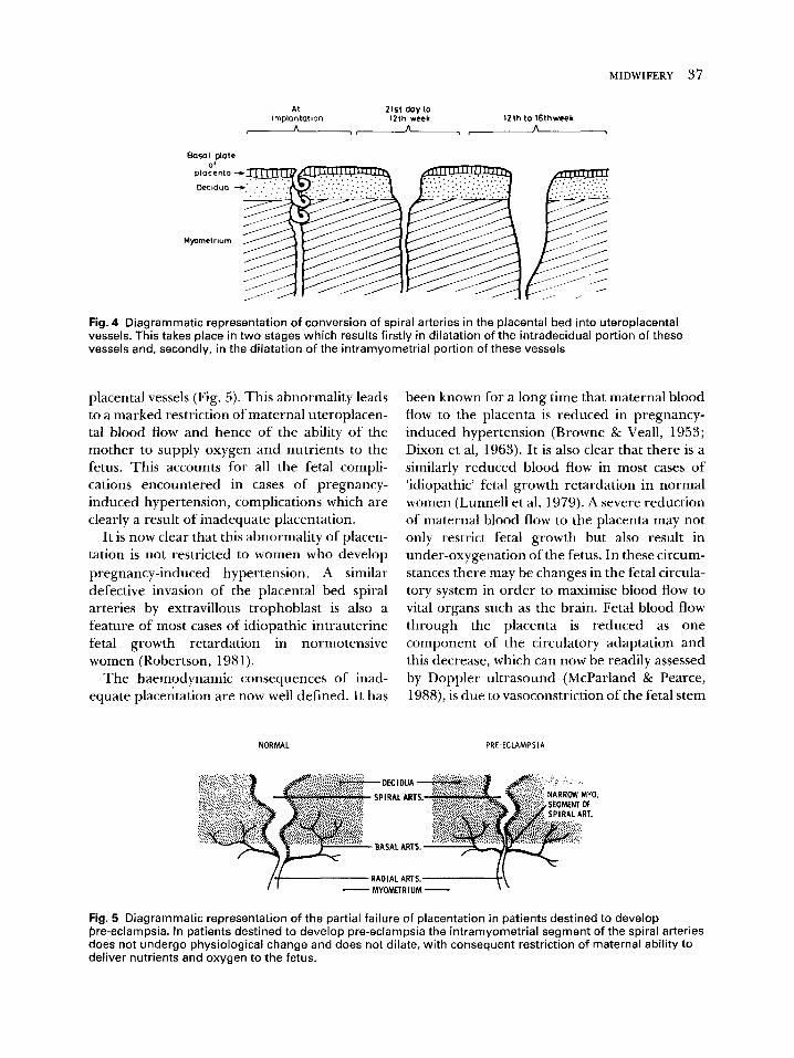
placental vessels (Fig. 5). This abnormality leads to a marked restriction of maternal uteroplacen- tal blood flow and hence of the ability of the mother to supply oxygen and nutrients to the fetus. This accounts for all the fetal compli- cations encountered in cases of pregnancy- induced hypertension, complications which are clearly a result of inadequate placentation.
It is now clear that this abnormali ty of placen- ration is not restricted to women who develop pregnancy-induced hypertension. A similar defective invasion of the placental bed spiral arteries by extravillous trophoblast is also a feature of most cases of idiopathic intrauterine fetal growth retardation in normotensive women (Robertson, 1981).
The haemodynamic consequences of inad- equate placentation are now well defined. It has
been known for a long time that maternal blood flow to the placenta is reduced in pregnancy- induced hypertension (Browne & Veall, 1953; Dixon et al, 1963). It is also clear that there is a similarly reduced blood flow in most cases of 'idiopathic' fetal growth retardation in normal women (Lunnell et al, 1979). A severe reduction of maternal blood flow to the placenta may not only restrict fetal growth but also result in under-oxygenation of the fetus. In these circum- stances there may be changes in the fetal circula- tory system in order to maximise blood flow to vital organs such as the brain. Fetal blood flow through the placenta is reduced as one component of the circulatory adaptation and this decrease, which can now be readily assessed by Doppler ultrasound (McParland & Pearce, 1988), is due to vasoconstriction of the fetal stem
NORMAL PRE-ECLAMPSIA
L
/
~..'."~.:.i. '~}~i~'...,~ ~'~!~ :! 9.~!~!i~:::';'.~": .̀ '::::"i. };.'.'-!t.."-~ 'ii "~ ~-~-.~.~i~" .~..';~.~' -.::!: ~ .:~:1 BASAL ARTS. ~¢: : f~ '~ ' :~: '~: ' : "
Fig. 5 Diagrammatic representation of the partial failure of placentation in patients destined to develop pre-eclampsia. In patients destined to develop pre-eclampsia the intramyometrial segment of the spiral arteries does not undergo physiological change and does not dilate, with consequent restriction of maternal ability to deliver nutrients and oxygen to the fetus.

38 MIDWIFERY
arteries of the placenta. P ro longed vasoconstric- t ion can lead to the obl i terat ion of some of these vessels (Giles et al, 1985; McCowan et al, 1987).
I t is thus clear that most cases of 'placental insufficiency' are, in reality, examples of mater- nal vascular insufficiency resul t ing f rom inad- equate placentat ion, It may appear pedant ic to draw this dist inction but the c o n t i n u i n g use of the term 'placental insufficiency' leads to a false emphasis be ing placed u p o n funct ional ly u n i m - po ran t abnormali t ies of the placenta, diverts research a long u n r e w a r d i n g pathways, and obscures the fact that many complicat ions of the later stages of p regnancy have their or ig in in the early months of gestation.
Acknowledgements Figure 3 is reproduced from Pathology of the Placenta by H Fox and is reproduced by kind permission of W B Saunders and Company.
Figures 1, 2 and 4 are reproduced from Basic Science in Obstetrics and Gyi~aecology byJ Dewhurst, M De Swiet and G V P Chamberlain and are reproduced by permission of
'the editors and Churchill Livingstone.
References Boyd P A 1984 Quantitative studies of the normal
human placenta from 10 weeks of gestation to term. Early Human Development 9:297-307
Brosens I, Robertson W B, Dixon H G 1967 The physiological response of the vessels of the placental bed in normal pregnancy. Journal of Pathology and Bacteriology 83:569-579
Browne JCMcC, Veall N 1953 The maternal placental blood flow in normotensive and hypertensive women. Journal of Obstetrics and Gynaecology of the British Empire 60:141-147
Burton G F, Palmer M E, Dalton'g KJ 1989 Morphometric differences between the placental vasculature of non-smokers, smokers and ex-smokers. British Journal of Obstetrics nd Gynaecology 96: 907- 915
Chellman V G, Rushton D I 1985 Chorioamnionitis and funinculitis in the placentas of 200 births weighing less than 2.5 kg. British Journal of Obstetrics and Gynaecology 92:808-814
de Wolf F, de Wolf-Peeters C, Brosens I 1973 Uhrastructure of the spiral arteries in the human placental bed at the end of normal pregnancy. American Journal of Obstetrics and Gynecology 117: 833-848
Di Sant' Agnese P A, de Meay Jensen K, Levin A, Miller R K 1983 Placental toxicity of cadmium in the rat: an uhrastructural study. Placenta 4: 149-164
Dixon H G, Browne J C McC, Davey D A 1963 Choriodecidual and myometrial blood flow. Lancet ii: 369-373
Drife J 1989 Infection and pre-term labour. British Journal of Obstetrics and Gynaecology 96:1128-1130
Fox H 1978 Pathology of the Placenta, Saunders, London
Fox H 1979 The placenta as a model for organ ageing In: Beaconsfield P, Villee C (eds) Placenta - A Neglected Experimental Animal. Pergamon, Oxford, p351
Fox H 1986 Pathology of the placenta. Clinics in Obstetrics and Gynaecology 13:501-519
Fox H 1988 The placenta in pregnancy hypertension, In: Rubin P C (ed) Hypertension in Pregnancy. Elsevier, Amsterdam, p16.
Fox H, Sen D K 1972 Placenta extrachorialis: a clinico- pathological study. Journal of Obstetrics and Gynaecology of the British Commonwealth 79:32-35
Galbraith R M, Fox H, Hsi B, Galbraith G M P, Bray R S, Faulk W P 1980 The human materno-fetal relationship in malaria II. Histological, ultrastructural and immunopathological studies of the placenta. Transactions of the Royal Society of Tropical Medicine and Hygiene 74:61-72
Gaunt M, Ockleford C D 1986 Microinjection of human placenta: 2. Biological application. Placenta 7: 325- 331
Geier G, Schuhmann R, Draus H 1975 Regional Unterschliedliche Zellproliferation innerhalb der Plazentateme reifer menschlicher Plazenten: autoradiographische Untersuchungen. Archiv fur Gynakologie 218:31-37
Giles W B, Trudinger B J, Baird PJ 1985 Fetal umbilical flow velocity waveforms and placental resistance: pathological correlation. British Journal of Obstetrics and Gynaecology 92:31-39
HustinJ, Foedart J M, Lambotte R 1984 Cellular proliferation in villi of normal and pathological pregnancies. Gynecologic and Obstetric Investigation 17:1-9
Iverson O E, Farsund T 1985 Flow cytometry in the assessment of human placental growth. Acta Obstetricia et Gynecologica Scandinavica 64:605-607
Knox W F, Fox H 1984 Villitis of unknown aetiology: its incidence and significance in placentae from a British population. Placenta 5:395-402
Khong T Y, de Wolf F, Robertson W B 1986 Inadequate maternal vascular response to placentation in pregnancies complicated by preeclampsia and by small for gestational age infants. British Journal of Obstetrics and Gynaecology 93: 1049-1059
Ladermacher D S, Vemeulen R C W, Harter J J, Arts N F T 1981 Circumvallate placenta and congenital malformation. Lancet i: 732
Lehtovirta P, Forss M 1978 The acute effect of smoking on intervillous blood flow of the placenta. British Journal of Obstetrics and Gynaecology 85:720-731
Lopaz-Bernal A, HanseU D J, Khong T Y et al 1989 Prostaglandin E production by the fetal membranes in unexplained preterm labour and preterm labour associated with chorioamnionitis. British Journal of Obstetrics and Gynaecology 9:1133-1139
Lorijn R H V, Longo L D 1980 Clinical and physiologic

MIDWIFERY 39
implications of increased fetal oxygen consumption. American Journal of Obstetrics and Gynecology 136: 451-457
Lunnell N O, Sarby B, Lavander R et al 1979 Comparison of uteroplacental blood flow in normal and in intrauterine growth retarded pregnancy: measurements with indium-113 m and a computer- linked gamma camera. Gynecologic and Obstetric Investigation 10:106-118
McCowan L M, Mullen B M, Ritchie K 1987 Umbilical artery flow velocity waveforms and the placental vascular bed. American Journal of Obstetrics and Gynecology 157:900-902
McParland P, Pearce J M 1988 Doppler blood flow in pregnancy. Placenta 9:427-450
Robertson W B 1981 Maternal blood supply in fetal growth retardation. In: van Asssche F A, Robertson W B (eds) Fetal Growth Retardation. Churchill Livingstone, Edinburgh p126
Robertson W B, Brosens I, Dixon H G 1967 The pathological response of the vessels of the placental bed in hypertensive pregnancy. Journal of Pathology and Bacteriology 93:581-592
Robertson W B, Brosens I, Dixon H G 1975 Utero- placental vascular pathology. European Journal.of Obstetrics, Gynecology and Reproductive Biology 5: 47-65
Robinson J S, Kingston EJ , Jomes C T et al 1979
Studies on experimental growth retardation of sheep: the effect of removal of endometrial caruncles on fetal size and metabolism. Journal of Developmental Physiology 1:379-398
Russell P 1980 Inflammatory lesions of the human placenta. III The histopathology of villitis of unknown aetiology. Placenta 1:227-244
Russell P 1987 Infections of the placental villi (villitis). In: Fox H (ed) Haines and Taylor: Obstetrical and Gynaecological Pathology. Churchill Livingstone, Edinburgh, p 1014
Sands J, Dobbing J 1985 Continuing growth and development of the third-trimester human placenta. Placenta 6:13-22
Uyanwah-Akpom P O, Fox H 1977 The clinical significance of marginal and velamentous insertion of the cord. British Journal of Obstetrics and Gynaecology 84: 941-943.
van der Veen F, Fox H 1982 The effects of cigarette smoking on the human placenta: a light and electron microscopic study. Placenta 3:243-256
Winick M, Coscia A, Noble A 1967 Cellular growth in human placenta. I Normal cellular growth, Pediatric 39:248-251
Woods D L, Malan A F 1978 The site of umbilical cord insertion and birthweight. British Journal of Obstetrics and Gynaecology 85:332-333