a comparison between totally laparoscopic hydrocelectomy ... · pdf filea comparison between...
TRANSCRIPT

A comparison between totally laparoscopic hydrocelectomyand scrotal incision hydrocelectomy with laparoscopic highligation for pediatric cord hydrocele
Byung Seo Choi1 • Geon Young Byun1 • Seong Bae Hwang1 • Bum Hwan Koo1 •
Sung Ryul Lee1
Received: 4 November 2016 / Accepted: 2 May 2017
� Springer Science+Business Media New York 2017
Abstract
Background The purpose of this study is to report clinical
characteristics and to investigate the feasibility and safety
of totally laparoscopic hydrocelectomy (TLH) compared to
scrotal incision hydrocelectomy with laparoscopic high
ligation (SIH) for pediatric cord hydrocele (CH).
Methods From September 2011 to February 2016, 148
patients underwent SIH, and 342 patients underwent TLH
for CH. In the TLH group, a large hydrocele that could not
pass through the internal ring was removed after percuta-
neous syringe aspiration. Age, laterality of hydrocele,
inguinal comorbidities, operation time, surgical complica-
tions, and recurrences were evaluated.
Results All the patients had spermatic cord cysts and
patent processus vaginalis in proximity to hydrocele
(mixed type). The mean age of CH patients was
34.1 ± 22.1 months. CHs are more common on the right
side (61.0%) than on the left (35.7%). Bilaterality occurred
in 3.3%. Comorbidities such as hernia (8.6%) and cryp-
torchidism (1.2%) were observed. There were no
complications except for two cases of wound hematoma in
SIH group. There was one (0.7%) case of recurrence
appeared in communicating hydrocele in SIH group. There
were no significant differences in the age, laterality of
hydrocele, inguinal comorbidities, operation time, com-
plications, and recurrences between TLH and SIH groups.
However, TLH for unilateral cord hydrocele had signifi-
cantly shorter operation time compared to SIH. The mean
operation time in TLH group was 15.6 ± 5.96 min and
there was no conversion to open surgery.
Conclusions TLH for pediatric CH is a feasible and safe
procedure without additional incisions. Therefore, TLH
can be one of the surgical options for pediatric CH espe-
cially in mixed type.
Keywords Hydrocele � Cord hydrocele � Pediatric �Laparoscopy
Cord hydrocele (CH) is a disease with fluid accumulation
along the spermatic cord above the testicle, resulting from
aberrant closure of the processus vaginalis [1]. The tradi-
tional method of hydrocele repair in children is the open
hydrocelectomy and additional high ligation procedure in
case of patent processus vaginalis (PPV) [2]. With the
advent of a minimally invasive surgery, laparoscopy has
become increasingly popular as the treatment for various
pediatric diseases. There are a lot of clinical studies on the
laparoscopic herniorrhaphy of pediatric inguinal hernia.
The benefits of laparoscopy compared to conventional
methods are reported by many authors [3–5]. Janetschek
et al. [6] first reported the application of laparoscopy for
hydrocele treatment. However, since then there have not
been many studies reported. Report regarding laparoscopy
for cord hydrocele is even rarer.
& Sung Ryul Lee
Byung Seo Choi
Geon Young Byun
Seong Bae Hwang
Bum Hwan Koo
1 Department of Surgery, Damsoyu Hospital,
213 Bongeunsa-ro, Gangnam-gu, Seoul, Korea
123
Surg Endosc
DOI 10.1007/s00464-017-5582-1
and Other Interventional Techniques

In our hospital, cord hydrocele is treated via laparo-
scopy. In the beginning of our study, scrotal incision
hydrocelectomy (SIH) with laparoscopic high ligation was
performed. Then we switched the method to totally
laparoscopic hydrocelectomy with high ligation (TLH).
The aim of this study is to report clinical characteristics of
pediatric cord hydroceles and compare TLH and SIH as a
treatment for pediatric cord hydrocele in terms of feasi-
bility and safety.
Materials and methods
This is a retrospective study for 490 male pediatric cord
hydrocele patients between September 2011 and February
2016. All operations were performed by a highly experi-
enced surgeon (Lee SR, over 1500 laparoscopic operation
per year). Operative indications in this study are as follows:
(1) If the hydrocele persists in patients older than two years
old, (2) If a child has other inguinal comorbidities (inguinal
hernias or cryptorchidism) on the same side of the hydro-
cele that require surgery regardless of age. Diagnosis of
cord hydrocele was determined by the patient’s history,
physical examination, ultrasonography, and was confirmed
via laparoscopy.
Prior to the surgery, parents of the patient were informed
about the advantages and disadvantages of the laparoscopic
surgery and gave consent to the surgery. Age, laterality,
comorbid ipsilateral inguinal diseases, operation time,
postoperative complication, and recurrence rate were
analyzed.
The operation was performed under general anesthesia
in supine position. For laparoscopic system, a 0�, 2.9 mm
laparoscopy (Stryker, Kalamazoo, MI, USA) and 2.7 mm
rigid instruments (Karl Storz, Tuttlingen, Germany) were
used. A transumbilical longitudinal incision was made and
a 3-mm Endopath� bladeless trocar (Ethicon, Cincinnati,
OH) was inserted. Pneumoperitoneum was established and
two stab incisions were made on the lateral abdominal wall
parallel to the umbilicus after laparoscopic inspection. Two
2.7 mm laparoscopic instruments were inserted through
separate stab incisions in the lateral abdomen without
trocars.
The procedure of SIH is as follows: After the visual-
ization of the internal ring, high ligation of patent proces-
sus vaginalis was performed with 3-0 Mersilk (Ethicon Ltd.
Edinburgh, UK). Unlike the TLH procedure, separate 1 cm
sized vertical incision was made on the scrotum, and then
hydrocelectomy was performed (Fig. 1)
The procedure of TLH is as follows (Figs. 2, 3): After
identifying the internal ring, we could find a bulging
hydrocele through the internal ring when we pressed the
skin of inguinal area externally. For large hydroceles that
were difficult to pass through the internal ring, percuta-
neous syringe aspiration was performed to reduce the size
before hydrocelectomy. When hydrocele was pulled into
the abdominal cavity with a grasper, distinct dissection
plane between the hydrocele, and the peritoneum can be
identified. As the hydrocele is continuously pulled into the
peritoneal cavity, the peritoneum which is attached to the
hydrocele pushes away in the opposite direction of pulling
the cyst. Through this process, the vas deferens and sper-
matic vessel become far enough away from the hydrocele
that they will not be damaged during surgery. As the dis-
section progresses, the hydrocele is gradually drawn into
the peritoneal cavity. Finally, the distal part of the hydro-
cele is resected within the abdominal cavity after con-
firming that the vas deferens or spermatic vessel is not
adjacent. After intracorporeal hydrocelectomy, high liga-
tion of processus vaginalis was performed with a 3-0
Mersilk (Ethicon Ltd. Edinburgh, UK). The needle was
inserted directly through the lower abdominal wall. The
procedure was performed with caution to avoid damaging
the vas deferens and spermatic vessels. The resected
hydrocele was removed through the right lateral incision.
After the incision was closed, the surgical bond (Der-
mabond; Ethicon Inc., Somerville, NJ, USA) was used for
covering the incision.
Unless there was any postoperative problem, the patients
could be discharged from hospital on the same day of the
surgery. Follow-up at 1 week, 1, and 3 months were per-
formed after surgery, and telephone interview was per-
formed to monitor for the three subsequent years.
Statistical analyses were performed using the SPSS
statistics 23 standard for medical service (IBM Co.,
Armonk, NY, USA). For categorical variables, the Fisher’s
exact test or v2 test was used. The t test was used to test for
the normality of the continuous variables. A p value
threshold of 0.05 was chosen; any p value below or equal to
0.05 was considered statistically significant.
Results
The data of whole cord hydrocele patients are shown in
Table 1. Cord hydrocele was mostly unilateral (96.7%),
and unilateral hydrocele occurred more often on the right
side (61%). Bilateral cord hydrocele (3.3%) was also
observed. Among the comorbidity of cord, inguinal hernia
was most common (8.6%) and cryptorchidism (1.2%) was
also observed. In our study, all the patients with cord
hydrocele detected by laparoscopy had proximally patent
processus vaginalis, and the cysts of hydroceles were not
communicating with the peritoneal cavity.
Comparison of results between SIH and TLH are shown
in Table 2. Demographic information such as age,
Surg Endosc
123
laterality, comorbidity showed no differences between
TLH and SIH group. Operative results such as operation
time, postoperative complication, and recurrence showed
no differences. However, complications and recurrences
were observed in SIH group and not in TLH group. There
was no complication other than two wound hematoma in
SIH group. The recurred patient in SIH group developed a
communicating hydrocele after cord hydrocele surgery. In
terms of operation time, there was no difference between
two groups for bilateral cord hydroceles, while TLH group
had significantly shorter operation time than SIH group for
unilateral cord hydroceles.
Both group required no conversion to open surgery, and
no intraoperative or immediate postoperative problem was
observed. All the patients in this study were discharged the
same day.
Discussion
The frequency of cord hydroceles in pediatric patients is
reported to be about 5.7% [7]. However, clinical features of
cord hydroceles are not readily available and reports about
laparoscopic approach to cord hydrocele are even rarer.
Martin et al. [1] categorized types of cord hydroceles into
funicular type (the peritoneal diverticulum communicating
with the peritoneal cavity at the internal inguinal ring) and
encysted type (the cyst not communicating with the peri-
toneal cavity or processus vaginalis). Chang et al. [8] cat-
egorized cord hydroceles that do not belong either
funicular or encysted type as mixed type. In mixed variety,
the cyst is not communicating with peritoneal cavity but
has a proximally patent processus vaginalis. However, all
the cord hydroceles we observed in this study were mixed
type, and neither funicular nor encysted type were
observed. One study of laparoscopic hydrocelectomy
reported 97.7% of hydroceles were patent around the
internal inguinal ring: 59.1% narrow patent processus
vaginalis covered with peritoneal veil, and 38.6% widely
open patent processus vaginalis [9]. Another study reported
that 75% of cord hydroceles have patent processus vagi-
nalis [8]. Our study is restricted to cord hydrocele, and the
sizes of PPVs were not recorded. However, all the pro-
cessus vaginalis observed in this study were patent.
Study regarding the laterality of cord hydrocele is rare
as well. One study for hydrocele reported that right uni-
lateral hydrocele is the most common (60%), but 7%
bilaterality is also observed [7]. In the case of mixed
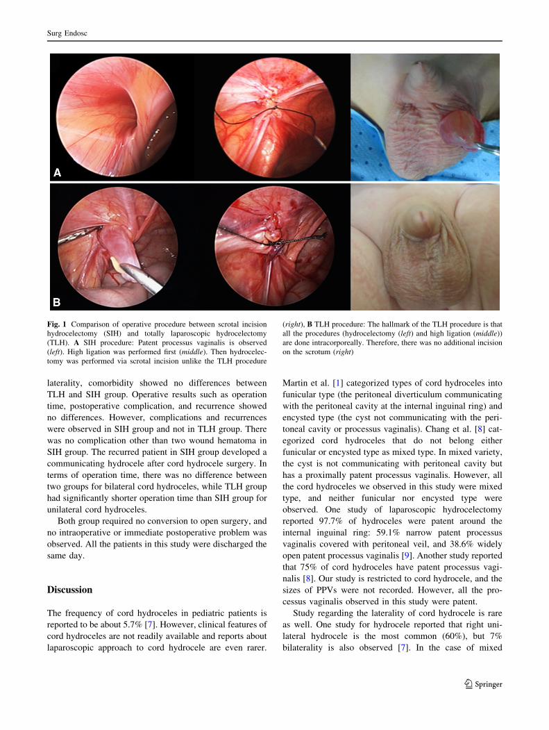
Fig. 1 Comparison of operative procedure between scrotal incision
hydrocelectomy (SIH) and totally laparoscopic hydrocelectomy
(TLH). A SIH procedure: Patent processus vaginalis is observed
(left). High ligation was performed first (middle). Then hydrocelec-
tomy was performed via scrotal incision unlike the TLH procedure
(right), B TLH procedure: The hallmark of the TLH procedure is that
all the procedures (hydrocelectomy (left) and high ligation (middle))
are done intracorporeally. Therefore, there was no additional incision
on the scrotum (right)
Surg Endosc
123

variety, a relatively small-sized study reported right and
left side cord hydroceles occur at the similar rate [8].
However, the results of our study revealed right unilateral
cord hydroceles (61%) were more common than left uni-
lateral cord hydrocele (35.7%). Bilaterality (3.3%) occur-
red as well.
The proper timing of the repair for cord hydrocele is
also controversial. The most PPV will spontaneously close
within 1–2 years. Therefore, most surgeons may avoid
hydrocele operation within 1–2 years of life unless hernia
cannot be excluded [10, 11]. One study reported the
hydrocele operation in the first year of life is only required
if it is huge in size or associated with inguinal hernia [12].
This is because most of infantile hydroceles (89%) are
spontaneously resolved within a year. In contrast, others
reported that in the case of cord hydrocele (especially for
mixed variety case), elective operation is recommended
regardless of age since there is a high risk of hernia
developed due to PPV [8]. However, surgical intervention
for cord hydrocele at our institution is performed for an
infant older than 2 year old. However, if comorbid ipsi-
lateral inguinal hernia or cryptorchidism that requires sur-
gery is observed, hydrocelectomy is performed at the same
time even if patients are under 2 years old. This study
included the patients only who received surgeries. So the
rate of naturally resolved cord hydrocele is not identified.
8.6% of cord hydrocele accompanying inguinal hernia was
observed, but complication such as incarceration was not
Fig. 2 External view of the totally laparoscopic hydrocelectomy
procedure: A Port placement, B Syringe aspiration before hydroc-
electomy, C Removal of the hydrocele through the right lateral
incision (yellow arrow: collapsed hydrocele), D Postoperative wound
(yellow arrow head: where the instrument inserted) (Color
figure online)
Surg Endosc
123

observed. Even though further studies are required, the
general 2 year-observation protocol for hydrocele patients
can be applied to cord hydrocele patients without hernia
and is considered safe.
There is no absolute laparoscopic treatment guideline
for cord hydrocele, but in general, open surgery through
inguinal incision is the standard procedure [2, 10, 11].
However, open approach has a risk to damaging vas def-
erens, spermatic vessel, or other inguinal structures [13].
Laparoscopic technique has been revolutionized, and
application of laparoscopy to pediatric surgical fields is
getting more and more popular. Benefits of laparoscopic
Fig. 3 Laparoscopic view of the totally laparoscopic hydrocelectomy
procedure: A The hydrocele was not observed, B After external
compression, a bulged hydrocele is observed through the internal ring
(short yellow arrow: dissection plane between the hydrocele and the
peritoneum), C As the hydrocele is continuously pulled into the
peritoneal cavity, the peritoneum just adjacent to the dissection plane
pushes away in the opposite direction (long yellow arrow: direction of
pulling the hydrocele), D Only the distal portion of the hydrocele is
attached just before dissection is complete. Vas deferens and
spermatic vessels (yellow arrow head) are far enough away from
the dissection site, E After removal of the hydrocele (blue arrow:
removed hydrocele), the damage of vas deferens and spermatic vessel
(yellow arrow head) is not seen, F High ligation (Color figure online)
Surg Endosc
123

approach to pediatric inguinal hernia include precise
diagnosis with direct visualization and lower risk of dam-
aging any important structures [4, 14]. However, in the
case of the TLH procedure, there may be a concern about
vas deferens or spermatic vessel damage, unlike hernia
surgery, during removal of the hydrocele cyst. Our surgical
procedure (As the hydrocele is continuously pulled into the
peritoneal cavity, the peritoneum just adjacent to the dis-
section plane pushes away in the opposite direction) can
avoid the damage of vas deferens or testicular vessel
because the dissection plane is not close to these structures.
Therefore, there was no damage of vas deferens or sper-
matic vessel during surgery.
Some authors claim that laparoscopy is an easier sur-
gical method for pediatric cord hydrocele since cord
hydrocele has a narrow PPV [13]. In this study, there was
no significant difference in operation time between TLH
and SIH group, while TLH group had significantly shorter
operation time than SIH group for unilateral cord hydro-
celes. The average operation time in the TLH group was
15.6 ± 5.96 min. While there are no specific reports about
the laparoscopic surgery of CH, there are some reports
about open and laparoscopic surgery for communicating
and non-communicating hydroceles [9, 13, 15]. The oper-
ation time of either TLH or SIH procedure was comparable
to these reports.
Table 1 Patient characteristics
N = 490
Age (month) 34.1 ± 22.1 (1.0–192.0)
Laterality of hydrocele
Bilateral 16 (3.3%)
Unilateral 474 (96.7%)
Left 175 (35.7%)
Right 299 (61.0%)
Inguinal comorbidity
None 442 (90.2%)
Inguinal hernia 42 (8.6%)
Cryptorchidism 6 (1.2%)
Complication
Hematoma 2 (0.4%)
None 488 (99.6%)
Recurrence
Yes 1 (0.2%)
No 489 (99.8%)
Operation time 15.9 ± 5.64 (8.0–50.0)
Bilateral 16.9 ± 3.86
Unilateral 15.8 ± 5.69
Follow-up period 17.5 ± 12.0 (3.0–54.0)
Categorical variables are represented as number (%) and continuous
variables as mean ± SD and ranges
Table 2 Patient characteristics
according to the surgical
method
TLH group (N = 342) SIH group (N = 148) P value
Age (month) 35.3 ± 23.3 (1.0–192.0) 31.3 ± 18.7 (1.0–101.0) 0.061
Laterality of hydrocele
Bilateral 11 (3.2%) 5 (3.4%) 0.965
Unilateral 331 (96.8%) 143 (96.6%)
Left 121 (35.4%) 54 (36.5%)
Right 210 (61.4%) 89 (60.1%)
Comorbidity
None 305 (89.2%) 137 (92.6%) 0.583
Inguinal hernia 32 (9.3%) 10 (6.7%)
Cryptorchidism 5 (1.5%) 1 (0.7%)
Complication
Hematoma 0 (0.0%) 2 (1.4%) 0.091
None 342 (100.0%) 146 (98.6%)
Recurrence 0.302
Yes 0 (0.0%) 1 (0.7%)
No 342 (100.0%) 147 (99.3%)
Operation time 15.6 ± 5.96 (8.0–50.0) 16.4 ± 4.80 (8.0–40.0) 0.176
Bilateral 16.9 ± 3.78 (13.0–25.0) 17.0 ± 4.47 (15.0–25.0) 0.913
Unilateral 15.6 ± 6.02 (8.0–50.0) 16.4 ± 4.82 (8.0–40.0) 0.003
Categorical variables are represented as number (%) and continuous variables as mean ± SD and ranges
TLH totally laparoscopic hydrocelectomy with high ligation, SIH scrotal incision hydrocelectomy with
laparoscopic high ligation
Surg Endosc
123

There were only 2 postoperative complications in SIH
group. These are wound hematomas that spontaneously
resolved without any treatment. The recurrence rate after
laparoscopic hydrocelectomy was reported to be 0–1.4%
[13, 15, 16]. In our study, there was one (0.7%) recurrence
in SIH group and the recurrence rate of the whole patients
was 0.2% which is not higher than reported rate for
hydrocele repair. In this study, follow-up was performed at
1 week, 1, and 3 months by visiting the hospital. After this
period, telephone interview was performed annually for
three years. However, the study period of this study is not
long, and some of the patients with follow-up period
shorter than 3 years were included. Hence, average follow-
up period was 17.5 ± 12.0 months. There will be addi-
tional reports on the long-term outcome of the surgery,
such as recurrence, through continued follow-up of these
patients. Also we have a separate team dedicated to follow-
up and management of patients after surgery, and maintain
ongoing contact via phone/e-mail. Therefore, there was no
follow-up loss among the subjects.
As it is mentioned earlier, the surgical methods in this
study were changed from SIH to TLH. Some may think
that learning curve of the procedure might influence the
difference in operation times or complications between two
groups. However, the operator is an expert in pediatric
laparoscopic surgery, and has many years of experience for
laparoscopic herniorrhaphy and hydrocelectomy. Also,
since the surgical methods of cord hydrocele are similar to
laparoscopic inguinal hernia repair which includes
intraperitoneal high ligation of hernia sac and occasional
removal of cord lipoma, the learning curve of the operator
is not thought to influence the operative outcomes.
However, this study has some limitations. Firstly, this is
a relatively short-term retrospective study at a single hos-
pital. Second, since our hospital only offers laparoscopic
hydrocelectomy as a treatment of cord hydrocele, the sur-
gical outcome of laparoscopic hydrocelectomy cannot be
compared with that of conventional open surgery.
Conclusion
All of cord hydroceles had PPV so high ligation was
required. The result of SIH was comparable with other
literature. The result of TLH was also comparable without
increased complication rate or operation time even though
there was no additional incision. TLH for unilateral cord
hydrocele, in particular, had significantly shorter operation
time compared to SIH. Hence, when performed by a sur-
geon, who is expert at laparoscopic surgery, TLH could be
one of the options for pediatric CH, especially for mixed
type.
Compliance with ethical standards
Disclosure Byung Seo Choi, Geon Young Byun, Seong Bae Hwang,
Bum Hwan Koo, and Sung Ryul Lee have no conflict of interest or
financial ties to disclose.
References
1. Martin LC, Share JC, Peters C, Atala A (1996) Hydrocele of the
spermatic cord: embryology and ultrasonographic appearance.
Pediatr Radiol 26:528–530
2. Wilson JM, Aaronson DS, Schrader R, Baskin LS (2008)
Hydrocele in the pediatric patient: inguinal or scrotal approach?
J Urol 180:1724–1727 discussion 1727-17283. Lao OB, Fitzgibbons RJ, Cusick RA (2012) Pediatric inguinal
hernias, hydroceles, and undescended testicles. Surg Clin North
Am 92:487–504
4. Schier F (2006) Laparoscopic inguinal hernia repair-a prospective
personal series of 542 children. J Pediatr Surg 41:1081–1084
5. Saranga Bharathi R, Arora M, Baskaran V (2008) Minimal access
surgery of pediatric inguinal hernias: a review. Surg Endosc
22:1751–1762
6. Janetschek G, Reissigl A, Bartsch G (1994) Laparoscopic repair
of pediatric hydroceles. J Endourol 8:415–417
7. Ein SH, Njere I, Ein A (2006) Six thousand three hundred sixty-
one pediatric inguinal hernias: a 35-year review. J Pediatr Surg
41:980–986
8. Chang YT, Lee JY, Wang JY, Chiou CS, Chang CC (2010)
Hydrocele of the spermatic cord in infants and children: its par-
ticular characteristics. Urology 76:82–86
9. Saka R, Okuyama H, Sasaki T, Nose S, Yoneyama C, Tsukada R
(2014) Laparoscopic treatment of pediatric hydrocele and the
evaluation of the internal inguinal ring. J Laparoendosc Adv Surg
Tech A 24:664–668
10. Lau ST, Lee YH, Caty MG (2007) Current management of her-
nias and hydroceles. Semin Pediatr Surg 16:50–57
11. Group IPE (2010) IPEG Guidelines for Inguinal Hernia and
Hydrocele. J Laparoendosc Adv Surg Tech A 20:10–14
12. Naji H, Ingolfsson I, Isacson D, Svensson JF (2012) Decision
making in the management of hydroceles in infants and children.
Eur J Pediatr 171:807–810
13. Wang Z, Xu L, Chen Z, Yao C, Su Z (2014) Modified single-port
minilaparoscopic extraperitoneal repair for pediatric hydrocele: a
single-center experience with 279 surgeries. World J Urol
32:1613–1618
14. Schier F, Montupet P, Esposito C (2002) Laparoscopic inguinal
herniorrhaphy in children: a three-center experience with 933
repairs. J Pediatr Surg 37:395–397
15. Yang XD, Wu Y, Xiang B, Wong K, Pei J, Li FY (2015) Ten year
experience of laparoscopic repair of pediatric hydrocele and the
long-term follow-up results. J Pediatr Surg 50:1987–1990
16. Peng Y, Li C, Lin W, Xu L (2015) Application of a laparoscopic,
single-port, double-needle technique for pediatric hydroceles
with multiple peritoneal folds: a trial from a single-center 5-year
experience. Urology 85:1466–1470
Surg Endosc
123