a class of gabaergic neurons in the prefrontal cortex sends long-range projections to the nucleus...
TRANSCRIPT
Brief Communications
A Class of GABAergic Neurons in the Prefrontal Cortex SendsLong-Range Projections to the Nucleus Accumbens andElicits Acute Avoidance Behavior
Anthony T. Lee,1,2,3 Daniel Vogt,1 X John L. Rubenstein,1 and Vikaas S. Sohal1,2,3
1Department of Psychiatry, 2Center for Integrative Neuroscience, and 3Sloan-Swartz Center for Theoretical Neurobiology, University of California, SanFrancisco, San Francisco, California 94143-0444
GABAergic projections from the neocortex to subcortical structures have been poorly characterized. Using Dlxi12b–Cre mice, we foundanatomical evidence for GABAergic neurons that project from the mouse medial prefrontal cortex (mPFC) to multiple subcortical targets.We used a combination of patch-clamp electrophysiology, optogenetics, and pharmacology to confirm that Dlxi12b-labeled projectionsfrom the mPFC to the nucleus accumbens (NAcc) release GABA and do not corelease glutamate. Furthermore, optogenetic stimulation ofthese GABAergic projections from mPFC to NAcc induces avoidance behavior in a real-time place preference task, suggesting that theselong-range projecting GABAergic neurons can transmit aversive signals. Finally, we found evidence for heterogeneous histochemicaland/or electrophysiological properties of long-range projecting GABAergic neurons in the mPFC. Some of these neurons were labeled inparvalbumin–Cre and vasoactive intestinal peptide–Cre mice. We also used a novel intersectional targeting strategy to label GABAergicneurons in the mPFC that project to NAcc and found that these neurons have fast-spiking properties and express parvalbumin. Theseresults define possible functions and properties for a class of long-range projecting GABAergic neurons in the neocortex.
Key words: aversion; GABA; nucleus accumbens; optogenetics; prefrontal cortex
IntroductionKnowledge about GABAergic neurons in the neocortex that sendlong-range projections to other structures remains sparse. Ana-tomical studies using retrograde tracing and immunohistochem-istry have estimated that, in mice, cats, and monkeys, �1–10% ofall neocortical GABAergic cells give rise to long-range projections(Peters et al., 1990; Tomioka et al., 2005; Tomioka and Rockland,2007; Higo et al., 2009; Tamamaki and Tomioka, 2010). Neocor-tical long-range projecting GABAergic neurons appear to consti-tute a heterogeneous population based on histochemical markers[parvalbumin-expressing(PV�),somatostatin-expressing(SOM�),neuropeptide Y-expressing (NPY�), neuronal nitric oxide synthase-expressing (nNOS�), NADPH�, and M2R-expressing] andmorphology (Tomioka et al., 2005; Jinno et al., 2007; Higo et al.,2009). Previous studies have not examined possible physiological orbehavioral functions for neocortical long-range GABAergic projec-tion neurons, but their connectivity onto GABAergic neurons in
distant cortical regions suggests that they may synchronize oscilla-tory activity (Caputi et al., 2013).
Recent applications of optogenetics have advanced our un-derstanding of GABAergic projection neurons in subcorticalstructures and the hippocampus. GABAergic neurons in the me-dial septum form reciprocal circuits with hippocampal GABAe-rgic neurons, signaling salient sensory events and controllinghippocampal theta oscillations (Jinno et al., 2007; Hangya et al.,2009; Kaifosh et al., 2013). Additional modulation of rhythmicoscillations occurs via a bidirectional GABAergic circuit com-prising the hippocampus and entorhinal cortex (Melzer et al.,2012; Caputi et al., 2013). GABAergic projections from the ventraltegmental area to the nucleus accumbens (NAcc) can enhance asso-ciative learning (Brown et al., 2012); similar projections also regulatestriatal output (Tritsch et al., 2012).
“Top-down” control by the prefrontal cortex (PFC) can influ-ence emotional valence and motivated actions, often by inhibitinginnate “bottom-up” processing (Miller, 2000; Shin and Liberzon,2010; Lammel et al., 2014). Thus, if subcortically projecting GABAe-rgic neurons exist in the PFC, they would be well positioned to exerttop-down inhibitory control on subcortical processes. Indeed, herewe describe evidence for long-range projecting GABAergic neuronsin the medial PFC (mPFC) and demonstrate that stimulation of theirprojections to NAcc elicits avoidance behavior.
Materials and MethodsAll experiments were conducted in accordance with procedures estab-lished by the Administrative Panels on Laboratory Animal Care at theUniversity of California, San Francisco.
Received March 21, 2014; revised June 8, 2014; accepted June 24, 2014.Author contributions: A.T.L., D.V., J.L.R., and V.S.S. designed research; A.T.L. and D.V. performed research; D.V.
and J.L.R. contributed unpublished reagents/analytic tools; A.T.L. analyzed data; A.T.L. and V.S.S. wrote the paper.This work was supported by the Staglin Family, the International Mental Health Research Organization, National
Institute of Mental Health Grants R00MH085946 and R37MH049428 (J.L.R.), National Institutes of Health/Office ofthe Director Grant 1DP2MH100011, and National Institute of General Medical Sciences Medical Scientist TrainingProgram Grant GM07618 (A.T.L.).
Correspondence should be addressed to Dr. Vikaas Sohal, University of California San Francisco, Box 0444, SanFrancisco, CA 94143-0444. E-mail: [email protected].
DOI:10.1523/JNEUROSCI.1157-14.2014Copyright © 2014 the authors 0270-6474/14/3411519-07$15.00/0
The Journal of Neuroscience, August 27, 2014 • 34(35):11519 –11525 • 11519
Cloning of viral constructs. To produce the inhibitory intersectionalretrograde tracer, we introduced MluI and BamHI compatible stickyends to the Dlxi12b–BG sequence with PCR. The AAV–EF1a–DIO–ChR2–EYFP (from Karl Deisseroth, Stanford University, Stanford, CA)was then cut with MluI/BamHI and ligated to the PCR insert to exchangethe EF1a promoter for Dlxi12b–BG. Virus was packaged by University ofNorth Carolina Vector Core with serotype AAV5.
Slice preparation. Slice preparation and intracellular recording fol-lowed our published protocol (Sohal and Huguenard, 2005). We cut 250�m coronal slices from 8- to 11-week-old mice of either sex. ACSF con-tained the following (in mM): 126 NaCl, 26 NaHCO3, 2.5 KCl, 1.25NaH2PO4, 1 MgCl2, 2 CaCl, and 10 glucose. We used the followingmouse lines: wild-type C57BL/6 mice (Charles River); B6;129P2–Pvalbtm1(cre)Arbr/J (line 008069; The Jackson Laboratory); Viptm1(cre)Zjh/J (line 010908; The Jackson Laboratory); Ssttm2.1(cre)Zjh/J (line 013044;The Jackson Laboratory); and Tg(I12b–cre)1Jlr.
Intracellular recording. We obtained somatic whole-cell patch record-ings using a Multiclamp 700A (Molecular Devices) and differential con-trast video microscopy on an upright microscope (BX51WI; Olympus).Patch electrodes (tip resistance, 2– 6 M�) were filled with the following(in mM): 130 K-gluconate, 10 KCl, 10 HEPES, 10 EGTA, 2 MgCl, 2MgATP, and 0.3 NaGTP (pH adjusted to 7.3 with KOH). All recordingswere at 32.5 � 1°C. Series resistance was usually 10 –20 M�, and exper-iments were discontinued above 30 M�.
Injection of opsin-containing virus or retrograde tracers. For Cre-dependent expression of channelrhodopsin 2 (ChR2) or enhanced yel-low fluorescent protein (EFYP), we injected 600 nl of a previouslydescribed adeno-associated virus (AAV) vector that drives Cre-dependent expression of a ChR2–EFYP fusion protein via previouslydescribed procedures (Sohal et al., 2009). Before stimulating ChR2-containing terminals in the NAcc, we first verified the absence of fluores-cent soma in the field of view. Coordinates for injection into mPFC wereas follows (in mm relative to bregma): 1.7 anteroposterior (AP), 0.3mediolateral (ML), and �2.50 to 2.7 dorsoventral (DV). Cholera toxinsubunit B-488 (CTb-488; Invitrogen) was injected as above with thefollowing NAcc coordinates (in mm): 0.90 AP, 0.65 ML, and �4.75 DV.We waited 2–3 d after injecting CTb before preparing brains slices.
ChR2 stimulation. We stimulated ChR2 in neurons using �5 mWflashes of light generated by a Lambda DG-4 high-speed optical switchwith a 300 W xenon lamp (Sutter Instruments) and an excitation filter setcentered around 470 nm, delivered through a 40� objective (Olympus).
Behavioral tests. For social and novel object exploration, a juvenilemouse (�4 weeks) or 50 ml Falcon tube cap, respectively, was placedwith the experimental mouse in its home cage for 5 min. Explorationtime was scored by the duration of nose–juvenile or nose–object contacts,and the reviewer was blinded to the virus identity and laser condition.
Real-time place preference (RTPP) occurred during three 20 min ses-sions over 3 d. On day 1, mice were habituated to the two-chamberapparatus. On day 2, mice were placed into one chamber and its move-ment was tracked by Anymaze (Stoelting). On day 3, mice were placed inthe chamber opposite to one that was randomly designated to trigger 20Hz laser pulses (470 nm, 15–20 mW, 5 ms) after entry. The sides of thestimulated chambers were counterbalanced across all mice, and the ex-perimenter was blind to the injected virus (control vs ChR2).
Drug application. Drugs were dissolved in water [DL-AP-5, CGP35348( p-3-aminopropyl-p-diethoxymethyl phosphoric acid)] or dimethyl-sulfoxide [CNQX, gabazine (GBZ)] before being diluted in ACSF.
Immunohistochemistry. After patch clamp, slices were fixed in 4% PFAfor at least 1 d. Primary antibody used was mouse monoclonal anti-PV(Millipore). Alexa Fluor 488 goat anti-mouse (Invitrogen) was first ap-plied and then washed for at least 2 d before addition of CF405 mono-clonal mouse anti-biotin (Biotium).
Statistical analysis. We used Student’s t tests to compare across condi-tions or n-way ANOVA unless noted otherwise. Error bars indicate � 1SEM.
ResultsTo visualize candidate long-range GABAergic projections origi-nating from the neocortex, we first injected a Dlxi12b–Cre mouse
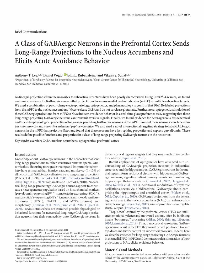
with AAV–DIO–ChR2–EYFP in the mPFC (Fig. 1A). Dlx1(Distal-less homeobox 1) and Dlx2 are expressed by developingGABAergic neurons as they mature and migrate out of the gan-glionic eminences in embryonic mice and are expressed predom-inately, if not exclusively, by GABAergic neurons (Anderson etal., 1997; Marin and Rubenstein, 2003). Indeed, many studieshave used Dlxi12b–Cre mice to selectively label GABAergic neu-rons (Potter et al., 2009; Flandin et al., 2011; Han et al., 2012;Arguello et al., 2013). More recently, our laboratory has used theDlxi12b enhancer to selectively express mCherry or ChR2 in cor-tical GABAergic neurons (Lee et al., 2014).
To confirm that, as in previous studies, Dlxi12b-labeled neu-rons are GABAergic and not glutamatergic, we recorded optoge-netically evoked synaptic responses (470 nm; 5 ms; �5 mW/mm 2) from mPFC pyramidal neurons of Dlxi12b–Cre miceinjected with AAV–DIO–ChR2–EYFP. Thus, we used patchedpyramidal neurons as “biosensors” to detect neurotransmittersreleased by nearby Dlxi12b-labeled neurons (Fig. 1C). Duringvoltage-clamp recordings at both �70 and �10 mV, applicationof GBZ (10 �M) plus CGP35348 (5 �M) essentially abolished alloptogenetically evoked synaptic currents (Fig. 1C; the small re-sidual outward current in one recording at �70 mV presumablyrepresents incompletely blocked GABABR-mediated current).This confirms that no glutamatergic neurons were labeled in themPFC of Dlxi12b–Cre mice.
Dlxi12b-labeled fibers project from mPFC to distantsubcortical brain regionsOur injection site spanned the mPFC, including the anterior cin-gulate, prelimbic, and infralimbic regions (Fig. 1A, left). Surpris-ingly, we observed labeled fibers several millimeters from theinjection site within corpus callosum and subcortical structures,including dorsal striatum, ventral striatum (NAcc), claustrum,and basolateral amygdala (Fig. 1A,B). Importantly, no labeledcell bodies were found in these distant sites, including fields ofview that were directly adjacent to the injection site (Fig. 1A,middle).
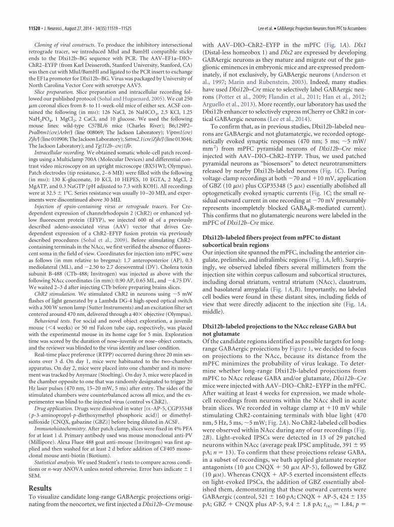
Dlxi12b-labeled projections to the NAcc release GABA butnot glutamateOf the candidate regions identified as possible targets for long-range GABAergic projections by Figure 1, we decided to focuson projections to the NAcc, because its distance from themPFC minimizes the probability of virus leakage. To deter-mine whether long-range Dlxi12b-labeled projections frommPFC to NAcc release GABA and/or glutamate, Dlxi12b–Cremice were injected with AAV–DIO–ChR2–EYFP in the mPFC.After waiting at least 4 weeks for expression, we made whole-cell recordings from neurons within the NAcc shell in acutebrain slices. We recorded in voltage clamp at �10 mV whilestimulating ChR2-containing terminals with blue light (470nm, 5 Hz, 5 ms, �5 mW; Fig. 2A). No ChR2-labeled cell bodieswere observed within NAcc during any of our recordings (Fig.2B). Light-evoked IPSCs were detected in 13 of 29 patchedneurons within NAcc (average peak IPSC amplitude, 391 � 95pA; n 13). To confirm that these projections release GABA,in a subset of recordings, we bath applied glutamate receptorantagonists (10 �M CNQX � 50 �M AP-5), followed by GBZ(10 �M). Whereas CNQX � AP-5 exerted inconsistent effectson light-evoked IPSCs, the addition of GBZ essentially abol-ished them, demonstrating that these outward currents wereGABAergic (control, 521 � 160 pA; CNQX � AP-5, 424 � 135pA; GBZ � CNQX plus AP-5, 9.4 � 1.8 pA; t(6) 1.84, p
11520 • J. Neurosci., August 27, 2014 • 34(35):11519 –11525 Lee et al. • GABAergic Projection Neurons from PFC to Accumbens

0.12 for control vs CNQX � AP-5; t(6) 3.19, p 0.02 for control vs GBZ �CNQX � AP-5; t(6) 3.09, p 0.02 forCNQX � AP-5 vs GBZ � CNQX �AP-5; n 7 cells; interaction by drugcondition, F(2,18) 9.52, p 0.003; Fig.2C,D). Furthermore, in separate exper-iments, GBZ alone abolished light-evoked currents recorded at either �70or �10 mV, demonstrating the absenceof glutamate corelease (currents at �70mV: control, 12 � 7 pA; GBZ, 2 � 1 pA;F(1,6) 8.21, p 0.001; currents at �10mV: control, 203 � 53 pA; GBZ, 12 � 4pA; F(1,6) 4.77, p 0.01; n 4 cells;Fig. 2 E, F ).
Stimulating GABAergic projectionsfrom mPFC to NAcc elicitsavoidance behaviorWe hypothesized that stimulating long-range GABAergic inputs to NAcc wouldmodulate motivational valence. To testthis, we delivered optogenetic stimulation(470 nm, 20 Hz, 5 ms, 15–20 mW/mm 2)into the NAcc of Dlxi12b–Cre mice thathad been injected at least 4 weeks earlierwith AAV to drive Cre-dependent ChR2expression in the mPFC (Fig. 3A). Wemeasured the effects of this stimulationon behavior within a two-chamber RTPPtask (Jennings et al., 2013; Fig. 3B). In theRTPP task, the two chambers are contex-tually identical and the stimulation occursin real time, i.e., the light turns on when-ever a mouse enters the designated stimu-lation chamber. Thus, in some ways, theRTPP task resembles self-stimulationmore than conditioned place preference,and the RTPP task can be used to assayacute behavioral effects of stimulation.Importantly, mice do not exhibit an in-herent place bias in the two-chamberRTPP task (Fig. 3F; time spent in placedside/total time: 0.50 � 0.03; t(18) 0.009,p 0.99; n 19 mice).
We habituated mice to the RTPPchambers on day 1, measured the timespent in each chamber in the absence ofstimulation on day 2 (baseline condition),and finally measured the time spent ineach chamber in the presence of stimula-tion on day 3 (test condition). Comparedwith the baseline condition (day 2), micespend significantly less time in the stimu-lated side on day 3 (test condition; Fig. 3C;fraction time spent in the stimulated sideat baseline, 0.51 � 0.04; in presence ofstimulation, 0.29 � 0.04; t(9) 4.246, p 0.002; n 10 mice). ChR2� mice alsospend less time on the stimulated sidecompared with control (ChR2-negative)mice that received light stimulation but
Figure 1. GABAergic neurons in the PFC project to subcortical targets. A, Left, AAV-DIO-ChR2-EYFP was injected into thecingulate, prelimbic (PL), and infralimbic (IL) cortices of mPFC in Dlxi12b–Cre mice. Middle, Magnified view of the region ofthe left panel indicated by the dashed box. No labeled cell bodies were present in areas adjacent to the injection site,suggesting limited viral spread. Right, Dlxi12b-labeled fibers were found in ventral striatum. Scale bars, 50 �m. B,Dlxi12b-labeled fibers from PFC are found in corpus callosum, dorsal striatum, claustrum, and basolateral amygdala (BLA).Scale bars, 30 �m. C, Dlxi12b-labeled cells are exclusively GABAergic. Left, Experimental design: we made voltage-clamp(VC) recordings from pyramidal cells (triangle) while optogenetically stimulating ChR2 � neurons (green) in Dlxi12b–Cremice injected with AAV–DIO–ChR2–EFYP. Right, Optogenetically evoked synaptic currents were abolished by GBZ �CGP35348 (CGP) (n 6 pyramidal neurons).
Lee et al. • GABAergic Projection Neurons from PFC to Accumbens J. Neurosci., August 27, 2014 • 34(35):11519 –11525 • 11521
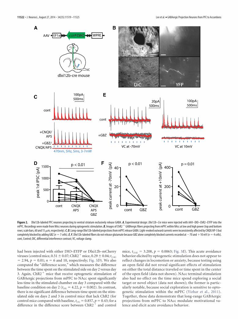
had been injected with either DIO–EYFP or Dlxi12b–mCherryviruses (control mice, 0.51 � 0.07; ChR2� mice, 0.29 � 0.04; t(14)
2.94, p 0.01; n 6 and 10, respectively; Fig. 3D). We alsocomputed the “difference score,” which measures the differencebetween the time spent on the stimulated side on day 2 versus day3. Again, ChR2� mice that receive optogenetic stimulation ofGABAergic projections from mPFC to NAcc spent significantlyless time in the stimulated chamber on day 3 compared with thebaseline condition on day 2 (t(9) 4.22, p 0.002). In contrast,there is no significant difference between time spent on the stim-ulated side on days 2 and 3 in control mice that lack ChR2 (forcontrol mice compared with baseline, t(5) 0.857, p 0.43; for adifference in the difference score between ChR2� and control
mice, t(14) 3.208, p 0.0063; Fig. 3E). This acute avoidancebehavior elicited by optogenetic stimulation does not appear toreflect changes in locomotion or anxiety, because testing usingan open field did not reveal significant effects of stimulationon either the total distance traveled or time spent in the centerof the open field (data not shown). NAcc terminal stimulationalso had no effect on the time mice spend exploring a socialtarget or novel object (data not shown); the former is partic-ularly notable, because social exploration is sensitive to opto-genetic stimulation within the mPFC (Yizhar et al., 2011).Together, these data demonstrate that long-range GABAergicprojections from mPFC to NAcc modulate motivational va-lence and elicit acute avoidance behavior.
Figure 2. Dlxi12b-labeled PFC neurons projecting to ventral striatum exclusively release GABA. A, Experimental design: Dlxi12b–Cre mice were injected with AAV–DIO–ChR2–EYFP into themPFC. Recordings were made from NAcc neurons during optogenetic stimulation. B, Images of ChR2 � GABAergic fibers projecting from mPFC within NAcc at low and high power (top and bottomrows; scale bars, 60 and 15 �m, respectively). C, D, Long-range Dlxi12b-labeled projections from mPFC release GABA. Light-evoked outward currents were inconsistently affected by CNQX/AP-5 butcompletely blocked by adding GBZ (n 7 cells). E, F, Dlxi12b-labeled fibers do not release glutamate because GBZ alone completely blocked currents recorded at �70 and �10 mV (n 4 cells).cont, Control; DIC, differential interference contrast; VC, voltage clamp.
11522 • J. Neurosci., August 27, 2014 • 34(35):11519 –11525 Lee et al. • GABAergic Projection Neurons from PFC to Accumbens
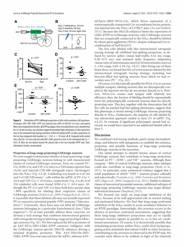
Properties of long-range projecting GABAergic neuronsWe next sought to determine whether at least some long-rangeprojecting GABAergic neurons belong to well characterizedclasses of cortical GABAergic neurons. First, we crossed PV–Cre, SOM–Cre, and VIP–Cre mice to a TdTomato reporter line(Ai14) and injected CTb-488 retrograde tracer (Invitrogen)into the NAcc (Fig. 4 A, B). Colabeling was found in 6 of 144and 5 of 100 TdTomato � cells within the mPFC of PV–Cre �Ai14 and VIP–Cre � Ai14 mice, respectively (Fig. 4 A, B); 0 of156 colabeled cells were found SOM–Cre � Ai14 mice. Al-though the PV–Cre and VIP–Cre lines both have greater than�90% specificity for labeling their respective classes ofGABAergic neurons (Sohal et al., 2009; Taniguchi et al., 2011),it is possible that these labeled cells may not actually representPV or vasoactive intestinal peptide (VIP) neurons (“false pos-itives”). Conversely, these lines may not label all GABAergicneurons belonging to a particular histochemical class (“falsenegatives”). To at least partially address these concerns, wedevised a viral strategy that combines intersectional geneticswith retrograde tracing to label long-range projecting GABAe-rgic neurons (Fig. 4C). We first replaced the EF1a promoter ofan AAV–EF1a–DIO–ChR2–EYFP construct (Addgene) withthe GABAergic neuron-specific Dlxi12b enhancer driving aminimal �-globin promoter. This AAV–Dlxi12b–DIO–ChR2–EYFP virus was injected into the mPFC, whereas AAV–
mCherry–IRES–WGA–Cre, which drives expression of atransynaptically transported Cre-recombinase fusion protein,was injected into the NAcc of C57BL/7 mice (Xu and Sudhof,2013). Because the Dlxi12b enhancer limits the expression ofChR2–EYFP to GABAergic neurons, only GABAergic neuronsthat are synaptically connected to the NAcc should be labeledby wheat-germ agglutinin (WGA), despite widespread Cre re-combination of loxP/lox2272.
The five cells labeled with this intersectional retrogradetracing strategy all exhibited fast-spiking properties, as de-fined by narrow spikes (mean half-width, 0.42 ms; range,0.28 – 0.71 ms) and minimal spike frequency adaptation(mean ratio of interstimulus interval 10/interstimulus interval1, 1.05; range, 0.85–1.19; Fig. 4 E, F ). After fixing one of thesebrain slices, we found that five of seven neurons labeled by thisintersectional retrograde tracing strategy, including twobiocytin-filled fast-spiking neurons from which we had re-corded, were PV � (Fig. 4G).
Of course, it is theoretically possible for WGA–Cre to traversemultiple synapses, labeling neurons that are disynaptically cou-pled to the injection site but do not project directly to it. How-ever, WGA–Cre crosses each synapse with relatively lowefficiency; thus, the fraction of labeling should be exponentiallylower for polysynaptically connected neurons than for directlyprojecting ones. This fact, together with the observation that allfive cells we patched had fast-spiking phenotypes, suggests thatthis phenotype is found among GABAergic neurons projectingdirectly to NAcc. Furthermore, the majority of cells labeled byour intersection approach resided in layer 2/3 of mPFC (Fig.4A,D). In contrast, if significant polysynaptic spread had oc-curred, we would have expected to see additional labeled cells inlayer 5.
DiscussionWe combined viral tracing methods, patch-clamp electrophysi-ology, and behavior with optogenetics to establish the existence,properties, and possible functions of long-range projectingGABAergic neurons in the neocortex.
Our initial attempts to identify known classes of corticalGABAergic neurons that contribute to long-range projectionsfocused on PV�, SOM�, and VIP� neurons. Although thesecomprise �80% of cortical GABAergic neurons, other subtypescould also contribute to long-range projections (Rudy et al.,2011). In particular, some anatomical evidence suggest that asmall population of nNOS�/NPY� neurons project callosallyand subcortically (Tomioka et al., 2005; Tomioka and Rockland,2007; Higo et al., 2009; Taniguchi et al., 2011). Similar to distinctclasses of neocortical pyramidal neurons, additional subtypes oflong-range projecting GABAergic neurons may target distinctsubcortical structures (Shepherd, 2013).
We focused our study on long-range inhibition of theNAcc, because of its well appreciated role in reward seekingand motivated behavior. We find that long-range prefrontalinhibition of the NAcc results in acute avoidance behavior inan RTPP paradigm. Interestingly, this aversion occurs in theabsence of previous contextual associations, suggesting thatthese long-range inhibitory projections may act to rapidlytransmit aversive signals in parallel to, or in lieu of, condi-tioned associations. Of course, it is theoretically possible thatstimulating projections within NAcc could elicit backpropa-gating action potentials that release GABA in other locations,contributing to the aversion we observed in the RTPP task. Weconsider such effects to be unlikely in light of the relatively
Figure 3. Stimulation of PFC GABAergic projections to NAcc elicits avoidance. A, Experimen-tal design: AAV–DIO–ChR2–EYFP was injected into mPFC of Dlxi12b–Cre mice, and opticalfibers were implanted in the NAcc. B, RTPP paradigm. Mice were placed into a two-chamber boxfor 3 d. On the test day, one chamber triggered immediate light stimulation. C, Mice spend lesstime in the stimulated side during activation of Dlxi12b-labeled mPFC-to-NAcc projections onthe test day compared with baseline ( p � 0.01, n 10 mice). D, E, Compared with controlmice, Dlxi12b–Cre mice injected with DIO–ChR2 spend less time in the stimulated side ( p �0.01). F, Mice are not biased toward the placed side in the two-chamber RTPP task. Stim,Stimulated; Unstim, unstimulated.
Lee et al. • GABAergic Projection Neurons from PFC to Accumbens J. Neurosci., August 27, 2014 • 34(35):11519 –11525 • 11523

long distance between mPFC and NAcc and the absence ofstimulation-induced effects on other behavioral assays that measurethe function of the mPFC, dorsal striatum, and amygdala. Regardlessof whether stimulation in the NAcc leads to GABA release else-where, our results clearly demonstrate that long-range projectingGABAergic neurons, despite being relatively small in number,can powerfully shape behavior.
Notably, we failed to observe effects of tonically stimulatinglong-range GABAergic projections to NAcc on social explora-tion. This may reflect (1) inadequate recruitment of mPFC–NAccGABAergic inputs attributable to unilateral stimulation, (2) adiscrepancy between the effects of tonic versus phasic patterns ofinput (i.e., during RTPP, stimulation may be effectively phasic,because the mouse is free to move away from the stimulatedchamber at any time), or (3) the absence of a role for PFC–NAcc
GABAergic projections in social exploration. Future experimentscould use real-time stimulation of these projections duringepochs of various behaviors, e.g., social interaction, to clarifypotential ways in which these projections could modulate addi-tional behaviors.
Together, we find evidence for long-range GABAergic projec-tions from the mPFC to subcortical targets, including the NAcc.This mPFC–NAcc projection releases GABA but not glutamateand can elicit acute avoidance behavior. Our results suggest thatthese long-range projecting GABAergic neurons may compriseheterogeneous subpopulations: some are labeled in VIP–Cremice, whereas others are fast spiking and express PV. Futurestudies may uncover additional subtypes of long-range project-ing GABAergic neurons in the neocortex.
Figure 4. Long-range GABAergic projection neurons in PFC to NAcc are heterogeneous. A, Experimental design: the retrograde tracer CTb-488 was injected into the NAcc of PV–Cre, SOM–Cre, orVIP-Cre mice crossed with Ai14 mice. B, PV–Cre- and VIP–Cre-labeled neurons in mPFC (red) colabel with the retrograde tracer (green) (scale bars, 15 �m). C, Experimental design of atransynaptic–intersectional GABAergic neuron marker: AAV–mCherry–IRES–WGA::Cre and AAV–Dlxi12b–DIO–ChR2–EYFP viruses were injected into NAcc and mPFC, respectively. D, Images ofGABAergic neurons labeled by a transynaptic–intersectional GABAergic marker in layers 2/3 at low and high power (top and bottom rows; scale bars, 60 and 15 �m, respectively). E, Cells labeledby the transynaptic–intersectional tracer were fast spiking. F, The adaption ratios and action potential half-widths for cells labeled in E. G, Transynaptically labeled cells stain for PV. Scale bar, 10 �m.DIC, differential interference contrast; IL, infralimbic cortex; PL, prelimbic cortex.
11524 • J. Neurosci., August 27, 2014 • 34(35):11519 –11525 Lee et al. • GABAergic Projection Neurons from PFC to Accumbens
ReferencesAnderson SA, Eisenstat DD, Shi L, Rubenstein JL (1997) Interneuron mi-
gration from basal forebrain to neocortex: dependence on Dlx genes.Science 278:474 – 476. CrossRef Medline
Arguello A, Yang X, Vogt D, Stanco A, Rubenstein JL, Cheyette BN (2013)Dapper antagonist of catenin-1 cooperates with Dishevelled-1 duringpostsynaptic development in mouse forebrain GABAergic interneurons.PLoS One 8:e67679. CrossRef Medline
Brown MT, Tan KR, O’Connor EC, Nikonenko I, Muller D, Luscher C(2012) Ventral tegmental area GABA projections pause accumbal cho-linergic interneurons to enhance associative learning. Nature 492:452–456. CrossRef Medline
Caputi A, Melzer S, Michael M, Monyer H (2013) The long and short ofGABAergic neurons. Curr Opin Neurobiol 23:179 –186. CrossRefMedline
Flandin P, Zhao Y, Vogt D, Jeong J, Long J, Potter G, Westphal H, RubensteinJL (2011) Lhx6 and Lhx8 coordinately induce neuronal expression ofShh that controls the generation of interneuron progenitors. Neuron 70:939 –950. CrossRef Medline
Han S, Tai C, Westenbroek RE, Yu FH, Cheah CS, Potter GB, Rubenstein JL,Scheuer T, de la Iglesia HO, Catterall WA (2012) Autistic-like behaviourin Scn1a�/� mice and rescue by enhanced GABA-mediated neurotrans-mission. Nature 489:385–390. CrossRef Medline
Hangya B, Borhegyi Z, Szilagyi N, Freund TF, Varga V (2009) GABAergicneurons of the medial septum lead the hippocampal network during thetaactivity. J Neurosci 29:8094 – 8102. CrossRef Medline
Higo S, Akashi K, Sakimura K, Tamamaki N (2009) Subtypes of GABAergicneurons project axons in the neocortex. Front Neuroanat 3:25. CrossRefMedline
Jennings JH, Sparta DR, Stamatakis AM, Ung RL, Pleil KE, Kash TL, StuberGD (2013) Distinct extended amygdala circuits for divergent motiva-tional states. Nature 496:224 –228. CrossRef Medline
Jinno S, Klausberger T, Marton LF, Dalezios Y, Roberts JD, Fuentealba P,Bushong EA, Henze D, Buzsaki G, Somogyi P (2007) Neuronal diversityin GABAergic long-range projections from the hippocampus. J Neurosci27:8790 – 8804. CrossRef Medline
Kaifosh P, Lovett-Barron M, Turi GF, Reardon TR, Losonczy A (2013)Septo-hippocampal GABAergic signaling across multiple modalities inawake mice. Nat Neurosci 16:1182–1184. CrossRef Medline
Lammel S, Tye KM, Warden MR (2014) Progress in understanding mooddisorders: optogenetic dissection of neural circuits. Genes Brain Behav13:38 –51. CrossRef Medline
Lee AT, Gee SM, Vogt D, Patel T, Rubenstein JL, Sohal VS (2014) Pyramidalneurons in prefrontal cortex receive subtype-specific forms of excitationand inhibition. Neuron 81:61– 68. CrossRef Medline
Marín O, Rubenstein JL (2003) Cell migration in the forebrain. Annu RevNeurosci 26:441– 483. CrossRef Medline
Melzer S, Michael M, Caputi A, Eliava M, Fuchs EC, Whittington MA,Monyer H (2012) Long-range-projecting GABAergic neurons modu-late inhibition in hippocampus and entorhinal cortex. Science 335:1506 –1510. CrossRef Medline
Miller EK (2000) The prefrontal cortex and cognitive control. Nat Rev Neu-rosci 1:59 – 65. CrossRef Medline
Potter GB, Petryniak MA, Shevchenko E, McKinsey GL, Ekker M, RubensteinJL (2009) Generation of Cre-transgenic mice using Dlx1/Dlx2 enhanc-ers and their characterization in GABAergic interneurons. Mol Cell Neu-rosci 40:167–186. CrossRef Medline
Rudy B, Fishell G, Lee S, Hjerling-Leffler J (2011) Three groups of interneu-rons account for nearly 100% of neocortical GABAergic neurons. DevNeurobiol 71:45– 61. CrossRef Medline
Shepherd GM (2013) Corticostriatal connectivity and its role in disease. NatRev Neurosci 14:278 –291. CrossRef Medline
Shin LM, Liberzon I (2010) The neurocircuitry of fear, stress, and anxietydisorders. Neuropsychopharmacology 35:169 –191. CrossRef Medline
Sohal VS, Huguenard JR (2005) Inhibitory coupling specifically generatesemergent gamma oscillations in diverse cell types. Proc Natl Acad SciU S A 102:18638 –18643. CrossRef Medline
Sohal VS, Zhang F, Yizhar O, Deisseroth K (2009) Parvalbumin neuronsand gamma rhythms enhance cortical circuit performance. Nature 459:698 –702. CrossRef Medline
Taniguchi H, He M, Wu P, Kim S, Paik R, Sugino K, Kvitsiani D, Fu Y, Lu J,Lin Y, Miyoshi G, Shima Y, Fishell G, Nelson SB, Huang ZJ (2011) Aresource of Cre driver lines for genetic targeting of GABAergic neurons incerebral cortex. Neuron [Erratum (2011) 72:1091] 71:995–1013.CrossRef Medline
Tomioka R, Rockland KS (2007) Long-distance corticocortical GABAergicneurons in the adult monkey white and gray matter. J Comp Neurol505:526 –538. CrossRef Medline
Tomioka R, Okamoto K, Furuta T, Fujiyama F, Iwasato T, Yanagawa Y, ObataK, Kaneko T, Tamamaki N (2005) Demonstration of long-rangeGABAergic connections distributed throughout the mouse neocortex.Eur J Neurosci 21:1587–1600. CrossRef Medline
Tritsch NX, Ding JB, Sabatini BL (2012) Dopaminergic neurons inhibit stri-atal output through non-canonical release of GABA. Nature 490:262–266.CrossRef Medline
Xu W, Sudhof TC (2013) A neural circuit for memory specificity and gen-eralization. Science 339:1290 –1295. CrossRef Medline
Yizhar O, Fenno LE, Prigge M, Schneider F, Davidson TJ, O’Shea DJ, SohalVS, Goshen I, Finkelstein J, Paz JT, Stehfest K, Fudim R, Ramakrishnan C,Huguenard JR, Hegemann P, Deisseroth K (2011) Neocortical excita-tion/inhibition balance in information processing and social dysfunction.Nature 477:171–178. CrossRef Medline
Lee et al. • GABAergic Projection Neurons from PFC to Accumbens J. Neurosci., August 27, 2014 • 34(35):11519 –11525 • 11525