a case of pediatric paraparesis secondary to an idiopathic
TRANSCRIPT
case RePORT OPeN access
www.edoriumjournals.com
International Journal of Case Reports and Images (IJCRI)International Journal of Case Reports and Images (IJCRI) is an international, peer reviewed, monthly, open access, online journal, publishing high-quality, articles in all areas of basic medical sciences and clinical specialties.
Aim of IJCRI is to encourage the publication of new information by providing a platform for reporting of unique, unusual and rare cases which enhance understanding of disease process, its diagnosis, management and clinico-pathologic correlations.
IJCRI publishes Review Articles, Case Series, Case Reports, Case in Images, Clinical Images and Letters to Editor.
Website: www.ijcasereportsandimages.com
A case of pediatric paraparesis secondary to an idiopathic acute transverse myelitis
Joana Teixeira, Susana Carvalho, Sofia Martins, Teresa Pontes, Álvaro Machado, Henedina Antunes
ABSTRACT
Introduction: Acute transverse myelitis (ATM) refers to a frequently idiopathic, segmental spinal cord inflammation. It is a rare condition, in particular in children, and not previously reported in a family retinitis pigmentosa (RP) clinical setting. Case Report: An 11-year-old previously healthy girl, with a family history of RP, presented with a subacute flaccid paraparesis, with bilateral, up to the fourth dorsal level, mixed sensory hypoesthesia and autonomic dysfunction. Brain and spinal cord magnetic resonance imaging (MRI) showed an extensive, T2-hyperintense, non-contrast enhancing lesion from the second to fifth dorsal levels. Cerebrospinal fluid (CSF) and lab studies were normal, as the ophthalmologic observation. Treated with high-dose corticosteroids and intensive physical therapy, a significant recovery could be seen.Conclusion: Early pharmacological and physical treatment is fundamental and may indeed change the prognosis of this disease ATM. The family history of RP, although probably incidental, brings nevertheless the issue of a possible etiological contribution, or pathologic common pathways.
(This page in not part of the published article.)
International Journal of Case Reports and Images, Vol. 5 No. 12, December 2014. ISSN – [0976-3198]
Int J Case Rep Images 2014;5(12):826–830. www.ijcasereportsandimages.com
Teixeira et al. 826
CASE REPORT OPEN ACCESS
A case of pediatric paraparesis secondary to an idiopathic acute transverse myelitis
Joana Teixeira, Susana Carvalho, Sofia Martins, Teresa Pontes, Álvaro Machado, Henedina Antunes
AbstrAct
Introduction: Acute transverse myelitis (AtM) refers to a frequently idiopathic, segmental spinal cord inflammation. It is a rare condition, in particular in children, and not previously reported in a family retinitis pigmentosa (rP) clinical setting. case report: An 11-year-old previously healthy girl, with a family history of rP, presented with a subacute flaccid paraparesis, with bilateral, up to the fourth dorsal level, mixed sensory hypoesthesia and autonomic dysfunction. brain and spinal cord magnetic resonance imaging (MrI) showed an extensive, t2-hyperintense, non-contrast enhancing lesion from the second to fifth dorsal levels. cerebrospinal fluid (csF) and lab studies were normal, as the ophthalmologic observation. treated with high-dose corticosteroids and intensive physical therapy, a significant recovery could be seen. conclusion:
Joana Teixeira1, Susana Carvalho2, Sofia Martins2, Teresa Pontes2, Álvaro Machado3, Henedina Antunes4
Affiliations: 1MD, Pediatric Resident, Pediatrics Department, Hospital de Braga, Portugal; 2MD, Pediatric Consultant, Pediatrics Department, Hospital de Braga, Portugal; 3MD, Neurology Consultant, Neurology Department, Hospital de Braga, Portugal; 4MD, PhD, Pediatric Gastroenterology Senior Consultant and Professor of Pediatrics, Pediatrics Gastrenterology, Hepatology and Nutrition Unit, Hospital de Braga and Life and Health Sciences Research Institute (ICVS), Health Sciences School of University of Minho, Associated Laboratory ICVS/3B ‘s, Braga / Guimarães, Portugal.Corresponding Author: Joana Isabel Teixeira, Hospital de Braga, Sete Fontes, 4710-243 São Victor, Braga, Portugal; Ph: 00351 91 847 2337; Email: [email protected]
Received: 10 September 2014Accepted: 18 September 2014Published: 01 December 2014
Early pharmacological and physical treatment is fundamental and may indeed change the prognosis of this disease AtM. the family history of rP, although probably incidental, brings nevertheless the issue of a possible etiological contribution, or pathologic common pathways.
Keywords: transverse myelitis, retinitis pigmen-tosa, Paraparesis, Neurogenic urinary bladder
How to cite this article
Teixeira J, Carvalho S, Martins S, Pontes T, Machado Á, Antunes H. A case of pediatric paraparesis secondary to an idiopathic acute transverse myelitis. Int J Case Rep Images 2014;5(12):826–830.
doi:10.5348/ijcri-2014142-CR-10453
INtrODUctION
Acute transverse myelitis (ATM) refers to a multiple-level segmental spinal cord injury, caused by an acute inflammatory process.
It is very rare, with an estimated incidence of 1–5 cases per million per year [1]. Of these, only 1/5 occur in children, mainly before the age of two (a bimodal incidence can be seen, with a low number of cases between two and five years) [1, 2].
Although commonly idiopathic, an autoimmune disturbance is frequently suspected, and a polyphasic demyelinating disorder can only be disregarded after a reasonable follow-up period.
Clinically, it is characterized by acute to subacute onset of a variable signs of motor, sensory and autonomic dysfunction, which can be localized to a certain level (commonly a series of adjacent levels) of the spinal cord.
It can have major consequences, with residual sensitive, autonomic and motor dysfunction in up to 20% of cases [3].
International Journal of Case Reports and Images, Vol. 5 No. 12, December 2014. ISSN – [0976-3198]
Int J Case Rep Images 2014;5(12):826–830. www.ijcasereportsandimages.com
Teixeira et al. 827
Retinitis pigmentosa (RP) refers to a heterogeneous group of inherited ocular diseases resulting in a progressive retinal degeneration. It affects 1 in 3,000– 5,000 people and occurs in isolation or in a syndromal manner [4, 5].
cAsE rEPOrt
An 11-year-old previously healthy gymnastics practitioner girl, with anisometropia and family history of RP (mother and maternal aunt), was seen for a 4-day-evolving lower-limb loss of strength and sensitivity, combined with dorsal pain and sphincter dysfunction.
There was no fever or recent relevant traumatic injury.Neurological examination revealed a left-
predominant flaccid paraparesis with normal myotactic and superficial reflexes, a mixed sensory disturbance with algic hypoesthesia up to the fourth dorsal level, and proprioceptive distal loss.
Magnetic resonance imaging (MRI) of the medulla showed slight dorsal high intensity signal in all T2- weighted sequences. Laboratory examination, including all virologies, relevant serologies and immunity screening, were found to be normal. Cerebrospinal fluid (CSF) was also completely normal (including absent oligoclonal bands), as it was the computed tomography (CT) angiography of chest and neck vessels.
Assuming the most likely diagnosis of ATM, she was admitted and started methylprednisolone bolus (30 mg/kg/day).
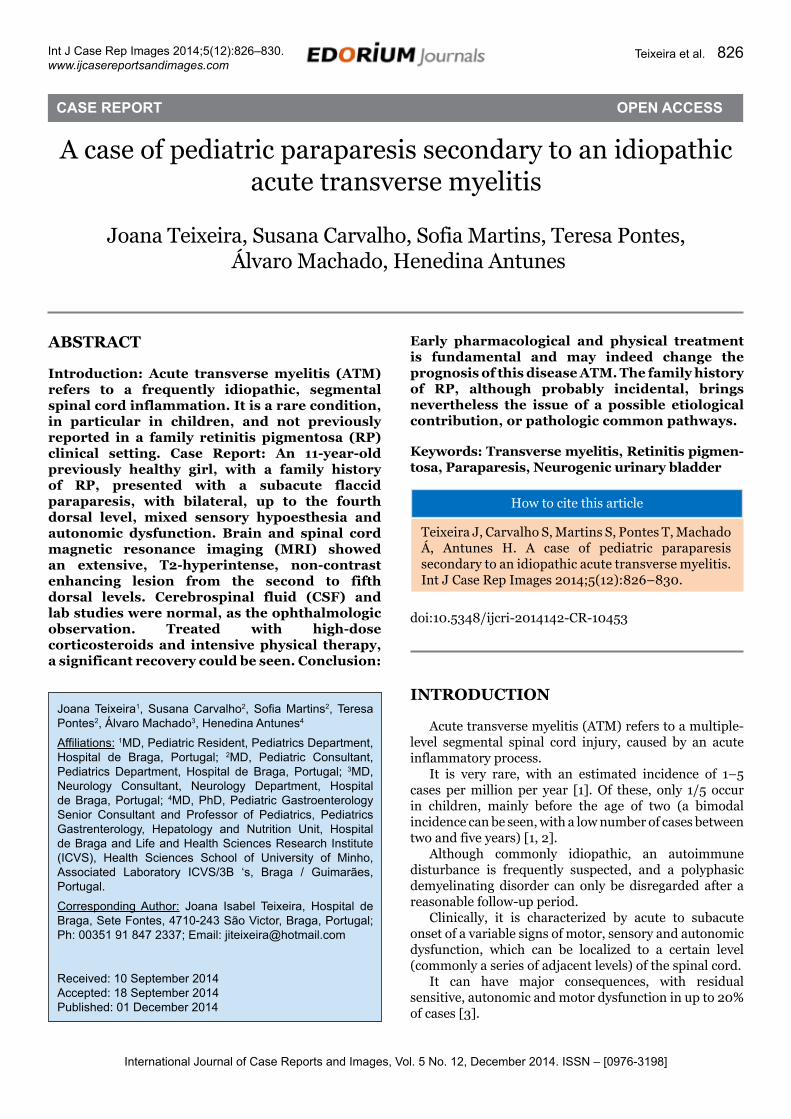
MRI was repeated at day-12 showing that the signal change extended from the first to fifth dorsal level, was bright in all T2-weighted sequences, did not uptake administered gadolinium, and was more clearly localized in the lateral and anterior columns (Figure 1).
At ophthalmologic evaluation there was no evidence of changes in visual acuity or ocular fundus.
Clinical improvement started at day-5 of methylprednisolone. There was increase muscle strength sufficient for autonomous, although limited, deambulation. It was decided to keep corticosteroid therapy (oral prednisolone 1 mg/kg/day). She also started physiatrist treatment with further muscle strength improvement and gait control. Bladder catheterization was needed because of high post-voiding residual volume.
She was discharged at day-23, with residual paraparesis (Medical Research Council Scale grade 4+), maintaining prednisolone, physiatrist treatment and intermittent bladder catheterization.
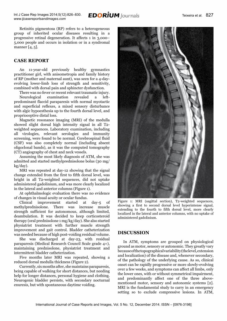
Five months later MRI was repeated, showing a reduced dorsal medulla thickness (Figure 2).
Currently, six months after, she maintains paraparesis, being capable of walking for short distances, but needing help for longer distances, personal hygiene and clothing. Neurogenic bladder persists, with secondary nocturnal enuresis, but with spontaneous daytime voiding.
DIscUssION
In ATM, symptoms are grouped on physiological ground as motor, sensory or autonomic. They greatly vary because of the topographical variability (the level, extension and localization) of the disease and, whenever secondary, of the pathology of the underlying cause. As so, clinical onset can be rapidly progressive or more slowly-evolving over a few weeks, and symptoms can affect all limbs, only the lower ones, with or without symmetrical impairment, and predominantly affect one of the three above-mentioned motor, sensory and autonomic systems [2]. MRI is the fundamental study to carry in an emergency setting so to exclude compressive lesions. In ATM,
Figure 1: MRI (sagittal section), T2-weighted sequences, showing a first to second dorsal level hyperintense signal, extending to the fourth to fifth dorsal level, more clearly localized in the lateral and anterior columns, with no uptake of administered gadolinium.
International Journal of Case Reports and Images, Vol. 5 No. 12, December 2014. ISSN – [0976-3198]
Int J Case Rep Images 2014;5(12):826–830. www.ijcasereportsandimages.com
Teixeira et al. 828
lesions are found mainly in the white matter surrounding the central medullar channel, usually involving several adjacent medullar segments, and are more easily seen in T2-wheighted sequences, where the edema appears bright. This usually precedes the latter medullar atrophy, the sole imagiological evidence of a past ATM [6–8].
In the majority cases, CSF has increased protein content as well as mild lymphocytosis. However, as spinal cord inflammation may not be evident at the beginning, some authors suggest that lumbar puncture should be repeated between second day and seventh day of the disease [6].
Oligoclonal bands should always be sought, because if they are found in the CSF and not in the blood, they raise the risk for multiple sclerosis [9]. An ophthalmologic evaluation is also recommended for all patients, as an additional finding of optic neuritis has a major implication on the diagnosis (multiple sclerosis or optic neuromyelitis) [6, 9].
Treatment is not consensual. The first line therapy is methylprednisolone for 5–7 days, followed by oral prednisolone (1 mg/kg/day) for 3–4 weeks. Non-pharmacological treatment includes intermittent bladder catheterization and physiotherapy [2, 9].
Etiological considerations in ATM should include viral/bacterial infections, autoimmune and connective tissue diseases, demyelinating diseases (multiple sclerosis, neuromyelitis optica), intra or extra-axial tumors and vascular diseases. Regarding the last etiological group idiopathic spontaneous dorsal spinal cord infarction is another possible unusual etiology of acute paraparesis in children. When it is possible to exclude all of these causes, ATM is then classified as idiopathic [1, 2, 6].
No etiologic cause of the ATM was found. Given the family history of (RP), a possible association was raised, although both the absence of prior descriptions and of RP signs in our patient, strongly reduce this possibility. There are few descriptions of RP associated with central nervous system infections. It was related to Creutzfeldt-Jakob disease and with congenital toxoplasmosis [4, 10]. There is also been described a close association between Human T-lymphotropic virus type I (HTLV-I) infection associated myelopathy and RP although the pathogenesis remains to be defined [5]. On this ground, we raise the possibility of an etiological contribution, or pathologic common pathways between ATM and RP.
cONcLUsION
Early pharmacological and physical treatment is fundamental and may indeed change the prognosis of this disease ATM. The family history of RP, although probably incidental, brings nevertheless the issue of a possible etiological contribution, or pathologic common pathways.
*********
Author contributionsJoana Teixeira – Substantial contributions to conception and design, Acquisition of data, Analysis and interpretation of data, Drafting the article, Revising it critically for important intellectual content, Final approval of the version to be publishedSusana Carvalho – Analysis and interpretation of data, Revising it critically for important intellectual content, Final approval of the version to be publishedSofia Martins – Analysis and interpretation of data, Revising it critically for important intellectual content, Final approval of the version to be publishedTeresa Pontes – Analysis and interpretation of data, Revising it critically for important intellectual content, Final approval of the version to be publishedÁlvaro Machado – Analysis and interpretation of data, Revising it critically for important intellectual content, Final approval of the version to be published
Figure 2: MRI (sagittal section), T2-weighted sequences, showing a hyperintense signal of anterior dominance in the middle dorsal region, associated with a reduced dorsal medulla thickness.
International Journal of Case Reports and Images, Vol. 5 No. 12, December 2014. ISSN – [0976-3198]
Int J Case Rep Images 2014;5(12):826–830. www.ijcasereportsandimages.com
Teixeira et al. 829
Henedina Antunes – Analysis and interpretation of data, Revising it critically for important intellectual content, Final approval of the version to be published
GuarantorThe corresponding author is the guarantor of submission.
conflict of InterestAuthors declare no conflict of interest.
copyright© 2014 Joana Teixeira et al. This article is distributed under the terms of Creative Commons Attribution License which permits unrestricted use, distribution and reproduction in any medium provided the original author(s) and original publisher are properly credited. Please see the copyright policy on the journal website for more information.
rEFErENcEs
1. Oñate Vergara E, Sota Busselo I, García-Santiago J, Gaztañaga Expósito R, Nogués Pérez A, Ruiz Benito MA. Transverse myelitis in immunocompetent children. An Pediatr (Barc) 2004 Aug;61(2):177–80. [Article in Spanish].
2. Wolf VL, Lupo PJ, Lotze TE. Pediatric Acute Transverse Myelitis Overview and Differential Diagnosis. J Child Neurol 2012 Nov;27(11):1426–36.
3. Thomas T, Branson HM, Verhey LH, et al. The demographic, clinical, and magnetic resonance imaging (MRI) features of transverse myelitis in children. J Child Neurol 2012 Jan;27(1):11–21.
4. Mitrovà E. A case of Creutzfeldt-Jakob disease related to familial retinitis pigmentosa patients. Eur J Epidemiol 1988 Mar;4(1):55–9.
5. Nakao K, Ohba N, Isashiki M, Isashiki Y, Unoki K, Osame M. Pigmentary retinal degeneration in patients with HTLV-I-associated myelopathy. Jpn J Ophthalmol 1989;33(4):383–91.
6. Transverse Myelitis Consortium Working Group. Proposed diagnostic criteria and nosology of acute transverse myelitis. Neurology 2002 Aug 27;59(4):499–505.
7. Alper G, Petropoulou KA, Fitz CR, Kim Y. Idiopathic acute transverse myelitis in children: an analysis and discussion of MRI findings. Mult Scler 2011 Jan;17(1):74–80.
8. Kim JY, Kim SJ, Bang MS. Spinal Cord Atrophy and Early Motor Recovery following Transverse Myelitis in Pediatric Patients. Ann Rehabil Med 2012 Jun;36(3):328–3.
9. Beh SC, Greenberg BM, Frohman T, Frohman EM. Transverse myelitis. Neurol Clin 2013 Feb;31(1):79-138.
10. Chhabra MS, Prakash G, Vashisht N, Garg SP. Retinitis pigmentosa and congenital toxoplasmosis: A rare coexistence. Indian J Ophthalmol 2007 Jul-Aug;55(4):303–4.
ABOUT THE AUTHORS
Article citation: Teixeira J, Carvalho S, Martins S, Pontes T, Machado Á, Antunes H. A case of pediatric paraparesis secondary to an idiopathic acute transverse myelitis. Int J Case Rep Images 2014;5(12):826–830.
Joana teixeira is Pediatric Resident at Hospital de Braga.
susana carvalho is a Pediatric Consultant at Hospital de Braga.
Sofia Martins is a Pediatric Consultant at Hospital de Braga.
International Journal of Case Reports and Images, Vol. 5 No. 12, December 2014. ISSN – [0976-3198]
Int J Case Rep Images 2014;5(12):826–830. www.ijcasereportsandimages.com
Teixeira et al. 830
teresa Pontes is a Pediatric Consultant at Hospital de Braga.
Àlvaro Machado is a Neurology Consultant at Hospital de Braga.
Henedina Antunes is a Pediatric Gastroenterology Senior Consultant and Professor of Pediatrics at Hospital de Braga Braga and Life and Health Sciences Research Institute (ICVS), Health Sciences School of University of Minho, Associated Laboratory ICVS/3B ‘s, Braga, Guimarães.
Access full text article onother devices
Access PDF of article onother devices
EDORIUM JOURNALS AN INTRODUCTION
Edorium Journals: On Web
About Edorium JournalsEdorium Journals is a publisher of high-quality, open ac-cess, international scholarly journals covering subjects in basic sciences and clinical specialties and subspecialties.
Edorium Journals www.edoriumjournals.com
Edorium Journals et al.
Edorium Journals: An introduction
Edorium Journals Team
But why should you publish with Edorium Journals?In less than 10 words - we give you what no one does.
Vision of being the bestWe have the vision of making our journals the best and the most authoritative journals in their respective special-ties. We are working towards this goal every day of every week of every month of every year.
Exceptional servicesWe care for you, your work and your time. Our efficient, personalized and courteous services are a testimony to this.
Editorial ReviewAll manuscripts submitted to Edorium Journals undergo pre-processing review, first editorial review, peer review, second editorial review and finally third editorial review.
Peer ReviewAll manuscripts submitted to Edorium Journals undergo anonymous, double-blind, external peer review.
Early View versionEarly View version of your manuscript will be published in the journal within 72 hours of final acceptance.
Manuscript statusFrom submission to publication of your article you will get regular updates (minimum six times) about status of your manuscripts directly in your email.
Our Commitment
Mentored Review Articles (MRA)Our academic program “Mentored Review Article” (MRA) gives you a unique opportunity to publish papers under mentorship of international faculty. These articles are published free of charges.
Favored Author programOne email is all it takes to become our favored author. You will not only get fee waivers but also get information and insights about scholarly publishing.
Institutional Membership programJoin our Institutional Memberships program and help scholars from your institute make their research accessi-ble to all and save thousands of dollars in fees make their research accessible to all.
Our presenceWe have some of the best designed publication formats. Our websites are very user friendly and enable you to do your work very easily with no hassle.
Something more...We request you to have a look at our website to know more about us and our services.
We welcome you to interact with us, share with us, join us and of course publish with us.
Browse Journals
CONNECT WITH US
Invitation for article submissionWe sincerely invite you to submit your valuable research for publication to Edorium Journals.
Six weeksYou will get first decision on your manuscript within six weeks (42 days) of submission. If we fail to honor this by even one day, we will publish your manuscript free of charge.
Four weeksAfter we receive page proofs, your manuscript will be published in the journal within four weeks (31 days). If we fail to honor this by even one day, we will pub-lish your manuscript free of charge and refund you the full article publication charges you paid for your manuscript.
This page is not a part of the published article. This page is an introduction to Edorium Journals and the publication services.