9 mri.ppt - chemistry.uoc.gr · magnetic resonance imaging (mri) is one of the newest diagnostic...
TRANSCRIPT
…MRI…
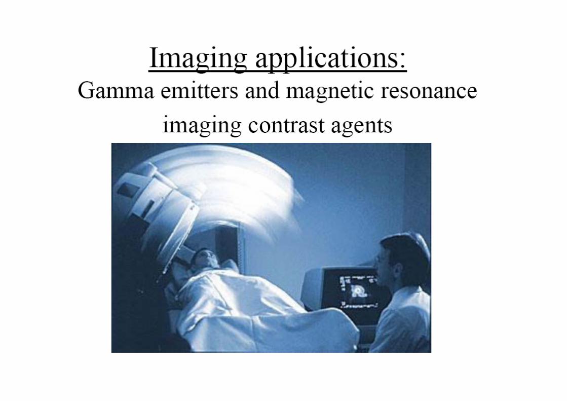
Magnetic resonance imaging (MRI) is one of the newest diagnostic
medical imaging technologies that uses strong magnets and pulses of
radio waves to manipulate the natural magnetic properties in the body
t t i ibl ito generate a visible image.
Magnetic Resonance Angiography: study blood flow
Magnetic Resonance Spectroscopy: chemical composition of diseased tissue
Magnetic Resonance CholangioPancreatography: a non-invasive potential alternative for the diagnostic procedure endoscopic retrograde
cholangiopancreatography
Advantages of MRI vs X-rays, Computed tomography scan (CT scan) and ultrasounds
1) G t t l t t
(CT scan) and ultrasounds
1) Greater natural contrast
2) Very minor fluctuations in chemical composition can be determined,
3) It can distinguish fine variations in tissues deep within the body,
4) Useful for spotting and distinguishing diseased tissues (tumors and other l i ) l i h i d llesions) early in their development,
5) The entire body can be scanned, from head to toe and from the skin to the deepest recesses of the brainthe deepest recesses of the brain,
6) MRI scans are not obstructed by bone, gas, or body waste,
) f d d d ll h f l d7) Safe…does not depend on potentially harmful ionizing radiation,
…BUT…rather complex procedure and…$$$...

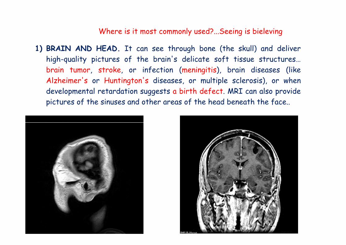
Where is it most commonly used?...Seeing is bieleving
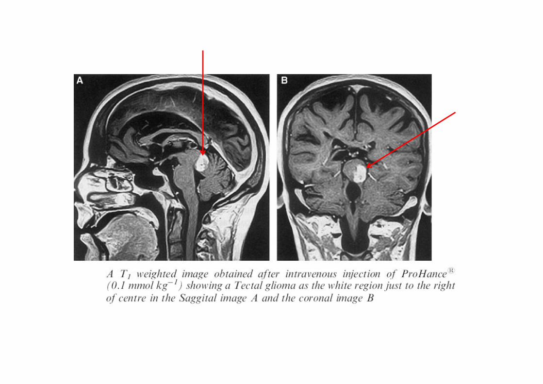
1) BRAIN AND HEAD. It can see through bone (the skull) and deliverhigh-quality pictures of the brain's delicate soft tissue structures…b i k i f i ( i i i ) b i di (likbrain tumor, stroke, or infection (meningitis), brain diseases (likeAlzheimer's or Huntington's diseases, or multiple sclerosis), or whendevelopmental retardation suggests a birth defect. MRI can also providep gg ppictures of the sinuses and other areas of the head beneath the face..
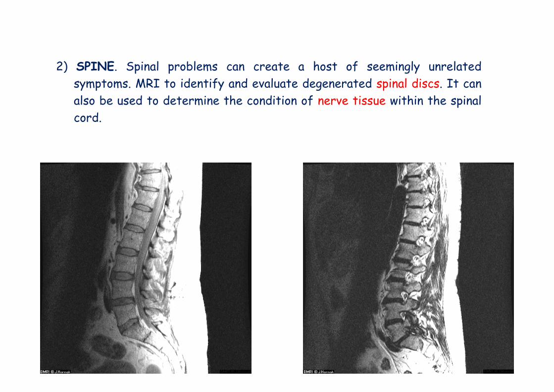
2) SPINE S i l bl t h t f i l l t d2) SPINE. Spinal problems can create a host of seemingly unrelatedsymptoms. MRI to identify and evaluate degenerated spinal discs. It canalso be used to determine the condition of nerve tissue within the spinalpcord.
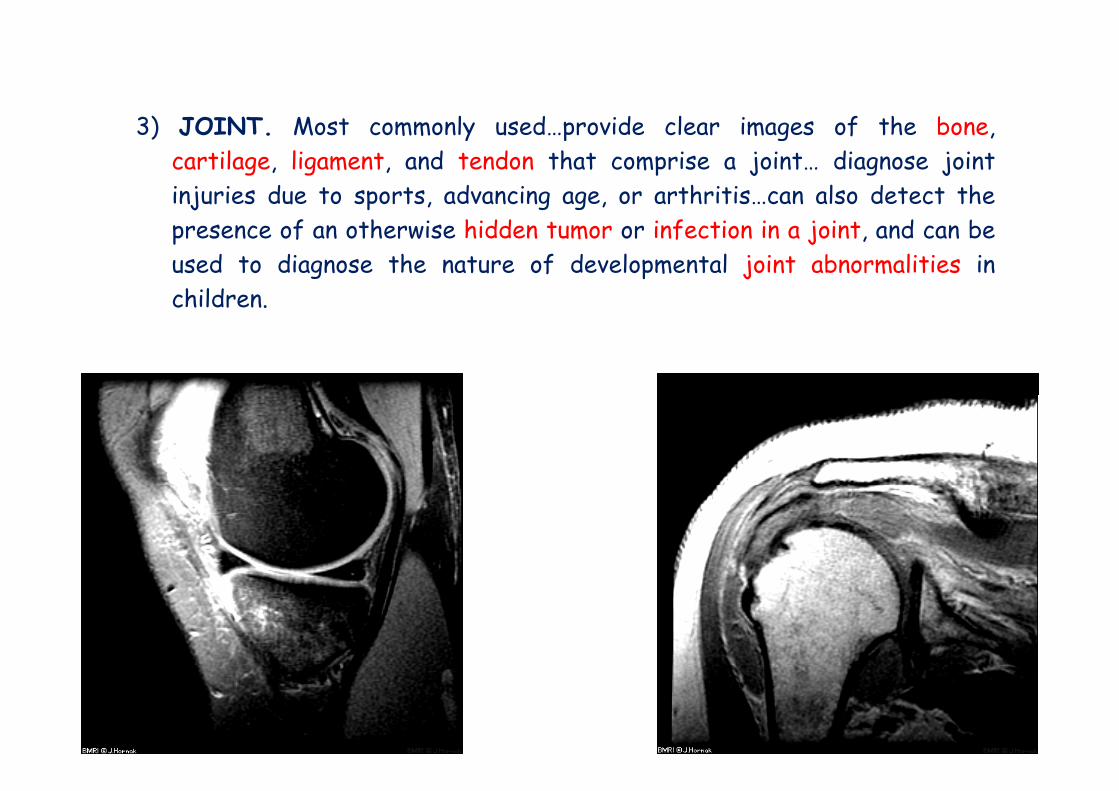
3) JOINT Most commonly used provide clear images of the bone3) JOINT. Most commonly used…provide clear images of the bone,cartilage, ligament, and tendon that comprise a joint… diagnose jointinjuries due to sports, advancing age, or arthritis…can also detect thepresence of an otherwise hidden tumor or infection in a joint, and can beused to diagnose the nature of developmental joint abnormalities inchildren.children.
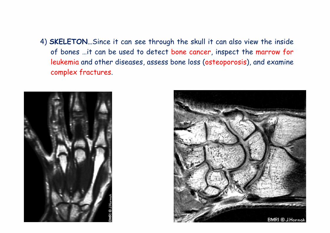
4) SKELETON…Since it can see through the skull it can also view the insideof bones …it can be used to detect bone cancer, inspect the marrow forleukemia and other diseases assess bone loss (osteoporosis) and examineleukemia and other diseases, assess bone loss (osteoporosis), and examinecomplex fractures.
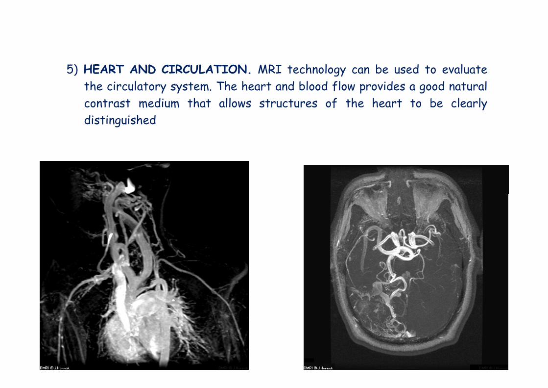
5) HEART AND CIRCULATION. MRI technology can be used to evaluatethe circulatory system. The heart and blood flow provides a good naturalcontrast medium that allows structures of the heart to be clearlycontrast medium that allows structures of the heart to be clearlydistinguished
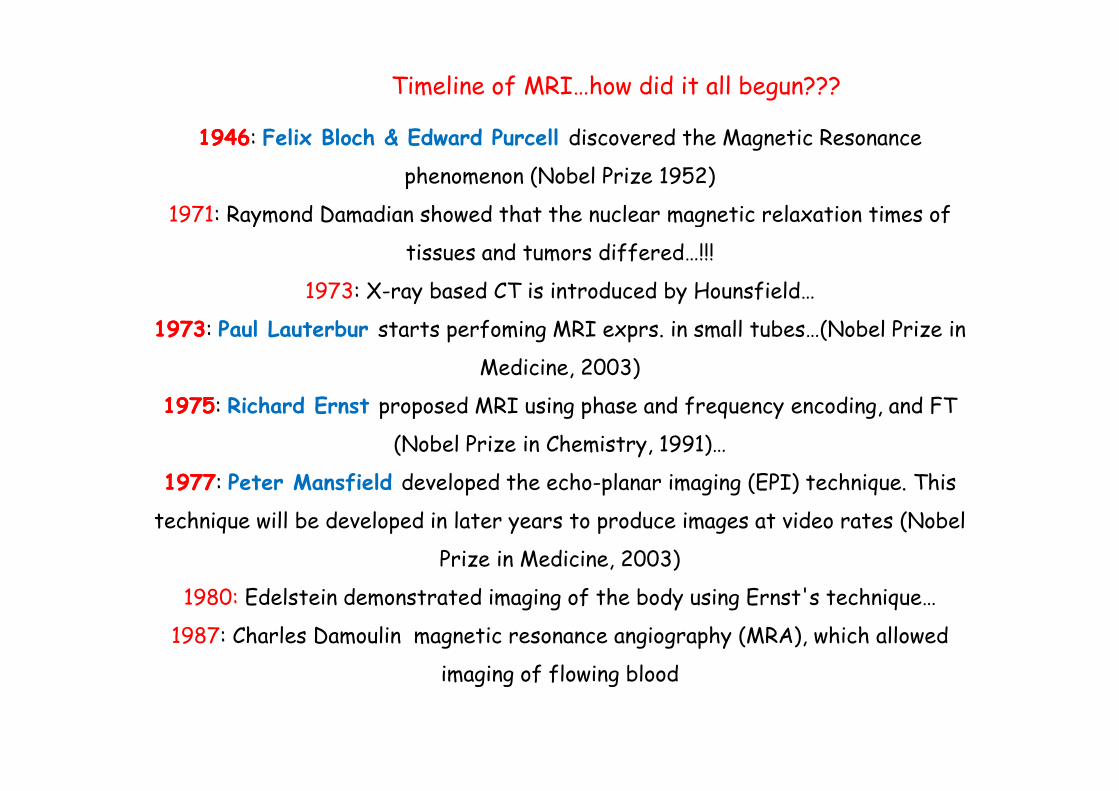
Timeline of MRI…how did it all begun???
19461946: Felix Bloch & Edward Purcell discovered the Magnetic Resonance
phenomenon (Nobel Prize 1952)
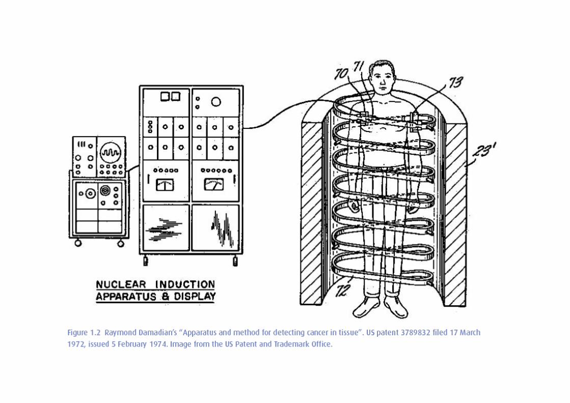
1971: Raymond Damadian showed that the nuclear magnetic relaxation times of 1971: Raymond Damadian showed that the nuclear magnetic relaxation times of
tissues and tumors differed…!!!
1973: X-ray based CT is introduced by Hounsfield…
19731973: Paul Lauterbur starts perfoming MRI exprs. in small tubes…(Nobel Prize in
Medicine, 2003)
19751975: Richard Ernst proposed MRI using phase and frequency encoding, and FT
(Nobel Prize in Chemistry, 1991)…
19771977: Peter Mansfield developed the echo-planar imaging (EPI) technique This 19771977: Peter Mansfield developed the echo planar imaging (EPI) technique. This
technique will be developed in later years to produce images at video rates (Nobel
Prize in Medicine, 2003)
1980: Edelstein demonstrated imaging of the body using Ernst's technique…
1987: Charles Damoulin magnetic resonance angiography (MRA), which allowed
f fl bl dimaging of flowing blood
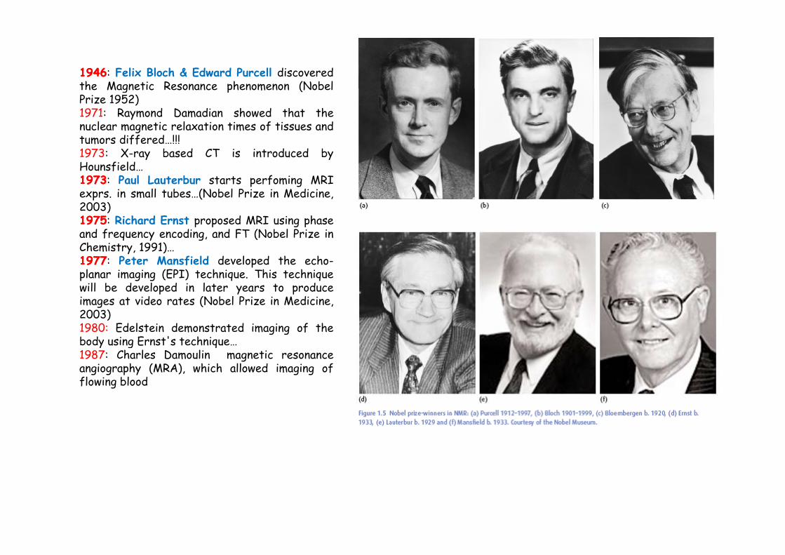
19461946: Felix Bloch & Edward Purcell discoveredthe Magnetic Resonance phenomenon (Nobelthe Magnetic Resonance phenomenon (NobelPrize 1952)1971: Raymond Damadian showed that thenuclear magnetic relaxation times of tissues andtumors differed…!!!1973: X ray based CT is introduced by1973: X-ray based CT is introduced byHounsfield…19731973: Paul Lauterbur starts perfoming MRIexprs. in small tubes…(Nobel Prize in Medicine,2003)19751975: Richard Ernst proposed MRI using phaseand frequency encoding, and FT (Nobel Prize inChemistry, 1991)…19771977: Peter Mansfield developed the echo-planar imaging (EPI) technique. This techniqueplanar imaging (EPI) technique. This techniquewill be developed in later years to produceimages at video rates (Nobel Prize in Medicine,2003)1980: Edelstein demonstrated imaging of thebody using Ernst's techniquebody using Ernst s technique…1987: Charles Damoulin magnetic resonanceangiography (MRA), which allowed imaging offlowing blood
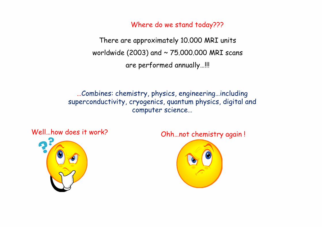
Where do we stand today???
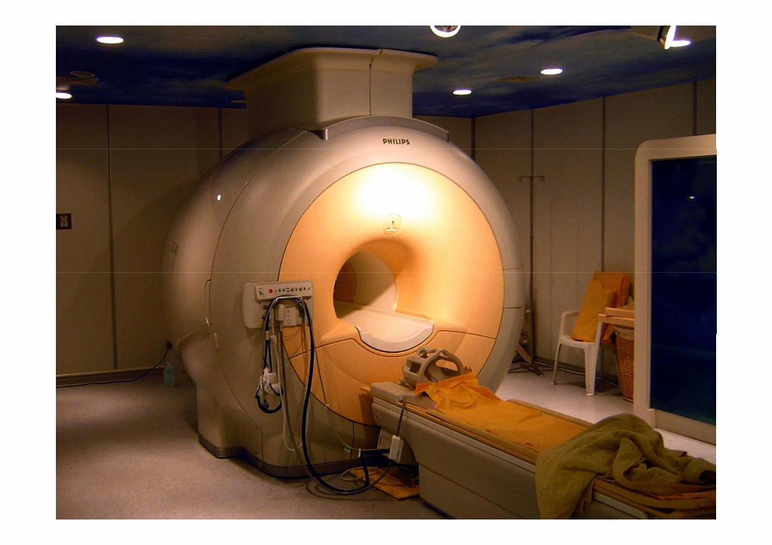
There are approximately 10.000 MRI units
worldwide (2003) and ~ 75.000.000 MRI scans
f d ll !!! are performed annually…!!!
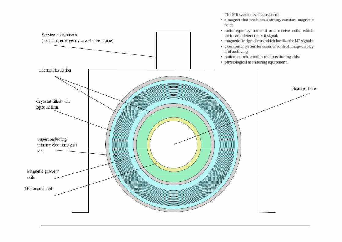
…Combines: chemistry, physics, engineering…including superconductivity, cryogenics, quantum physics, digital and
computer science…
Well…how does it work? Ohh…not chemistry again !
p
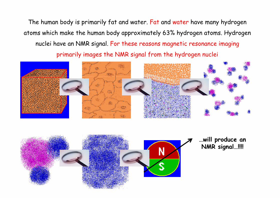
The human body is primarily fat and water. Fat and water have many hydrogen
t ms hi h m k th h m b d xim t l 63% h d t ms H d atoms which make the human body approximately 63% hydrogen atoms. Hydrogen
nuclei have an NMR signal. For these reasons magnetic resonance imaging
primarily images the NMR signal from the hydrogen nucleiprimarily images the NMR signal from the hydrogen nuclei
…will produce an NMR signal…!!!!
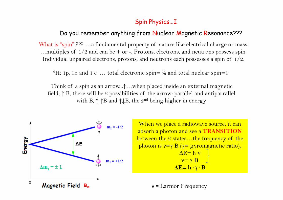
Spin Physics…I
D m mb n thin f m N l M n ti R s n n ???Do you remember anything from Nuclear Magnetic Resonance???
What is “spin” ??? …a fundamental property of nature like electrical charge or mass. …multiples of 1/2 and can be + or -. Protons, electrons, and neutrons possess spin. p p pIndividual unpaired electrons, protons, and neutrons each possesses a spin of 1/2.
2H: 1p, 1n and 1 e- … total electronic spin= ½ and total nuclear spin=1
Think of a spin as an arrow...↑…when placed inside an external magnetic field, ↑ B, there will be 2 possibilities of the arrow: parallel and antiparrallel
with B ↑ ↑B and ↑↓B the 2nd being higher in energywith B, ↑ ↑B and ↑↓B, the 2nd being higher in energy.
Wh l di it When we place a radiowave source, it can absorb a photon and see a TRANSITIONbetween the 2 states…the frequency of the photon is ν=γ. Β (γ= gyromagnetic ratio) photon is ν=γ. Β (γ= gyromagnetic ratio).
ΔE= h. νν= γ. Β
ΔΕ= h . γ . ΒΔΕ= h γ Β
ν = Larmor Frequency
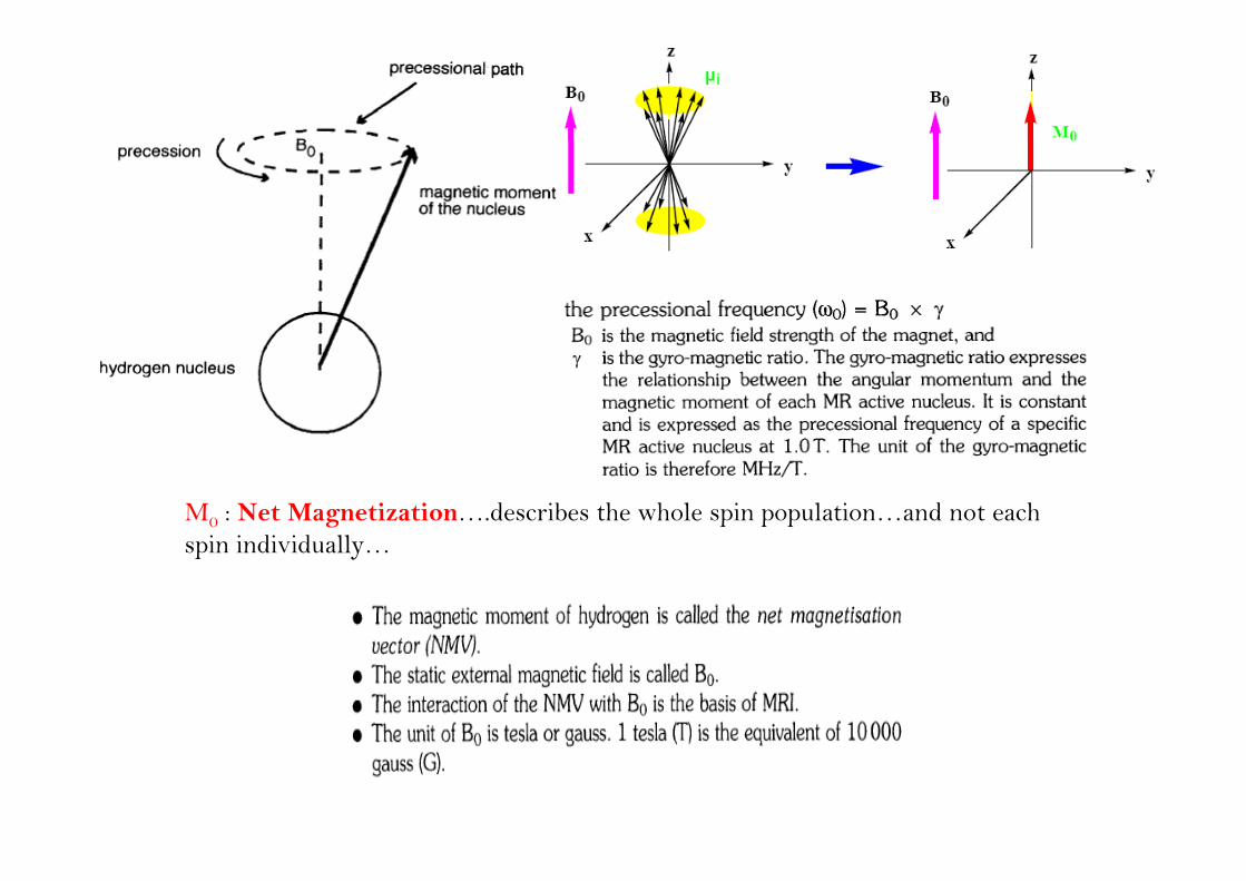
M0 : Net Magnetization….describes the whole spin population…and not each i i di id llspin individually…
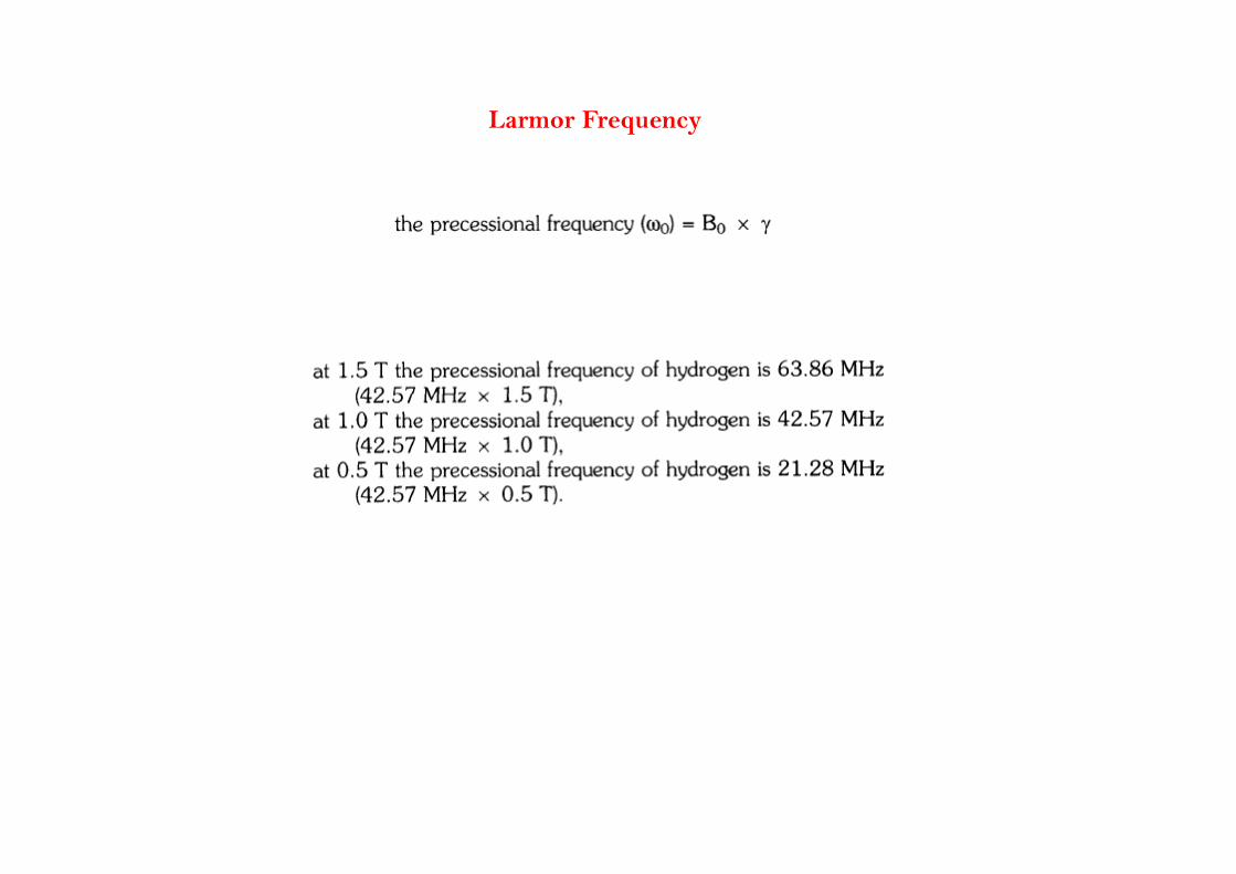
Larmor Frequency

CW NMR Experiment
The simplest NMR experiment is the continuous wave (CW) experiment. There are two ways of performing this experiment In the first a constant frequency which is continuously on of performing this experiment. In the first, a constant frequency, which is continuously on, probes the energy levels while the magnetic field is varied.
The CW experiment can also be performed with a constant magnetic field and a frequency The CW experiment can also be performed with a constant magnetic field and a frequency which is varied. The magnitude of the constant magnetic field is represented by the position of the vertical blue line in the energy level diagram.
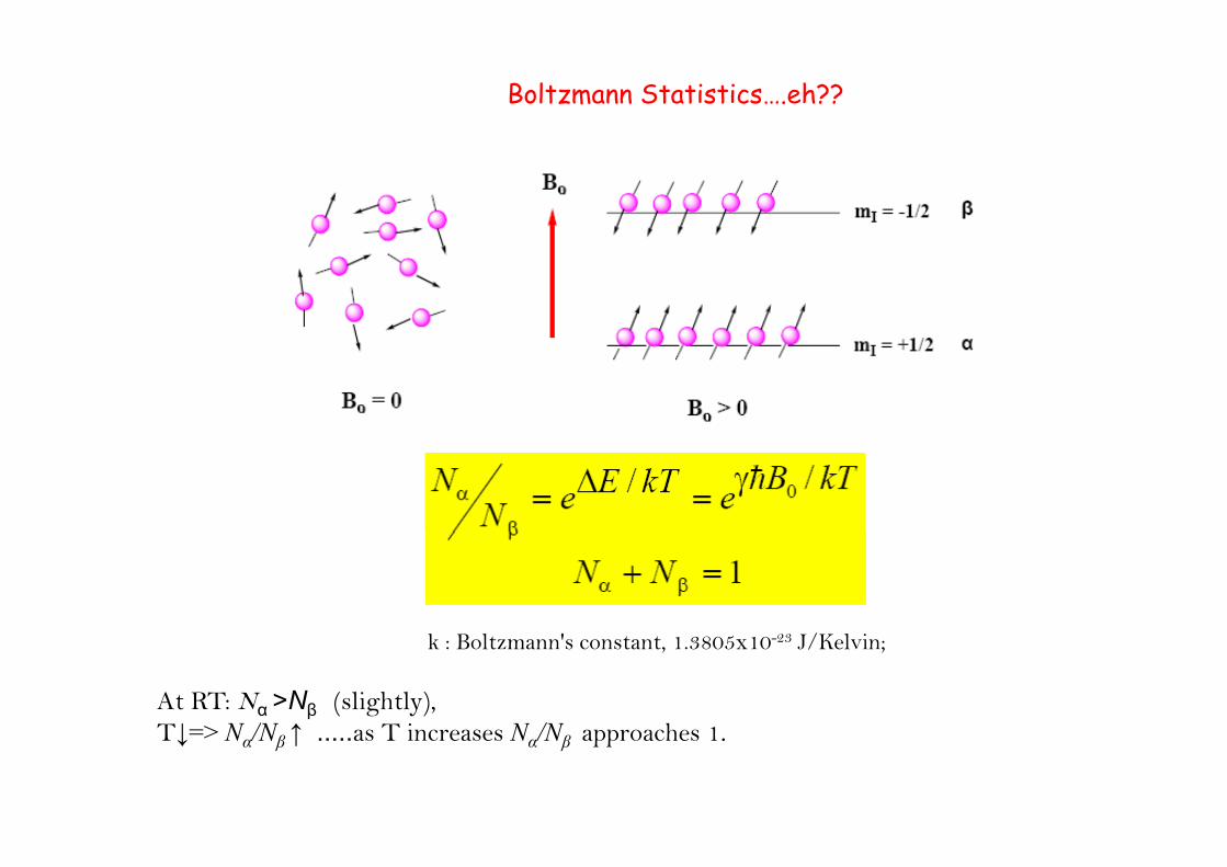
Boltzmann Statistics….eh??
At RT: Nα >Νβ (slightly), T↓ > N /Ν ↑ T i N /Ν h 1
k : Boltzmann's constant, 1.3805x10⁻²³ J/Kelvin;
T↓=> Nα/Νβ ↑ .....as T increases Nα/Νβ approaches 1.
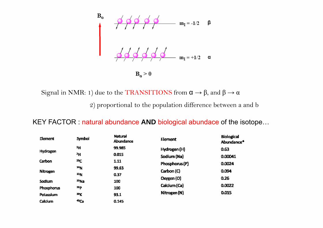
Signal in NMR: 1) due to the TRANSITIONS from α → β, and β → α
2) proportional to the population difference between a and b
KEY FACTOR : natural abundance AND biological abundace of the isotope…
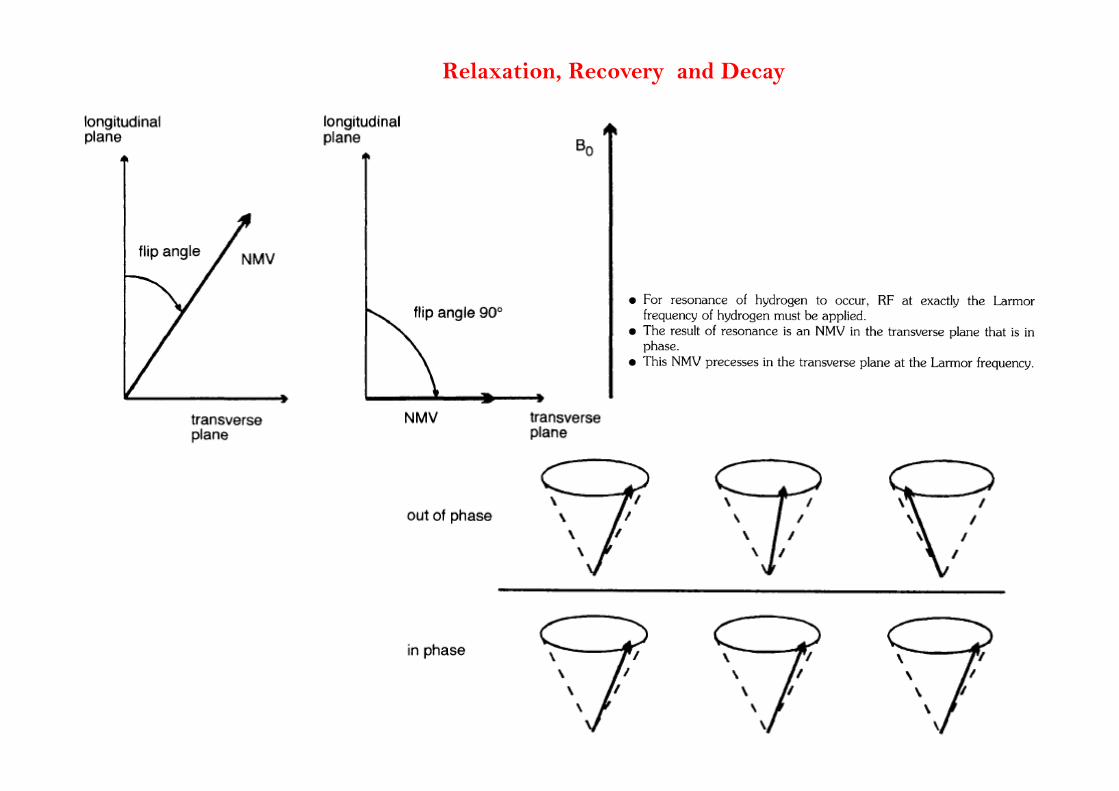
Relaxation, Recovery and Decay
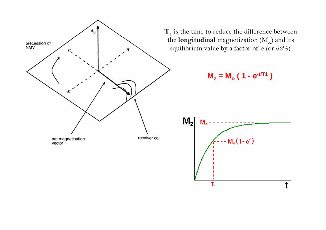
T1 is the time to reduce the difference between the longitudinal magnetization (MZ) and its equilibrium value by a factor of e (or 63%).
Mz = Mo ( 1 - e-t/T1 )
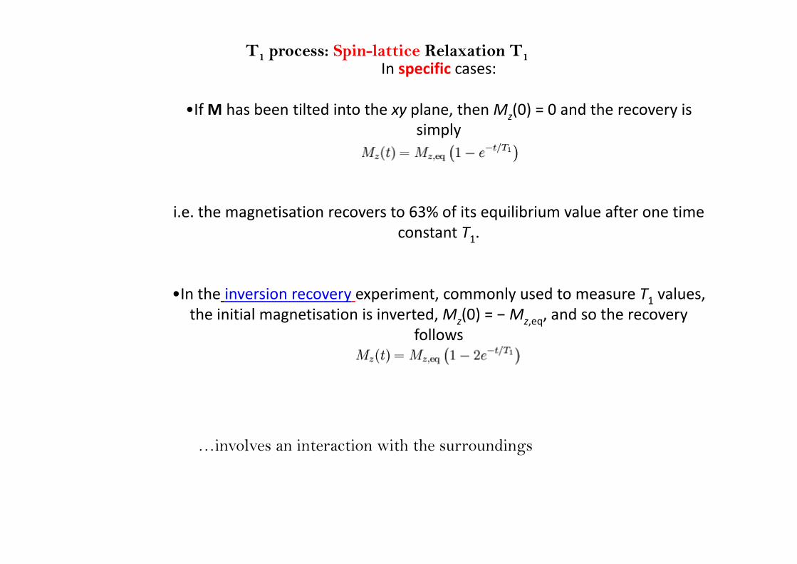
In specific cases:T1 process: Spin-lattice Relaxation T1
•If M has been tilted into the xy plane, then Mz(0) = 0 and the recovery is simply
i.e. the magnetisation recovers to 63% of its equilibrium value after one time g qconstant T1.
•In the inversion recovery experiment, commonly used to measure T1 values, the initial magnetisation is inverted, Mz(0) = − Mz,eq, and so the recovery
follows
…involves an interaction with the surroundings
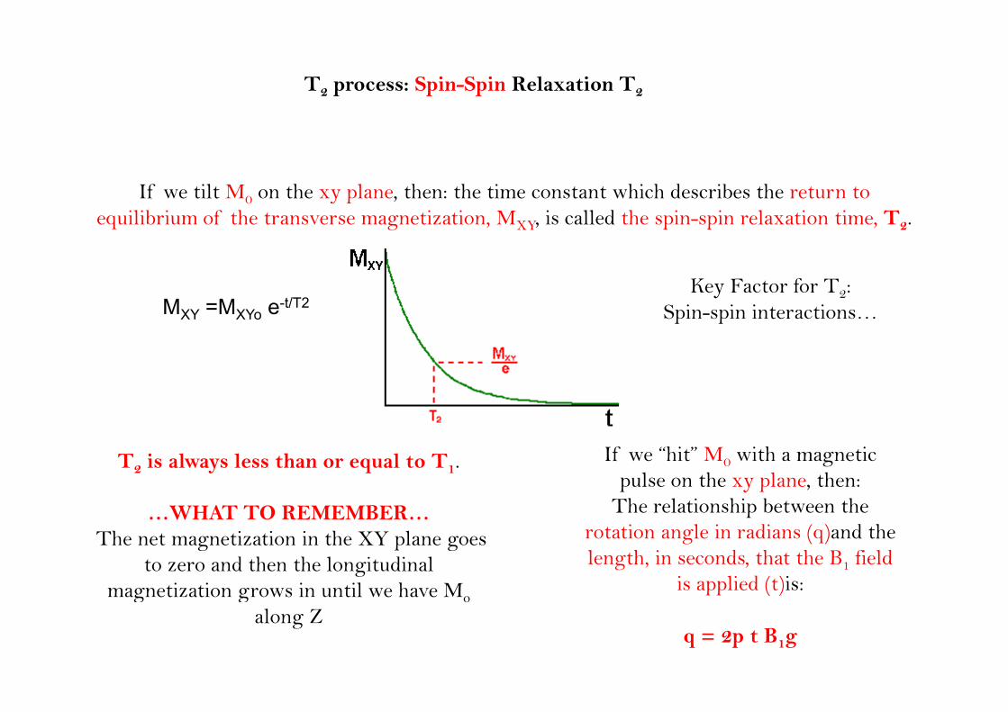
T2 process: Spin-Spin Relaxation T2
If we tilt M on the xy plane then: the time constant which describes the return to If we tilt M0 on the xy plane, then: the time constant which describes the return to equilibrium of the transverse magnetization, MXY, is called the spin-spin relaxation time, T2.
MXY =MXYo e-t/T2Key Factor for T2:
Spin-spin interactions…
T2 is always less than or equal to T1.
…WHAT TO REMEMBER…
If we “hit” M0 with a magnetic pulse on the xy plane, then:
The relationship between the …WHAT TO REMEMBER…The net magnetization in the XY plane goes
to zero and then the longitudinal magnetization grows in until we have Mo
protation angle in radians (q)and the length, in seconds, that the B1 field
is applied (t)is:g g oalong Z
q = 2p t B1g
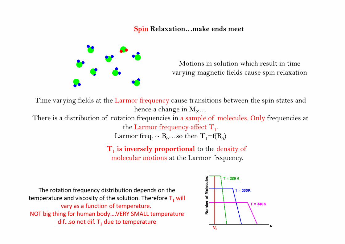
Spin Relaxation…make ends meet
Motions in solution which result in time varying magnetic fields cause spin relaxation
Time varying fields at the Larmor frequency cause transitions between the spin states and hence a change in MZ…
There is a distribution of rotation frequencies in a sample of molecules. Only frequencies at p ythe Larmor frequency affect T1.
Larmor freq. ~ B0…so then T1=f(B0)
T is inversely proportional to the density of T1 is inversely proportional to the density of molecular motions at the Larmor frequency.
The rotation frequency distribution depends on the temperature and viscosity of the solution. Therefore T1 will
vary as a function of temperaturevary as a function of temperature.NOT big thing for human body….VERY SMALL temperature
dif…so not dif. T1 due to temperature
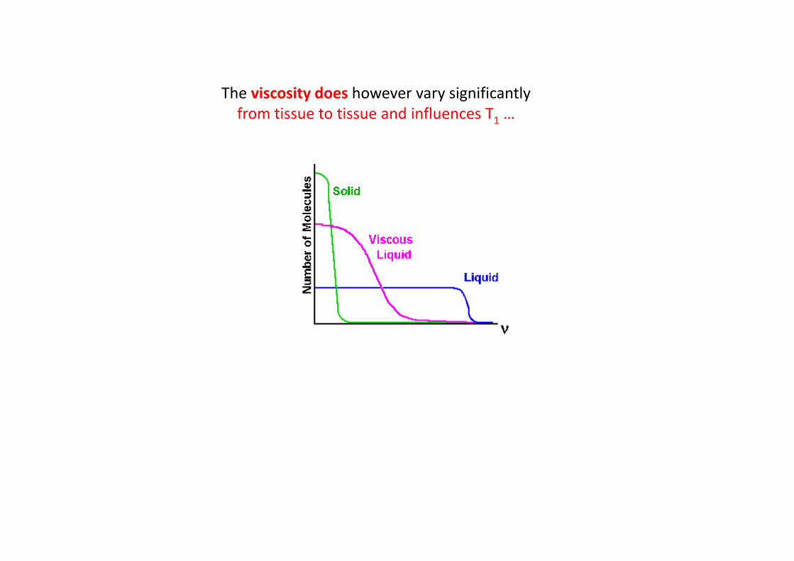
Th i it d h i ifi tlThe viscosity does however vary significantly from tissue to tissue and influences T1 …
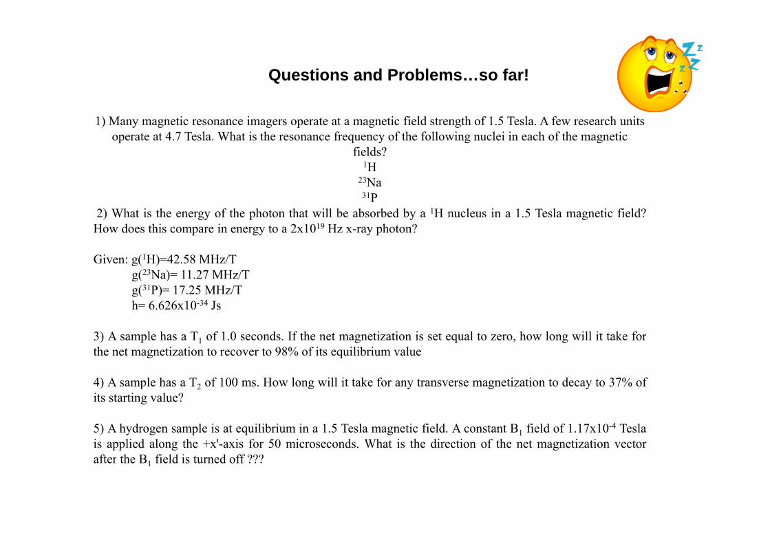
Questions and Problems…so far!
1) Many magnetic resonance imagers operate at a magnetic field strength of 1.5 Tesla. A few research units operate at 4.7 Tesla. What is the resonance frequency of the following nuclei in each of the magnetic
fi ld ?fields? 1H
23Na31P
2) Wh t i th f th h t th t ill b b b d b 1H l i 1 5 T l ti fi ld?2) What is the energy of the photon that will be absorbed by a 1H nucleus in a 1.5 Tesla magnetic field?How does this compare in energy to a 2x10¹⁹ Hz x-ray photon?
Given: g(1H)=42.58 MHz/T(23N ) 11 27 MH /Tg(23Na)= 11.27 MHz/T
g(31P)= 17.25 MHz/Th= 6.626x10⁻³⁴ Js
3) A l h T f 1 0 d If th t ti ti i t l t h l ill it t k f3) A sample has a T1 of 1.0 seconds. If the net magnetization is set equal to zero, how long will it take forthe net magnetization to recover to 98% of its equilibrium value
4) A sample has a T2 of 100 ms. How long will it take for any transverse magnetization to decay to 37% ofit t ti l ?its starting value?
5) A hydrogen sample is at equilibrium in a 1.5 Tesla magnetic field. A constant B1 field of 1.17x10⁻⁴ Teslais applied along the +x'-axis for 50 microseconds. What is the direction of the net magnetization vectorafter the B field is t rned off ???after the B1 field is turned off ???
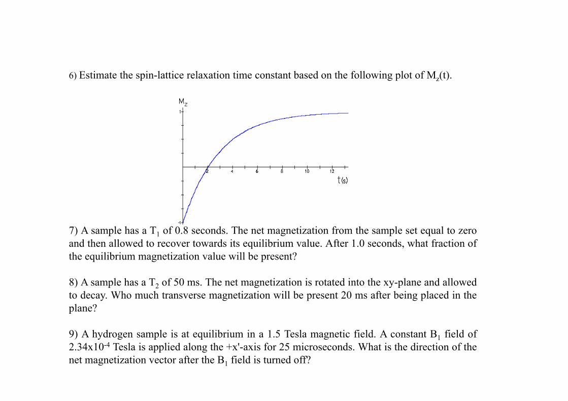
6) Estimate the spin-lattice relaxation time constant based on the following plot of Mz(t).
7) A sample has a T1 of 0.8 seconds. The net magnetization from the sample set equal to zeroand then allowed to recover towards its equilibrium value. After 1.0 seconds, what fraction ofand then allowed to recover towards its equilibrium value. After 1.0 seconds, what fraction ofthe equilibrium magnetization value will be present?
8) A sample has a T2 of 50 ms. The net magnetization is rotated into the xy-plane and allowedd Wh h i i ill b 20 f b i l d i hto decay. Who much transverse magnetization will be present 20 ms after being placed in the
plane?
9) A hydrogen sample is at equilibrium in a 1.5 Tesla magnetic field. A constant B1 field of9) A hydrogen sample is at equilibrium in a 1.5 Tesla magnetic field. A constant B1 field of2.34x10-4 Tesla is applied along the +x'-axis for 25 microseconds. What is the direction of thenet magnetization vector after the B1 field is turned off?
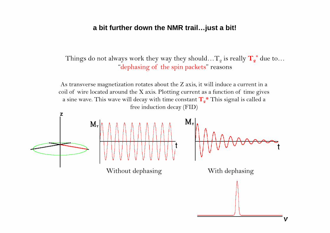
a bit further down the NMR trail…just a bit!
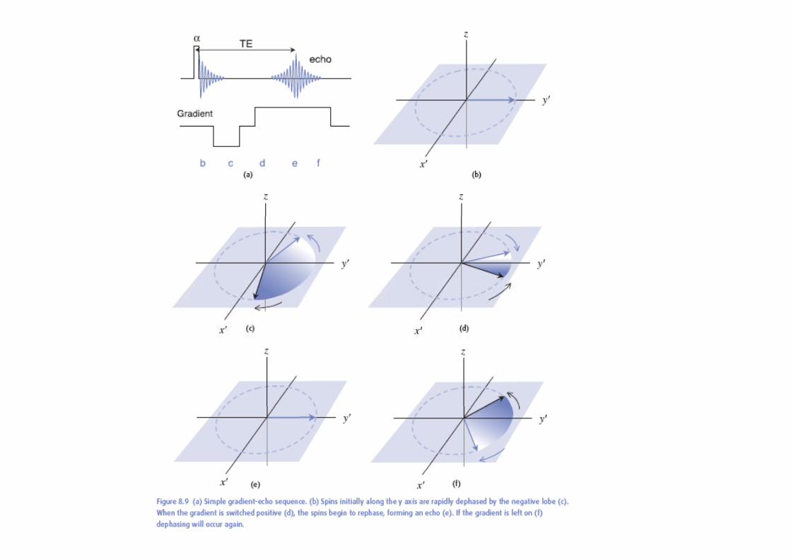
Things do not always work they way they should…T2 is really T2* due to…
“dephasing of the spin packets” reasons
As transverse magnetization rotates about the Z axis, it will induce a current in a il f i l d d h X i Pl i f i f i i coil of wire located around the X axis. Plotting current as a function of time gives
a sine wave. This wave will decay with time constant T2* This signal is called a free induction decay (FID)
With t d h i With d h iWithout dephasing With dephasing
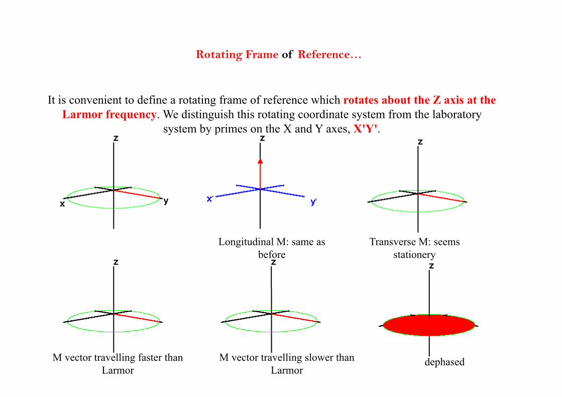
Rotating Frame of Reference…
It is convenient to define a rotating frame of reference which rotates about the Z axis at the Larmor frequency. We distinguish this rotating coordinate system from the laboratory
system by primes on the X and Y axes, X'Y'.
Longitudinal M: same as Transverse M: seemsLongitudinal M: same as before
Transverse M: seems stationery
M vector travelling faster than Larmor
M vector travelling slower than Larmor
dephased
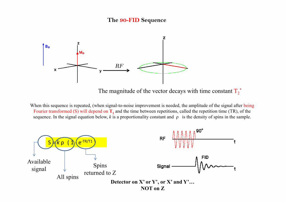
The 90-FID Sequence
RF
The magnitude of the vector decays with time constant T2*
When this sequence is repeated, (when signal-to-noise improvement is needed, the amplitude of the signal after being Fourier transformed (S) will depend on T1 and the time between repetitions, called the repetition time (TR), of the sequence. In the signal equation below, k is a proportionality constant and ρ is the density of spins in the sample.
S = k ρ ( 1 ‐ e‐TR/T1 )
Available signal Spins signal
All spinsreturned to Z
Detector on X’ or Y’, or X’ and Y’…NOT on Z
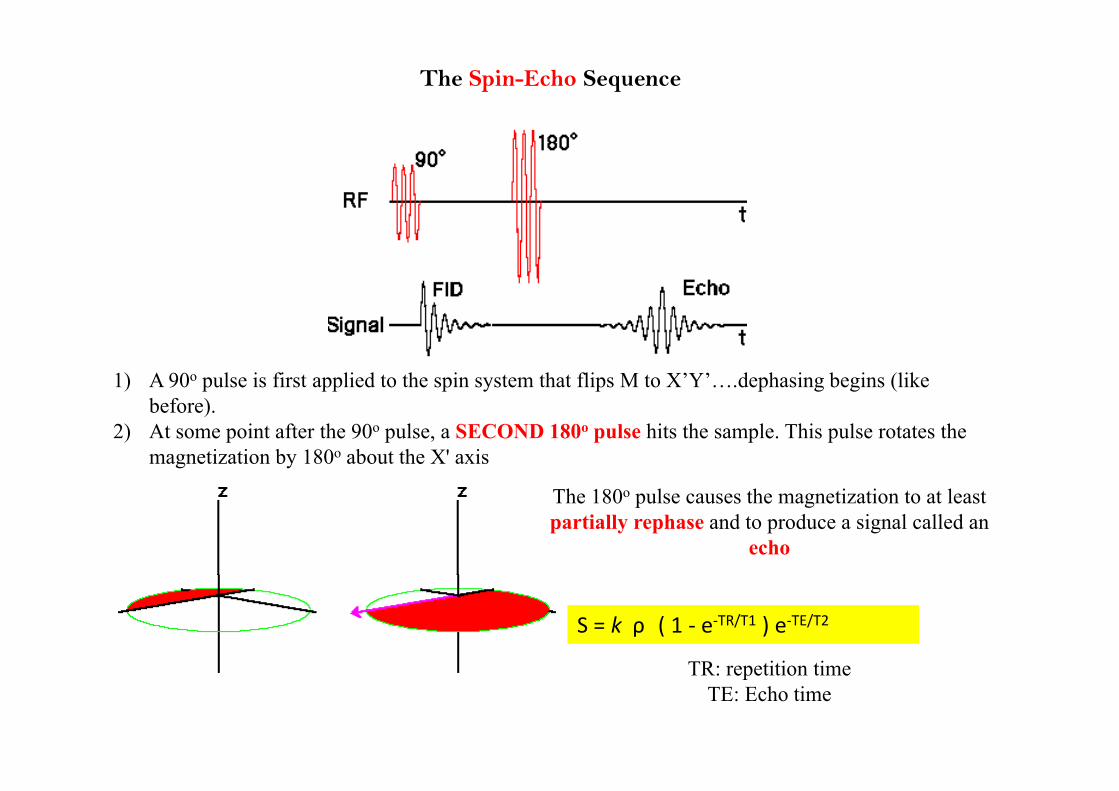
The Spin-Echo Sequence
1) A 90o pulse is first applied to the spin system that flips M to X’Y’….dephasing begins (like before).
2) At some point after the 90o pulse, a SECOND 180o pulse hits the sample. This pulse rotates the magnetization by 180o about the X' axis
The 180o pulse causes the magnetization to at least partially rephase and to produce a signal called an
echo
S = k ρ ( 1 ‐ e‐TR/T1 ) e‐TE/T2
TR titi tiTR: repetition timeTE: Echo time
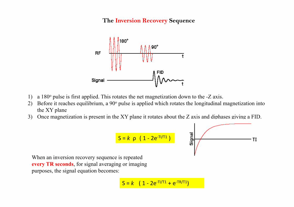
The Inversion Recovery Sequence
1) a 180o pulse is first applied. This rotates the net magnetization down to the -Z axis.2) Before it reaches equilibrium a 90o pulse is applied which rotates the longitudinal magnetization into2) Before it reaches equilibrium, a 90 pulse is applied which rotates the longitudinal magnetization into
the XY plane3) Once magnetization is present in the XY plane it rotates about the Z axis and diphases giving a FID.
S = k ρ ( 1 ‐ 2e‐TI/T1 )
When an inversion recovery sequence is repeated every TR seconds, for signal averaging or imaging purposes, the signal equation becomes:
S = k ( 1 ‐ 2e‐TI/T1 + e‐TR/T1)
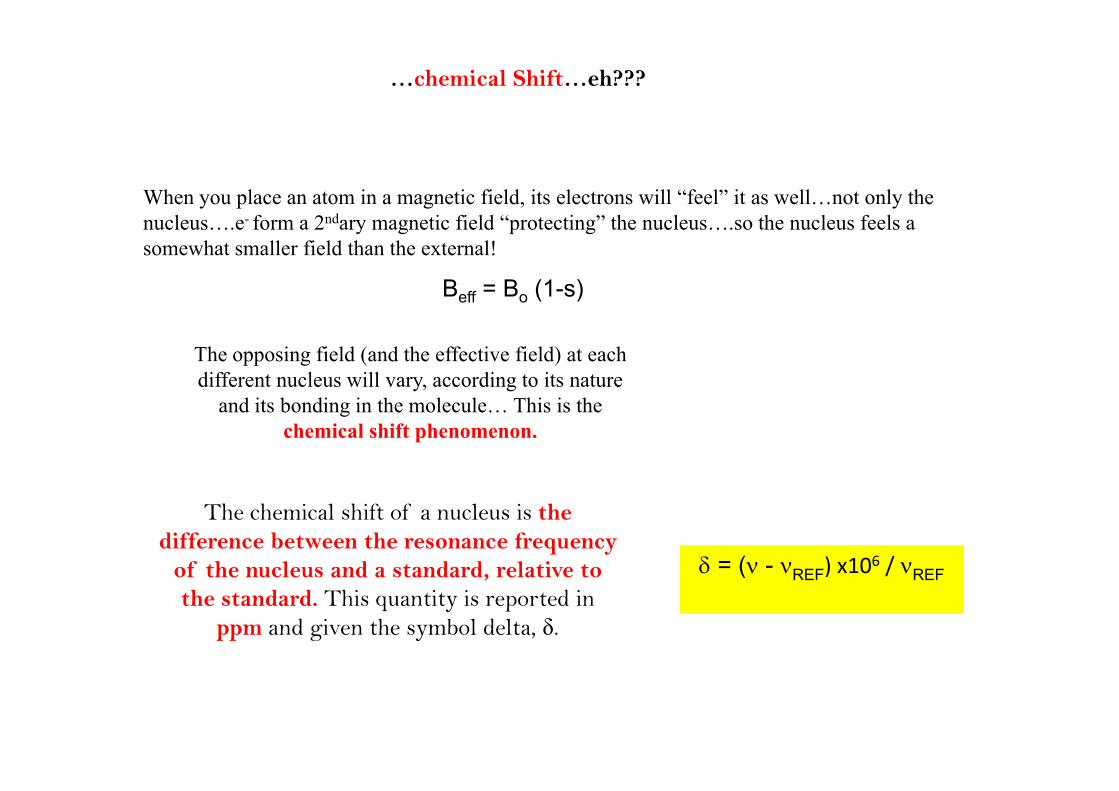
…chemical Shift…eh???
When you place an atom in a magnetic field, its electrons will “feel” it as well…not only the l f 2 d i fi ld “ i ” h l h l f lnucleus….e- form a 2ndary magnetic field “protecting” the nucleus….so the nucleus feels a
somewhat smaller field than the external!
Beff = Bo (1-s)
The opposing field (and the effective field) at each different nucleus will vary, according to its nature
and its bonding in the molecule… This is the chemical shift phenomenon.
The chemical shift of a nucleus is the difference between the resonance frequency
of the nucleus and a standard, relative to = ( - REF) x106 / REFof the nucleus and a standard, relative to
the standard. This quantity is reported in ppm and given the symbol delta, δ.
( REF) / REF
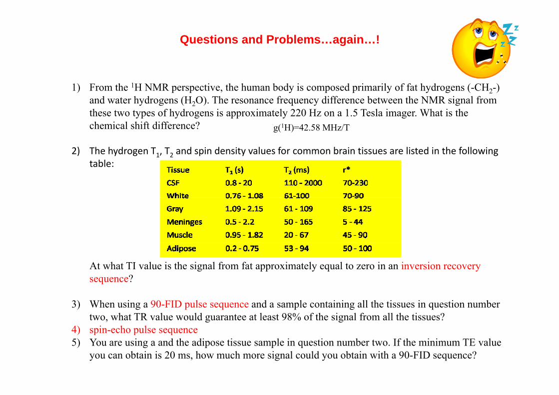
Questions and Problems…again…!
1) From the 1H NMR perspective, the human body is composed primarily of fat hydrogens (-CH2-) and water hydrogens (H2O). The resonance frequency difference between the NMR signal from these two types of hydrogens is approximately 220 Hz on a 1.5 Tesla imager. What is the chemical shift difference?
2) The hydrogen T T2 and spin density values for common brain tissues are listed in the following
g(1H)=42.58 MHz/T
2) The hydrogen T1, T2 and spin density values for common brain tissues are listed in the following table:
At what TI value is the signal from fat approximately equal to zero in an inversion recovery sequence?
3) When using a 90-FID pulse sequence and a sample containing all the tissues in question number two, what TR value would guarantee at least 98% of the signal from all the tissues?
4) spin-echo pulse sequence4) spin echo pulse sequence5) You are using a and the adipose tissue sample in question number two. If the minimum TE value
you can obtain is 20 ms, how much more signal could you obtain with a 90-FID sequence?
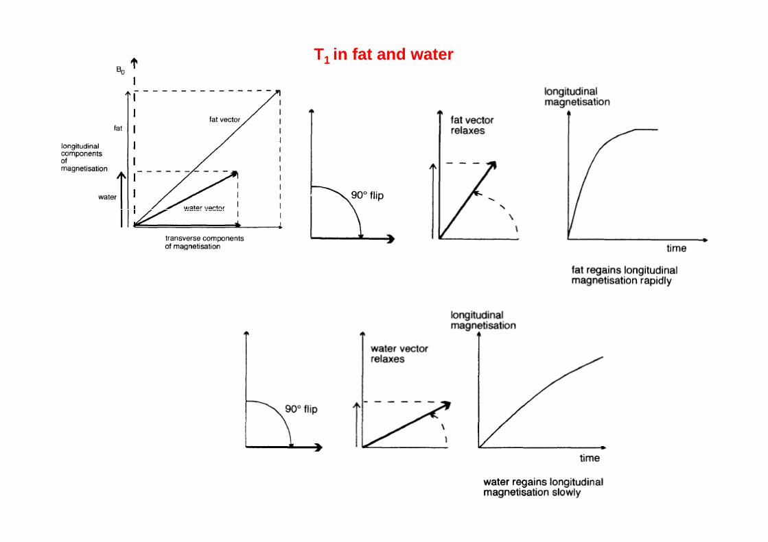
T1 in fat and water
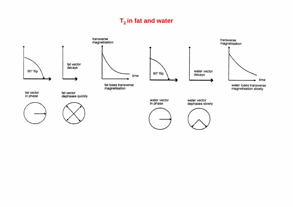
T2 in fat and water
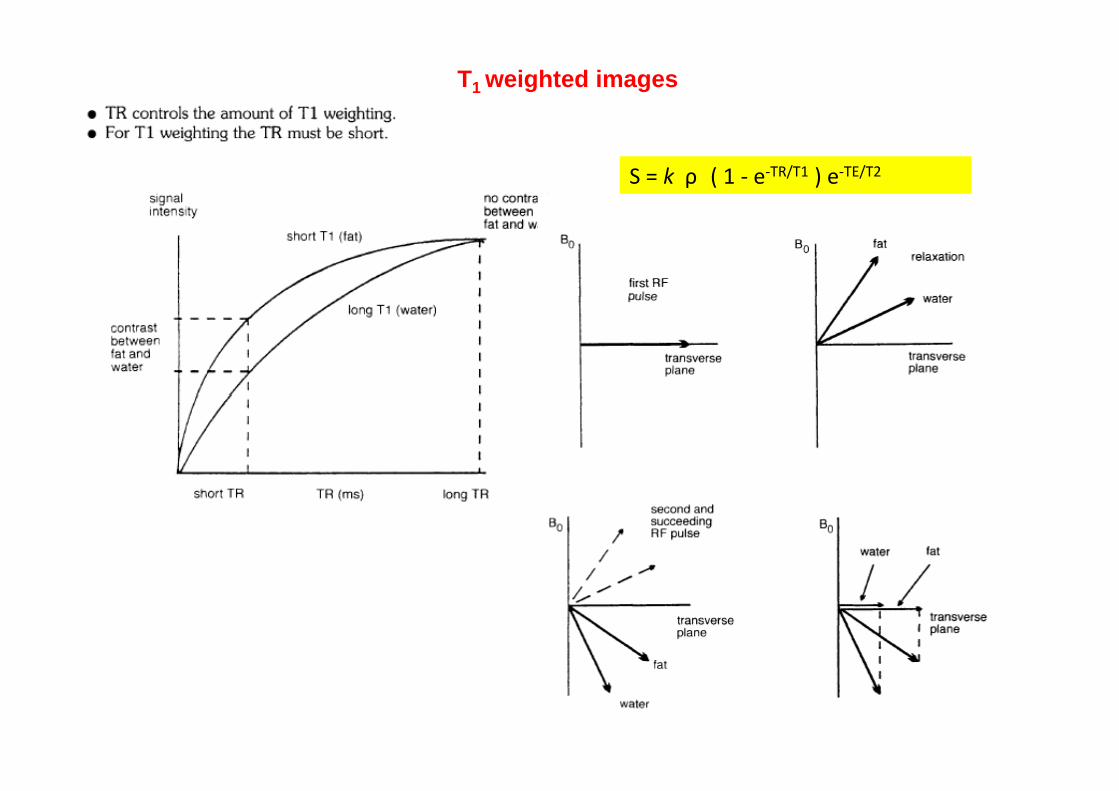
T1 weighted images
S = k ρ ( 1 ‐ e‐TR/T1 ) e‐TE/T2
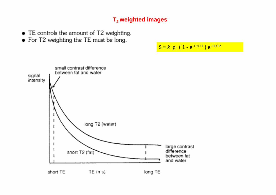
T2 weighted images
S = k ρ ( 1 ‐ e‐TR/T1 ) e‐TE/T2
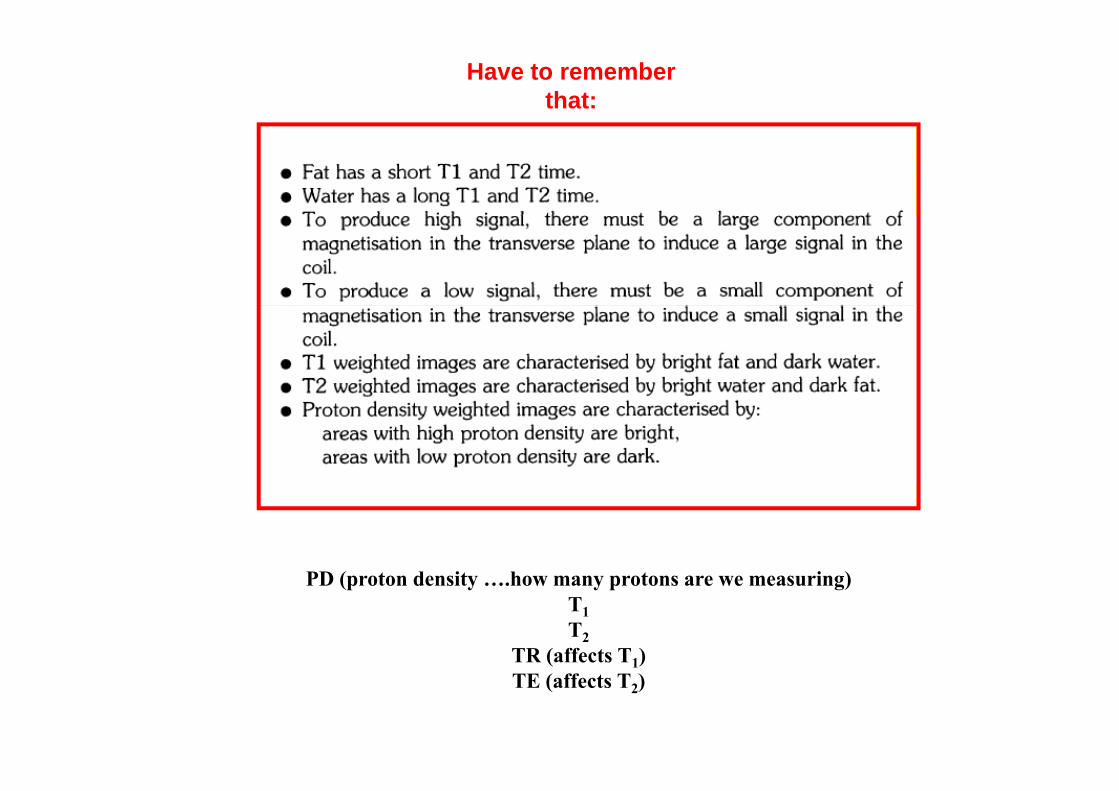
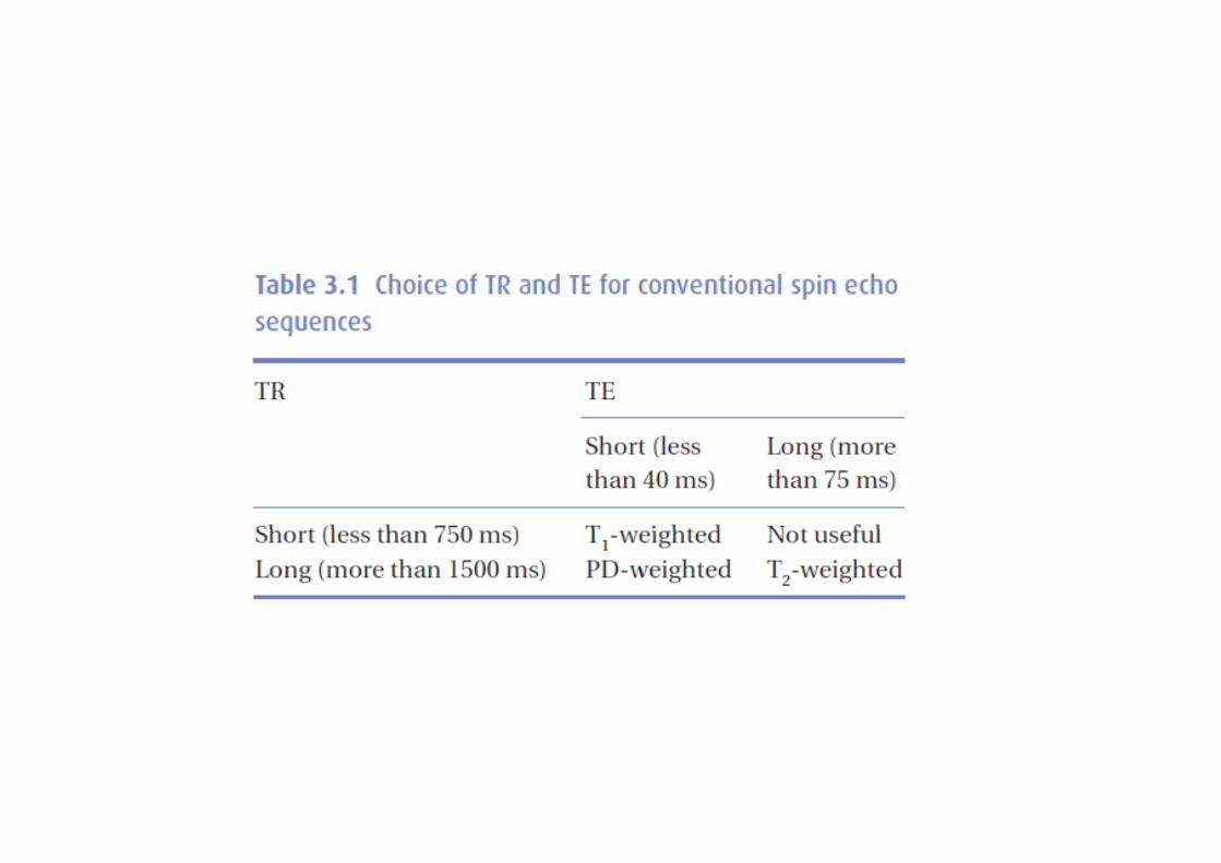
Have to remember that:
PD ( t d it h t i )PD (proton density ….how many protons are we measuring) T1T2
TR (affects T1) ( 1)TE (affects T2)
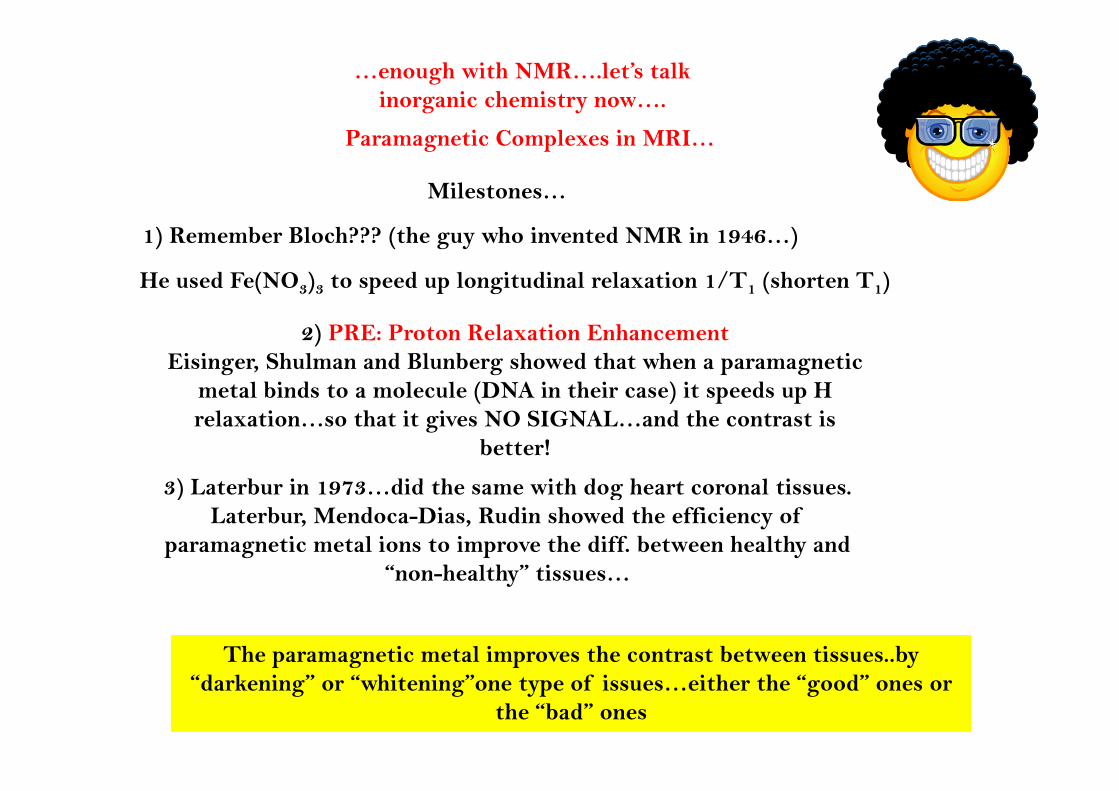
…enough with NMR….let’s talk inorganic chemistry now….
Paramagnetic Complexes in MRI…
Milestones…
1) Remember Bloch??? (the guy who invented NMR in 1946…)
He used Fe(NO3)3 to speed up longitudinal relaxation 1/T1 (shorten T1)
2) PRE: Proton Relaxation EnhancementEisinger, Shulman and Blunberg showed that when a paramagnetic
metal binds to a molecule (DNA in their case) it speeds up H metal binds to a molecule (DNA in their case) it speeds up H relaxation…so that it gives NO SIGNAL…and the contrast is
better!
3) Laterbur in 1973 did the same with dog heart coronal tissues 3) Laterbur in 1973…did the same with dog heart coronal tissues. Laterbur, Mendoca-Dias, Rudin showed the efficiency of
paramagnetic metal ions to improve the diff. between healthy and “non-healthy” tissuesnon-healthy tissues…
The paramagnetic metal improves the contrast between tissues..by p g p y“darkening” or “whitening”one type of issues…either the “good” ones or
the “bad” ones
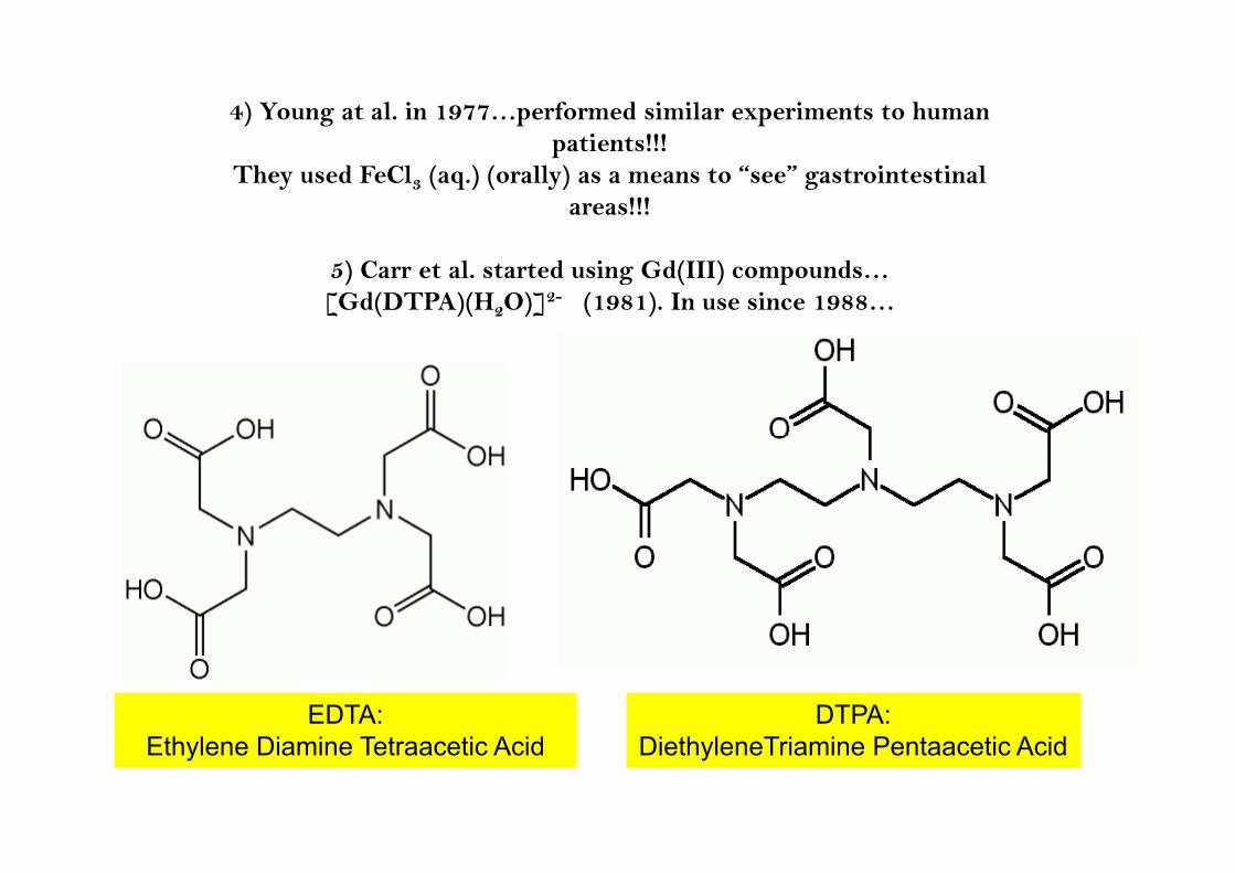
4) Young at al. in 1977…performed similar experiments to human ti t !!!patients!!!
They used FeCl3 (aq.) (orally) as a means to “see” gastrointestinal areas!!!
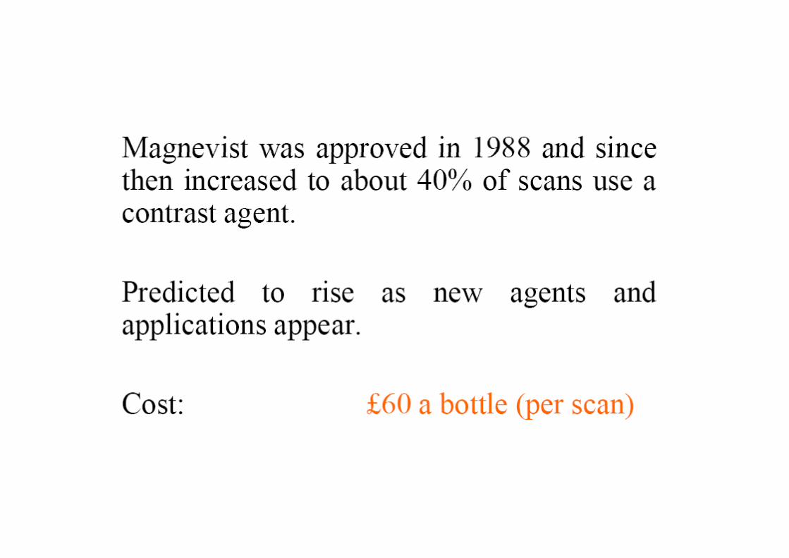
5) Carr et al. started using Gd(III) compounds…[Gd(DTPA)(H2O)]2- (1981). In use since 1988…
EDTA: DTPA: Ethylene Diamine Tetraacetic Acid DiethyleneTriamine Pentaacetic Acid
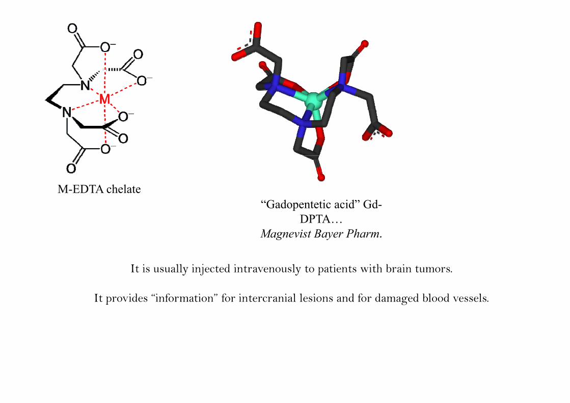
M-EDTA chelate“G d t ti id” Gd“Gadopentetic acid” Gd-
DPTA…Magnevist Bayer Pharm.
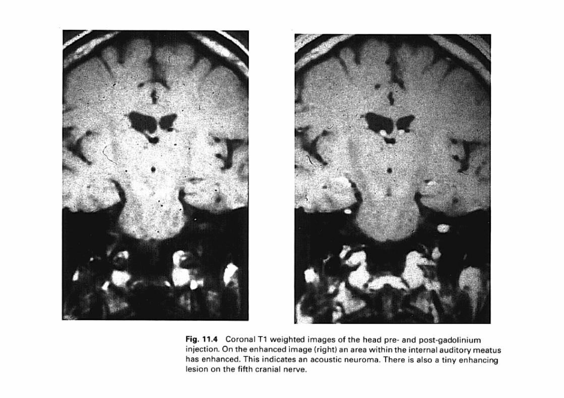
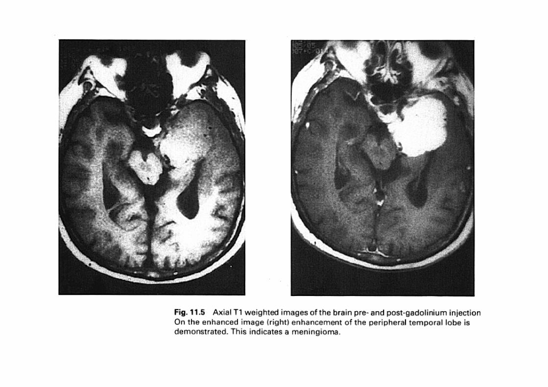
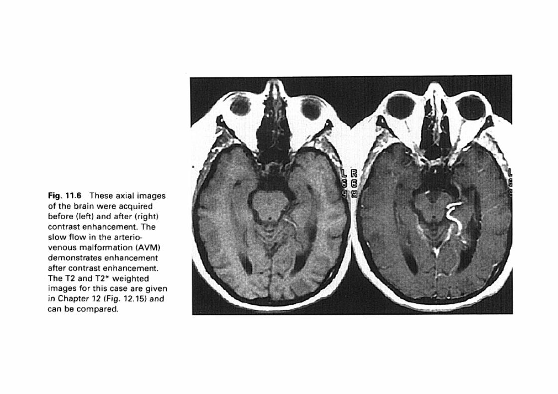
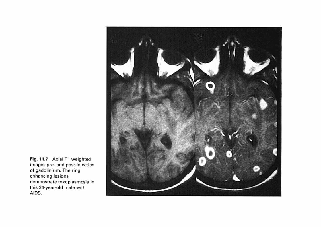
It is usually injected intravenously to patients with brain tumors.
It provides “information” for intercranial lesions and for damaged blood vessels.
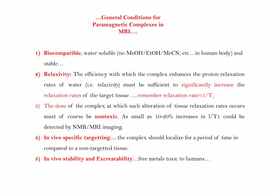
…General Conditions for Paramagnetic Complexes in
MRIMRI….
1) Biocompatible water soluble (no MeOH/EtOH/MeCN etc in human body) and 1) Biocompatible, water soluble (no MeOH/EtOH/MeCN, etc…in human body) and
stable…
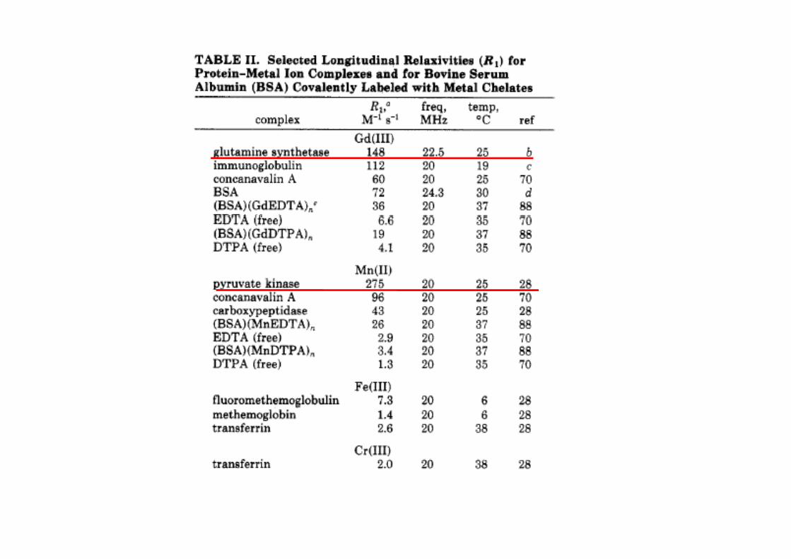
2) Relaxivity: The efficiency with which the complex enhances the proton relaxation2) Relaxivity: The efficiency with which the complex enhances the proton relaxation
rates of water (i.e. relaxivity) must be sufficient to significantly increase the
relaxation rates of the target tissue ….remember relaxation rate=1/T1ela ation ates of the ta get tissue …. e e be ela ation ate 1/ 1
3) The dose of the complex at which such alteration of tissue relaxation rates occurs
must of course be nontoxic. As small as 10-20% increases in l/T1 could be
detected by NMR/MRI imaging.
4) In vivo specific targetting… the complex should localize for a period of time in
compared to a non-targetted tissue.
5) In vivo stability and Excreatability…free metals toxic to humans…
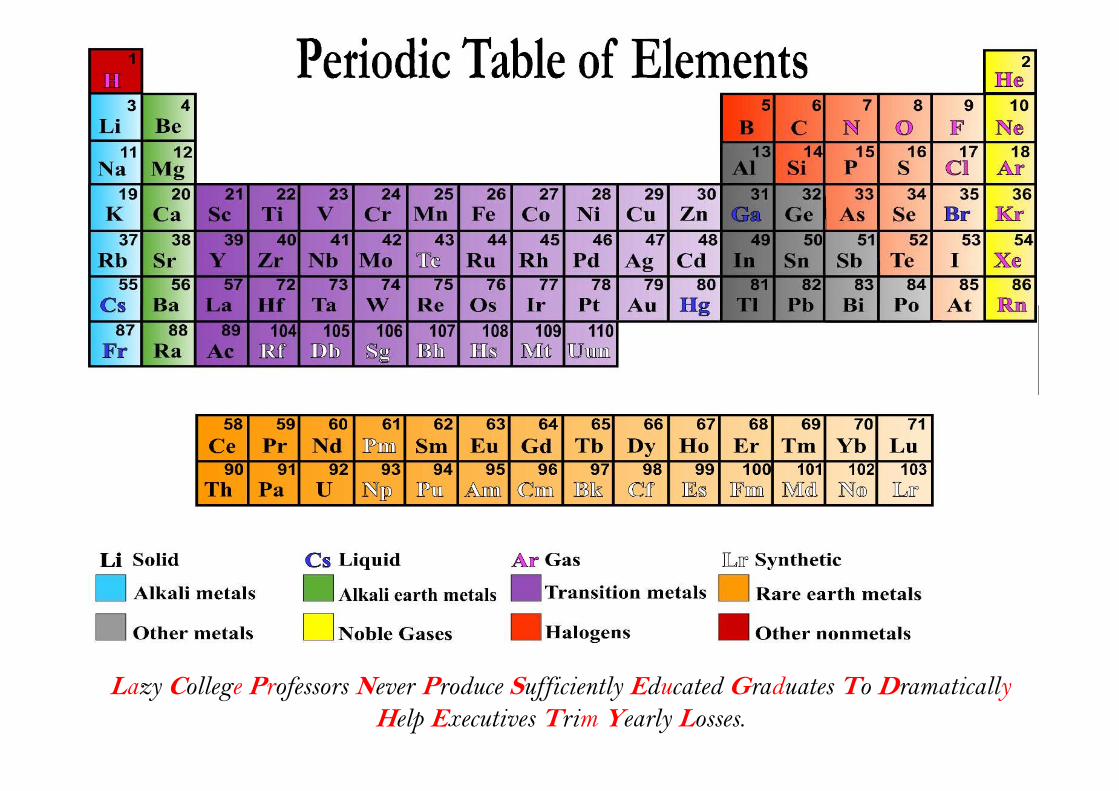
Lazy College Professors Never Produce Sufficiently Educated Graduates To DramaticallyHelp Executives Trim Yearly Losses.
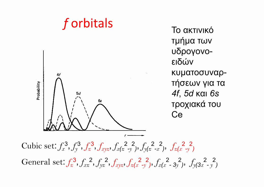
f orbitals Το ακτινικόf Το ακτινικό τμήμα των υδρογονουδρογονο-ειδών κυματοσυναρκυματοσυναρ-τήσεων για τα 4f 5d και 6s4f, 5d και 6sτροχιακά του CeCe
Cubic set: fx3, fy
3, fz3, fxyz, fx(z
2-y
2), fy(z
2-x
2), fz(x
2-y
2)
General set: fz3, fxz
2, fyz2, fxyz, fz(x
2-y
2), fx(x
2- 3y
2), fy(3x
2- y
2)
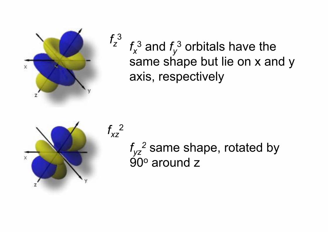
f 3fz3fx3 and fy3 orbitals have the same shape but lie on x and ysame shape but lie on x and y axis, respectively
fxz2
2fyz2 same shape, rotated by
90ο around z
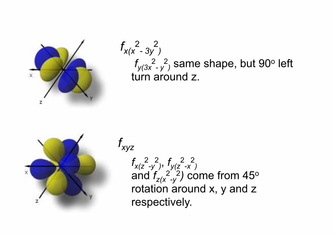
fx(x2
- 3y2
)f 2 2 same shape but 90ο leftfy(3x - y ) same shape, but 90 left turn around z.
ffxyz
fx(z2
-y2
), fy(z2
-x2
)2 2and fz(x2
-y2) come from 45ο
rotation around x, y and z respectively.
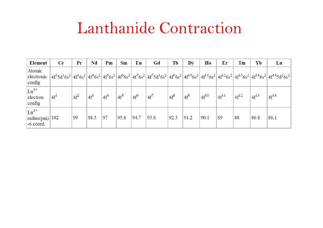
Lanthanide Contraction
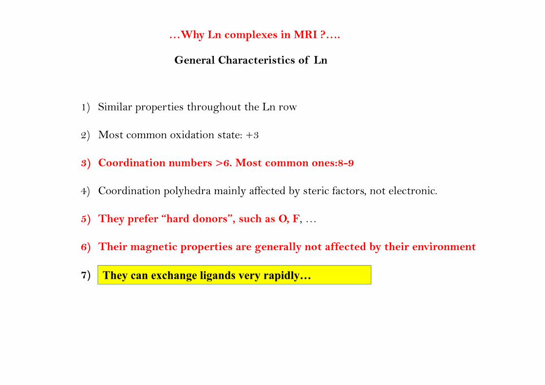
…Why Ln complexes in MRI ?….
General Characteristics of LnGeneral Characteristics of Ln
1) Si il ti th h t th L 1) Similar properties throughout the Ln row
2) Most common oxidation state: +3
3) Coordination numbers >6. Most common ones:8-9
4) Coordination polyhedra mainly affected by steric factors not electronic4) Coordination polyhedra mainly affected by steric factors, not electronic.
5) They prefer “hard donors”, such as O, F, …
6) Their magnetic properties are generally not affected by their environment
7) They can exchange ligands very rapidly…) y g g y p y
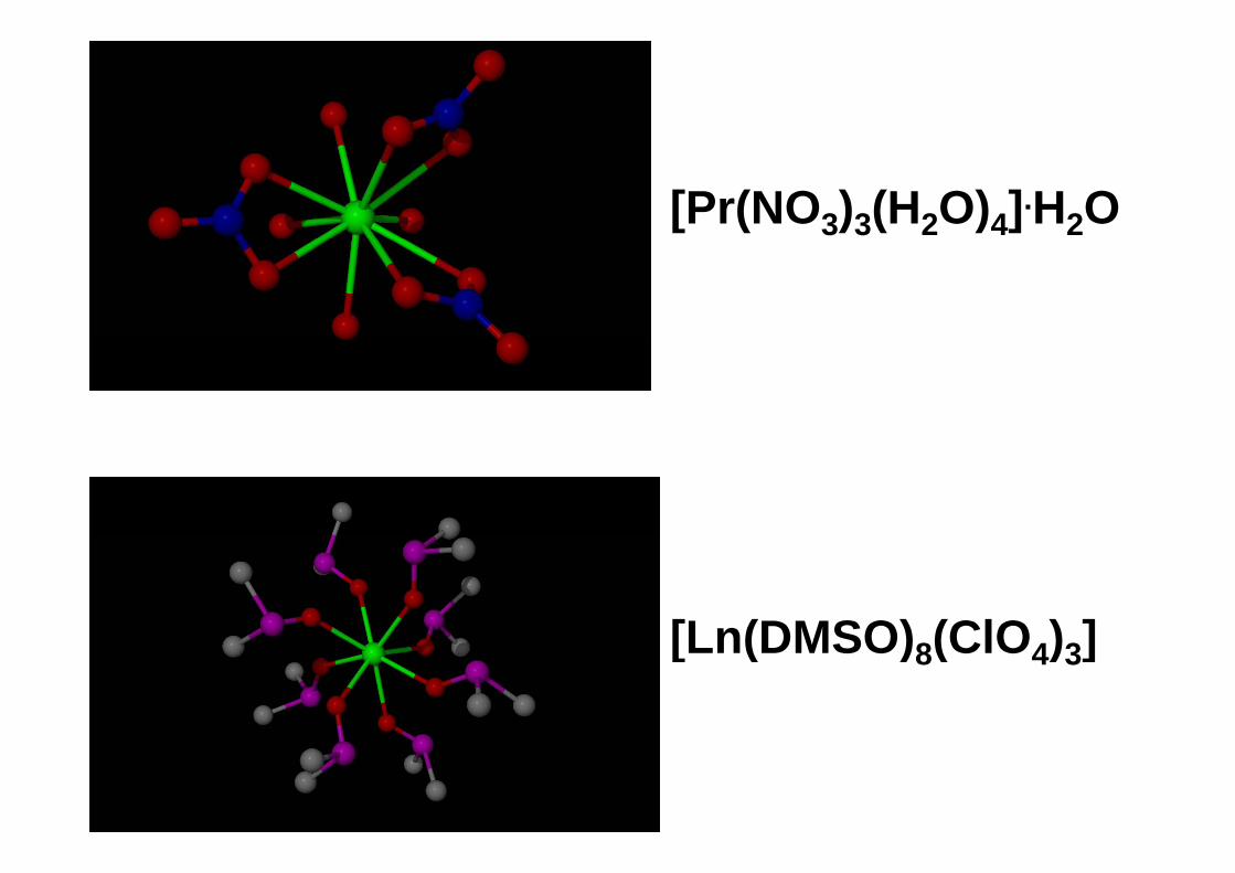
[Pr(NO3)3(H2O)4].H2O
[Ln(DMSO) (ClO ) ][Ln(DMSO)8(ClO4)3]
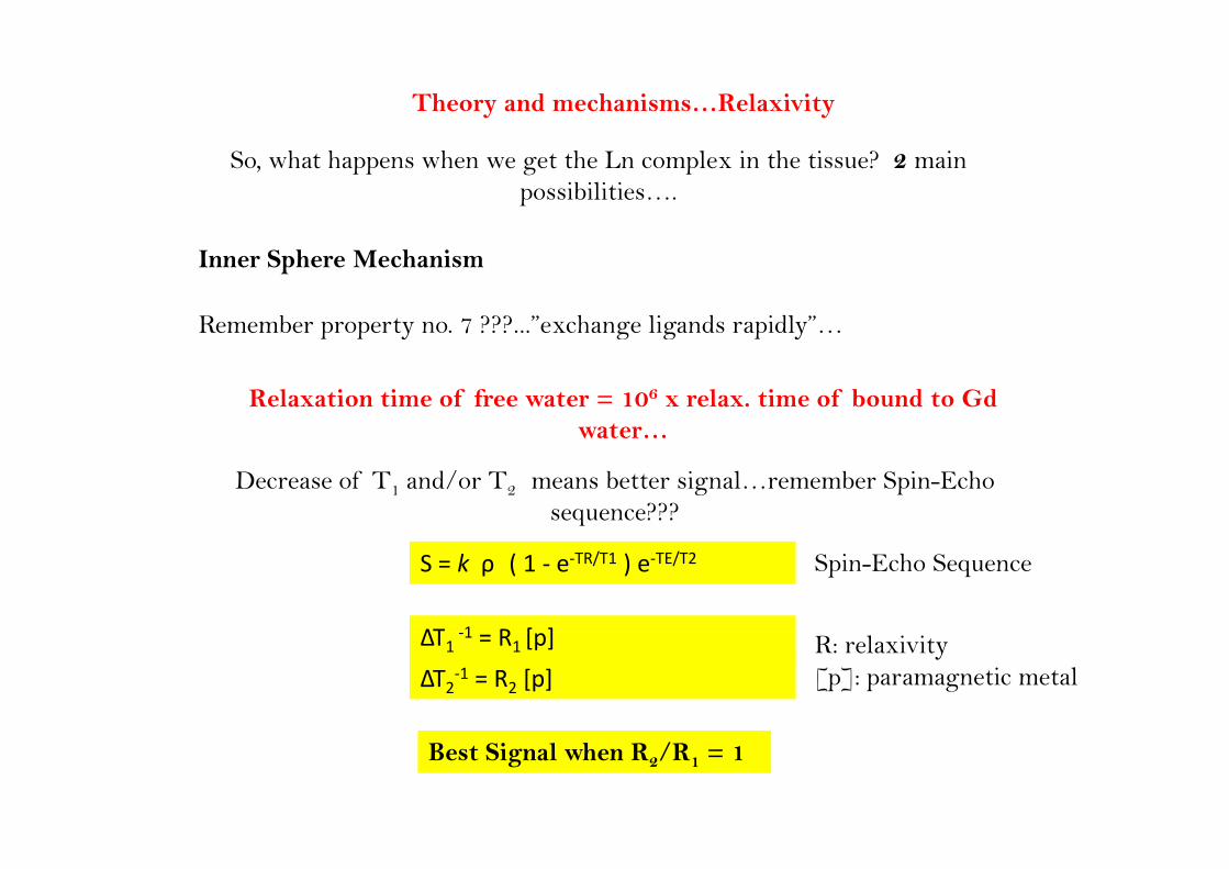
Theory and mechanisms…Relaxivity
So, what happens when we get the Ln complex in the tissue? 2 main possibilities….
Remember property no. 7 ???...”exchange ligands rapidly”…
Inner Sphere Mechanism
p p y g g p y
Relaxation time of free water = 106 x relax. time of bound to Gd waterwater…
Decrease of T1 and/or T2 means better signal…remember Spin-Echo sequence???
S = k ρ ( 1 ‐ e‐TR/T1 ) e‐TE/T2 Spin-Echo Sequence
ΔΤ 1 R [ ]ΔΤ1 ‐1 = R1 [p] R: relaxivity[p]: paramagnetic metalΔΤ2‐1 = R2 [p]
Best Signal when R2/R1 = 1
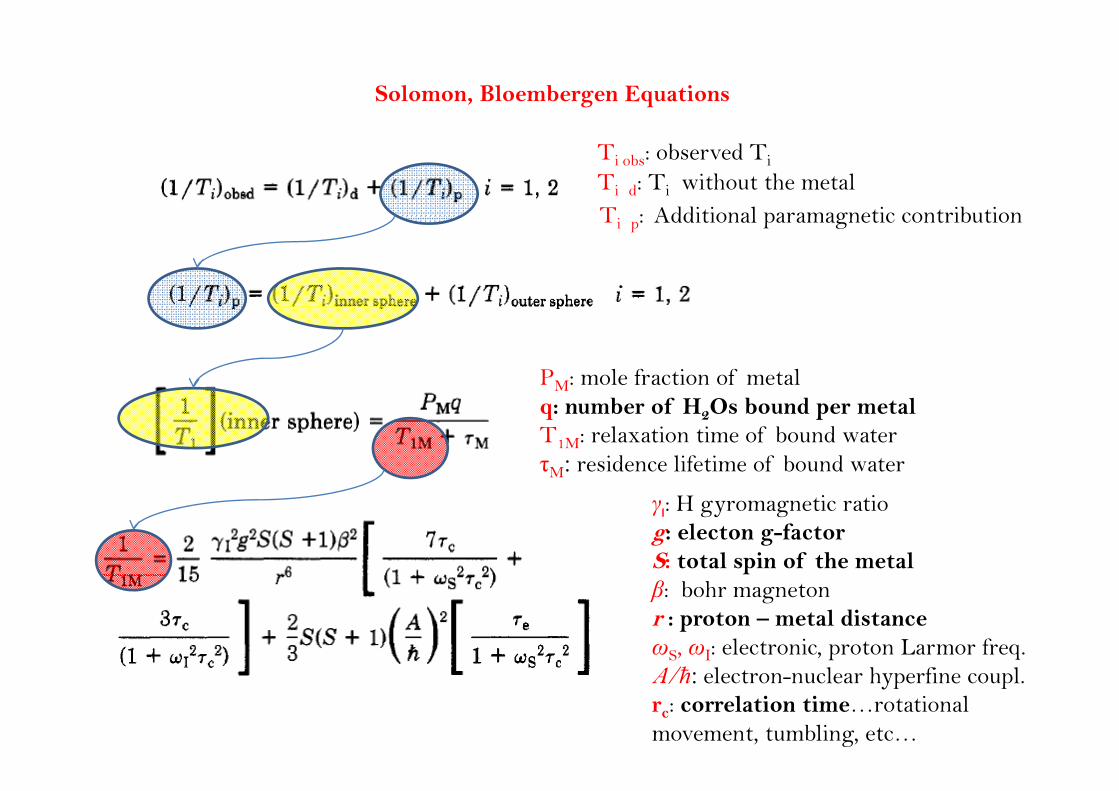
Solomon, Bloembergen Equations
Ti obs: observed Ti
Ti d: Ti without the metalT : Additional paramagnetic contributionTi p: Additional paramagnetic contribution
PM: mole fraction of metalq: number of H2Os bound per metalT1M: relaxation time of bound waterτM: residence lifetime of bound water
γι: H gyromagnetic ratiog: electon g-factorS: total spin of the metalβ: bohr magnetonr : proton – metal distanceωS, ωΙ: electronic, proton Larmor freq.A/ħ: electron-nuclear hyperfine coupl.rc: correlation time…rotational movement, tumbling, etc…
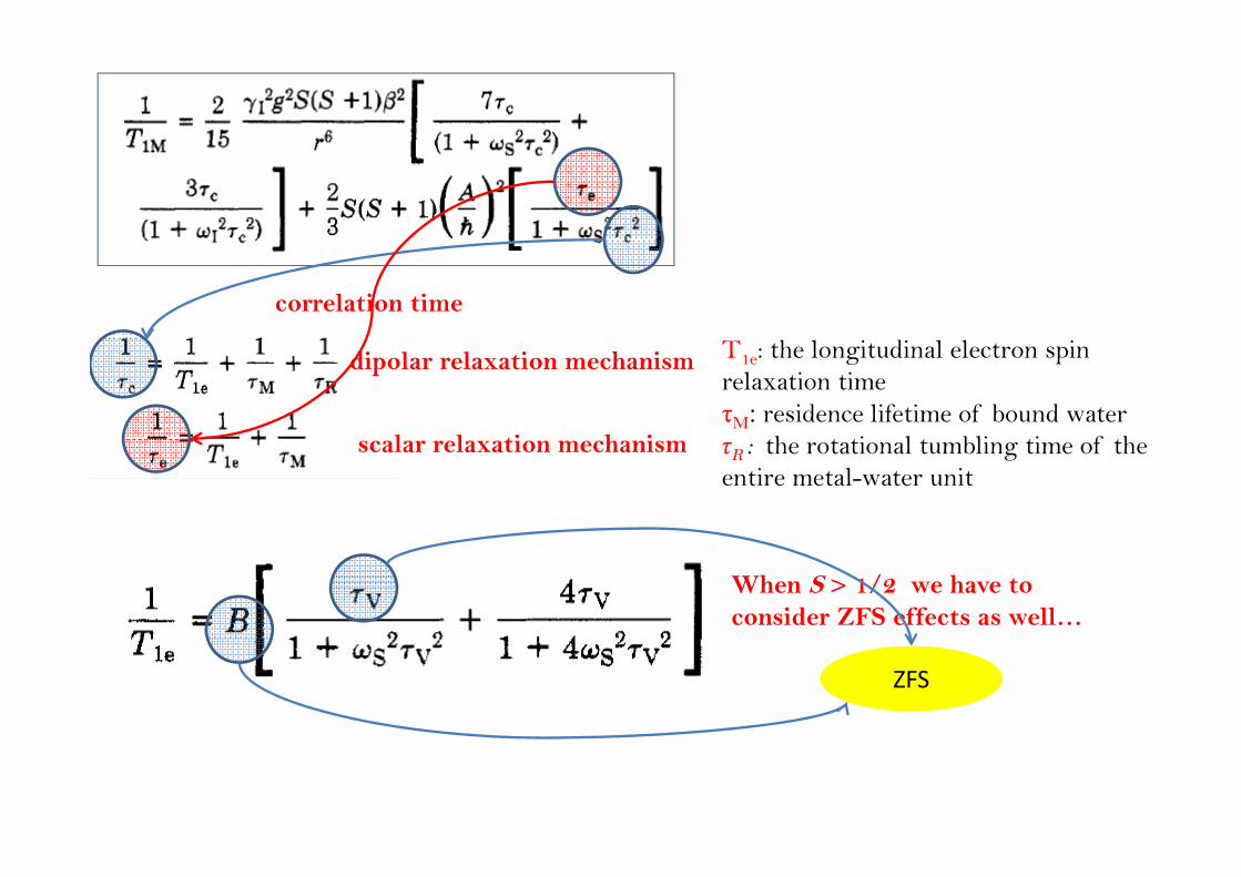
correlation time
T1e: the longitudinal electron spin relaxation timeτM: residence lifetime of bound water
h i l bli i f h
dipolar relaxation mechanism
l l ti h i τR : the rotational tumbling time of the entire metal-water unit
scalar relaxation mechanism
When S > 1/2 we have to consider ZFS effects as well…
ZFS
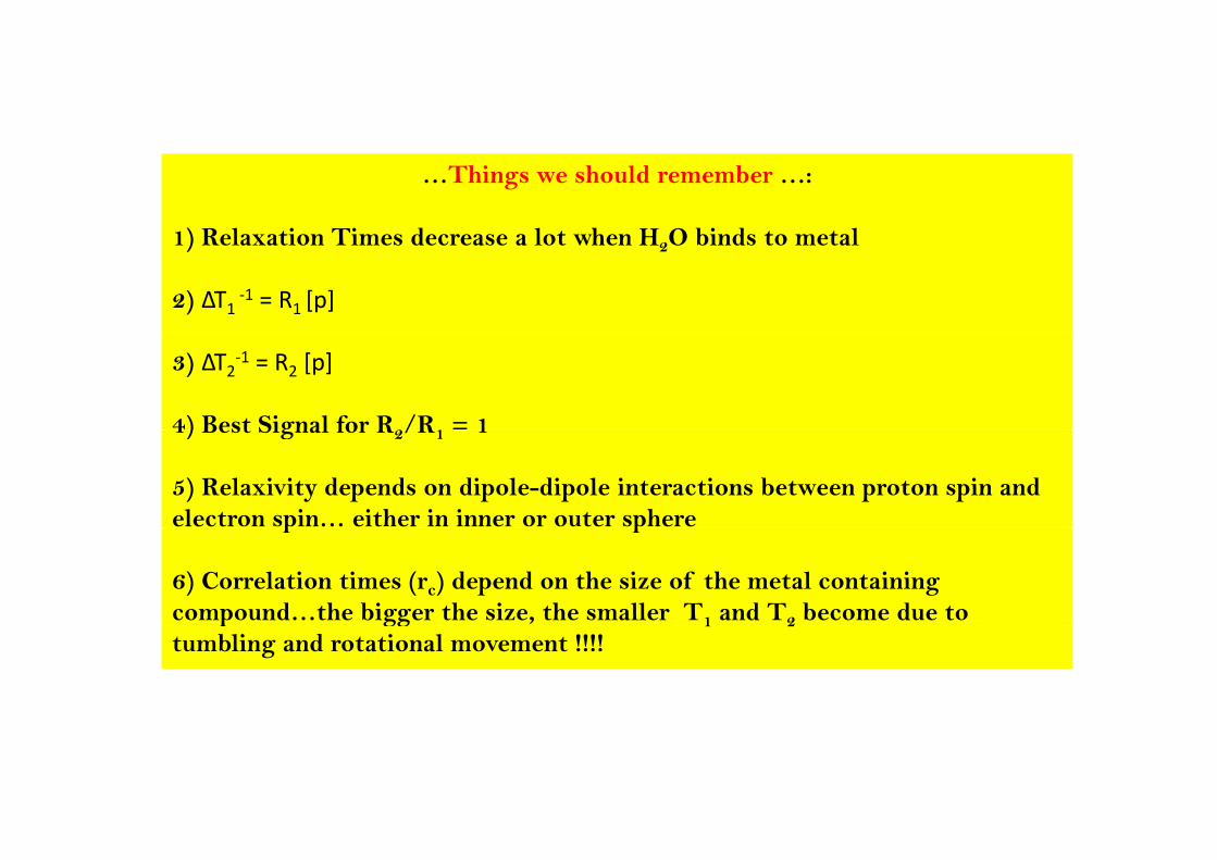
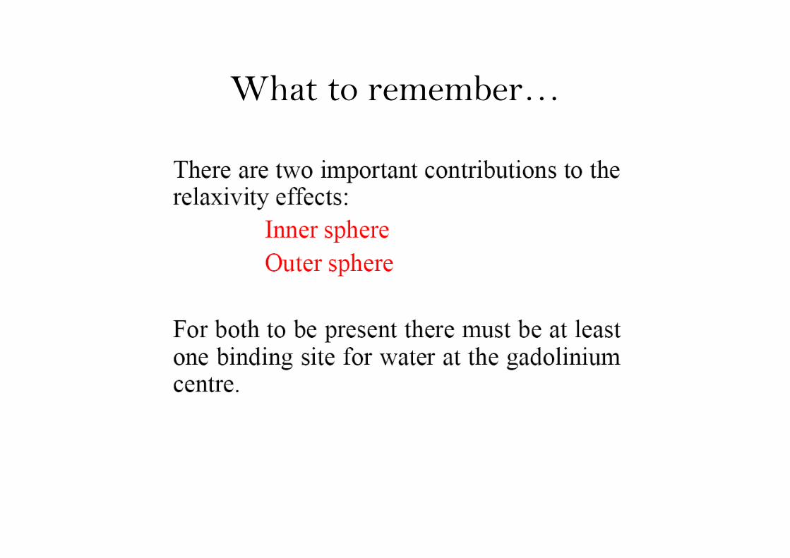
…Things we should remember …:
1) Relaxation Times decrease a lot when H O binds to metal1) Relaxation Times decrease a lot when H2O binds to metal
2) ΔΤ1 ‐1 = R1 [p]
3) ΔΤ2‐1 = R2 [p]
4) Best Signal for R2/R1 = 14) Best Signal for R2/R1 1
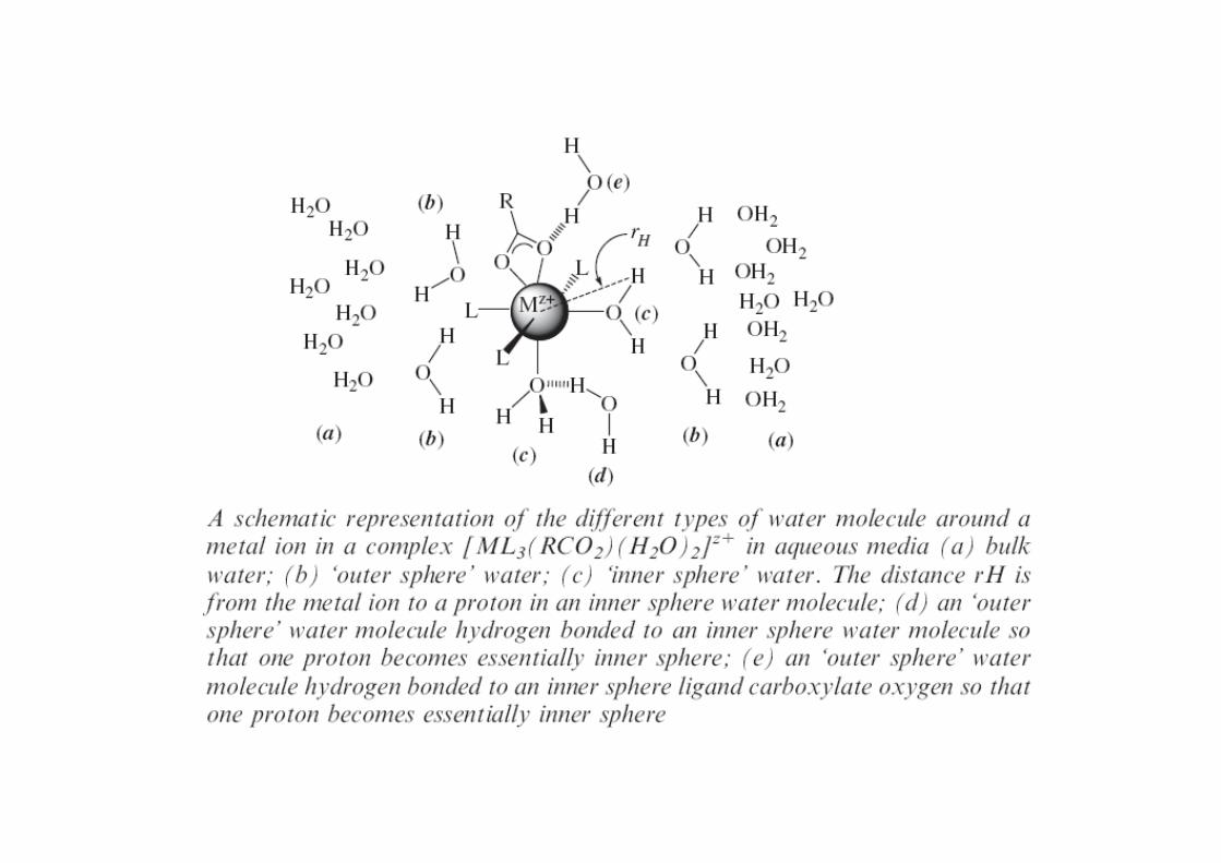
5) Relaxivity depends on dipole-dipole interactions between proton spin and electron spin… either in inner or outer spherep p
6) Correlation times (rc) depend on the size of the metal containing compound…the bigger the size, the smaller T1 and T2 become due to p gg 1 2tumbling and rotational movement !!!!
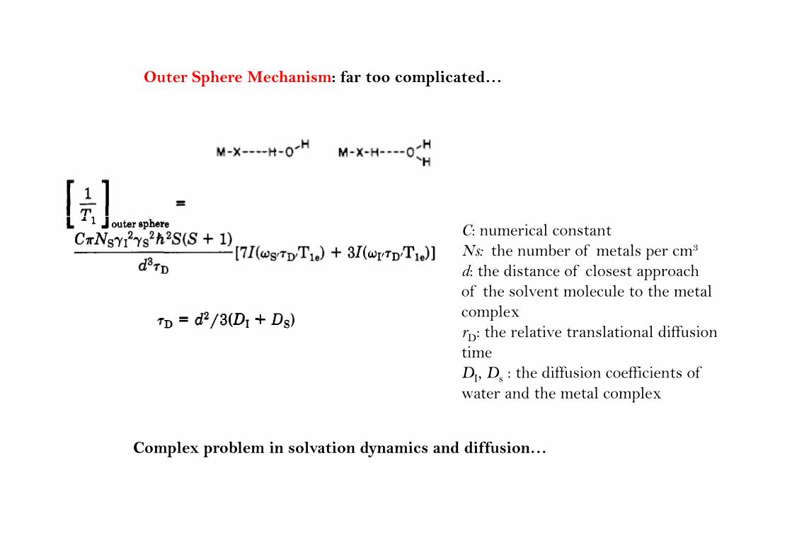
Outer Sphere Mechanism: far too complicated…
C: numerical constantNs: the number of metals per cm3
d: the distance of closest approachppof the solvent molecule to the metalcomplexrD: the relative translational diffusiontimeDI, Ds : the diffusion coefficients of water and the metal complex
Complex problem in solvation dynamics and diffusion…
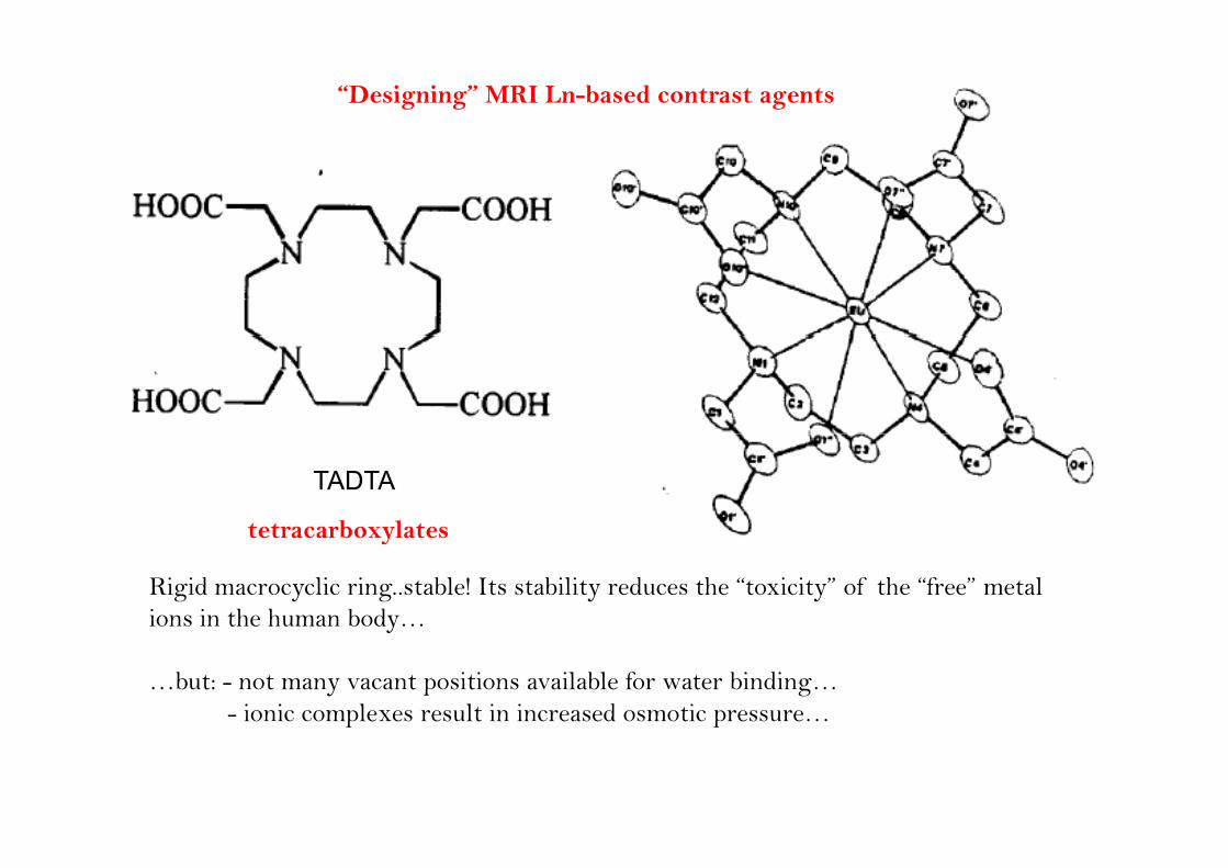
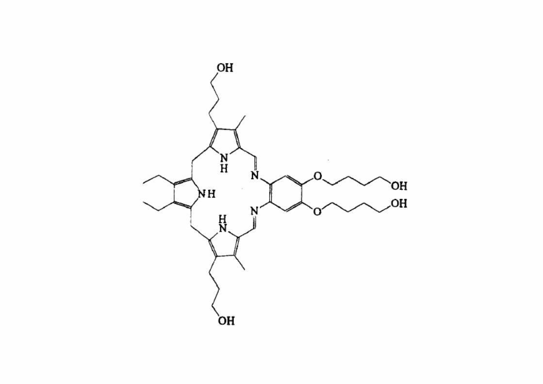
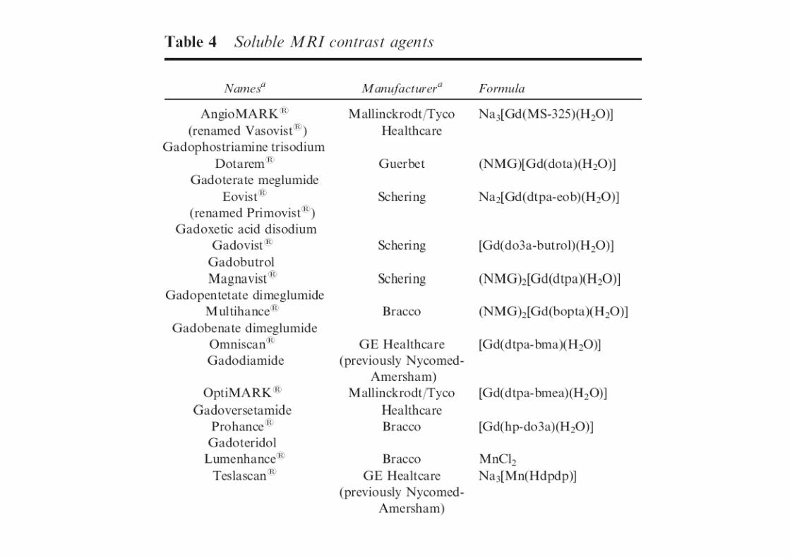
“Designing” MRI Ln-based contrast agents
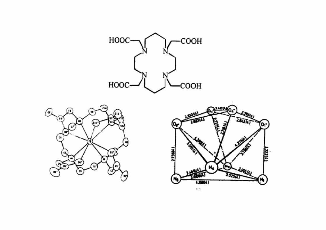
TADTA
tetracarboxylates
Rigid macrocyclic ring..stable! Its stability reduces the “toxicity” of the “free” metal ions in the human body…
tetracarboxylates
y
…but: - not many vacant positions available for water binding…- ionic complexes result in increased osmotic pressure…
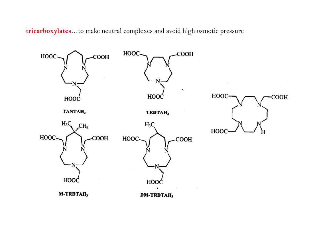
tricarboxylates…to make neutral complexes and avoid high osmotic pressure
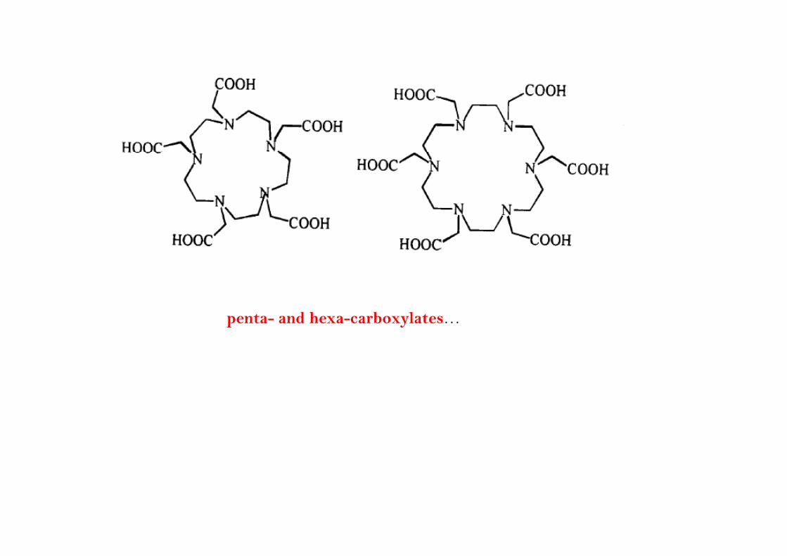
penta- and hexa-carboxylates…
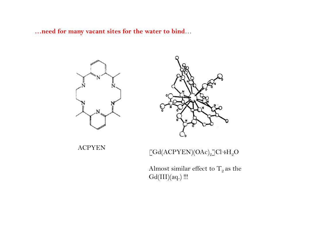
…need for many vacant sites for the water to bind…
ACPYEN[Gd(ACPYEN)(OAc)2]Cl.4H2O
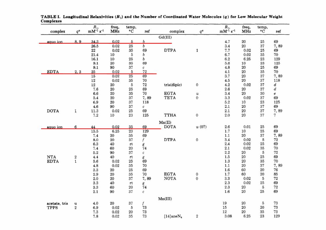
Almost similar effect to T2 as the Gd(III)(aq.) !!!
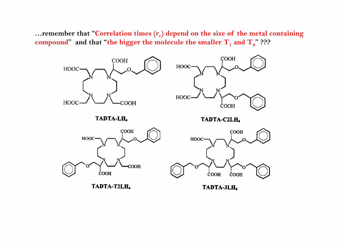
…remember that “Correlation times (rc) depend on the size of the metal containing compound” and that “the bigger the molecule the smaller T1 and T2” ???
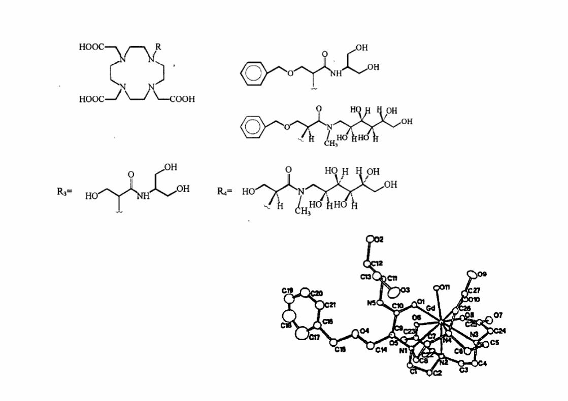
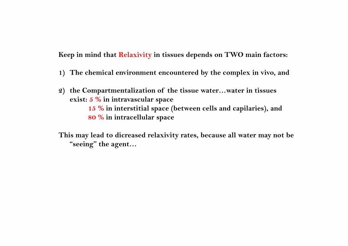
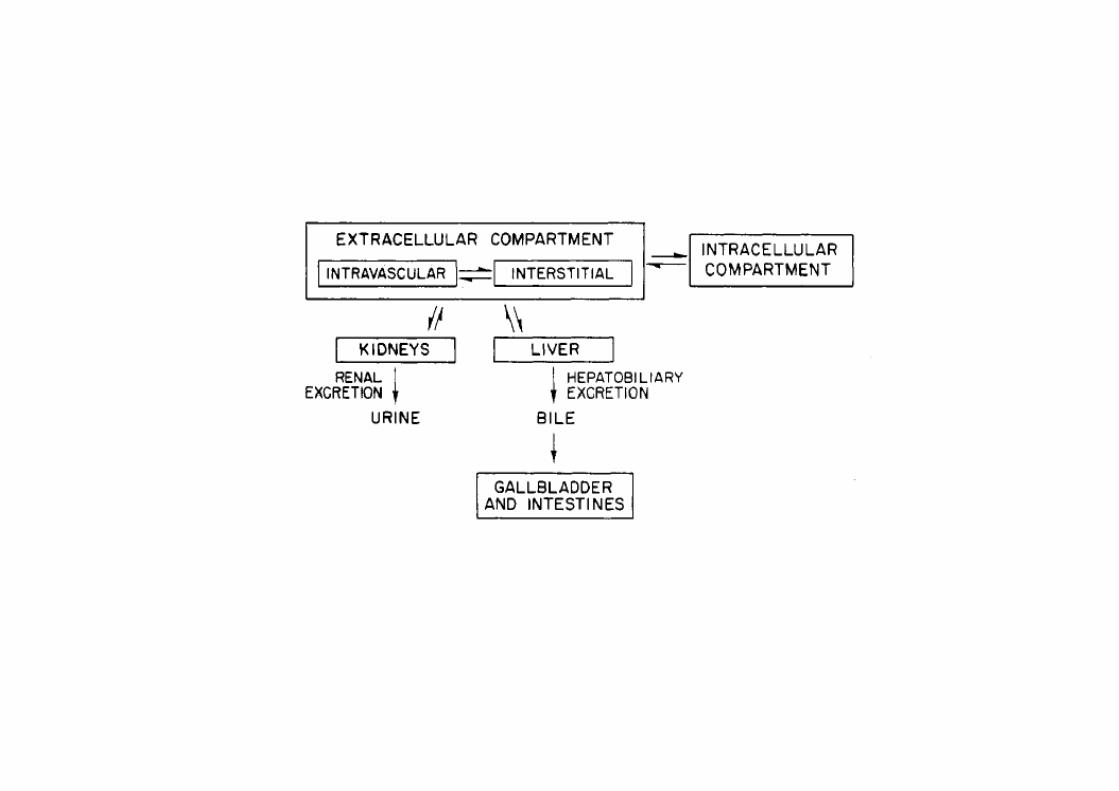
Keep in mind that Relaxivity in tissues depends on TWO main factors:
1) The chemical environment encountered by the complex in vivo, and
2) the Compartmentalization of the tissue water…water in tissues exist: 5 % in intravascular space
15 % in interstitial space (between cells and capilaries), and80 % in intracellular space
This may lead to dicreased relaxivity rates, because all water may not be “seeing” the agent…
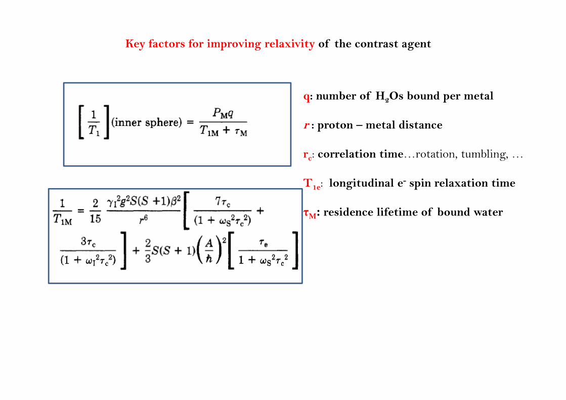
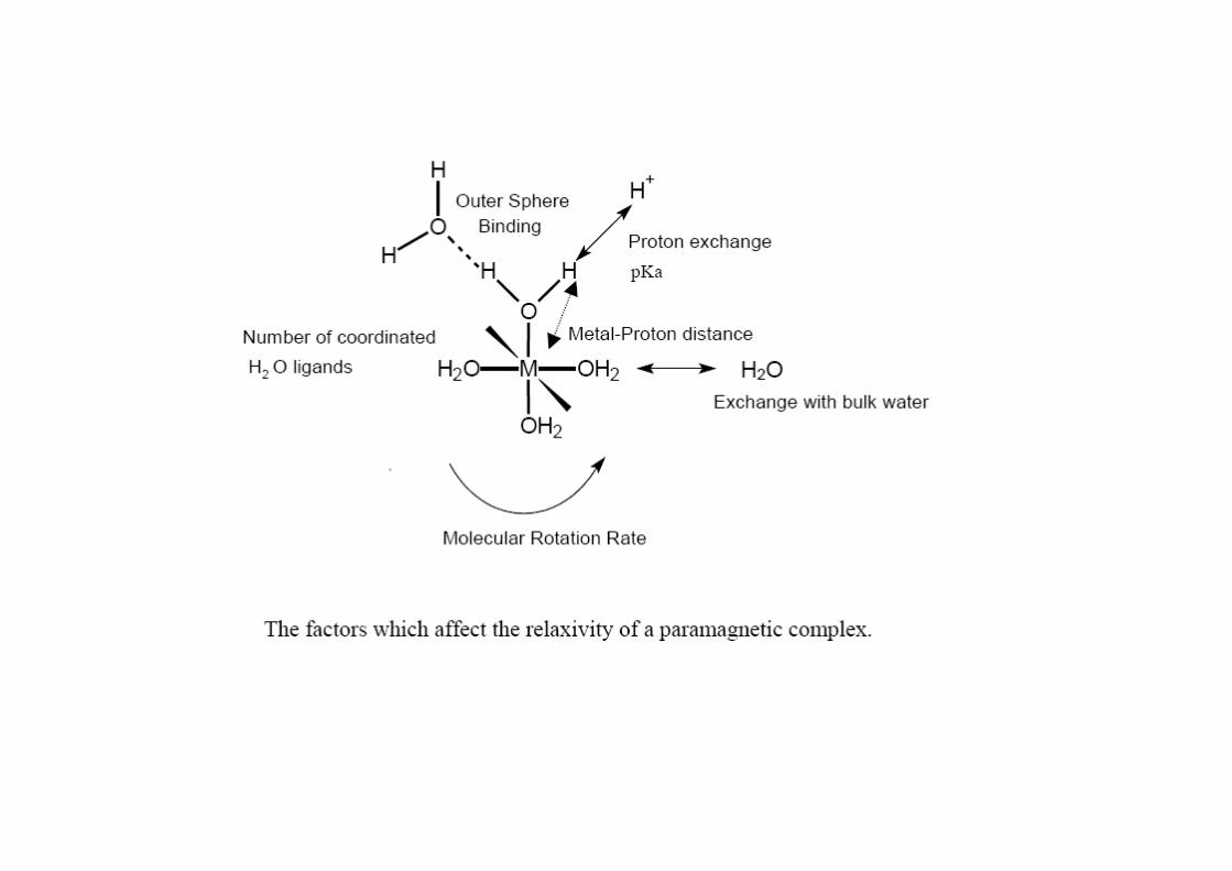
Key factors for improving relaxivity of the contrast agent
q: number of H2Os bound per metal
r : proton – metal distance
r : correlation time rotation tumbling rc: correlation time…rotation, tumbling, …
T1e: longitudinal e- spin relaxation time
τM: residence lifetime of bound water
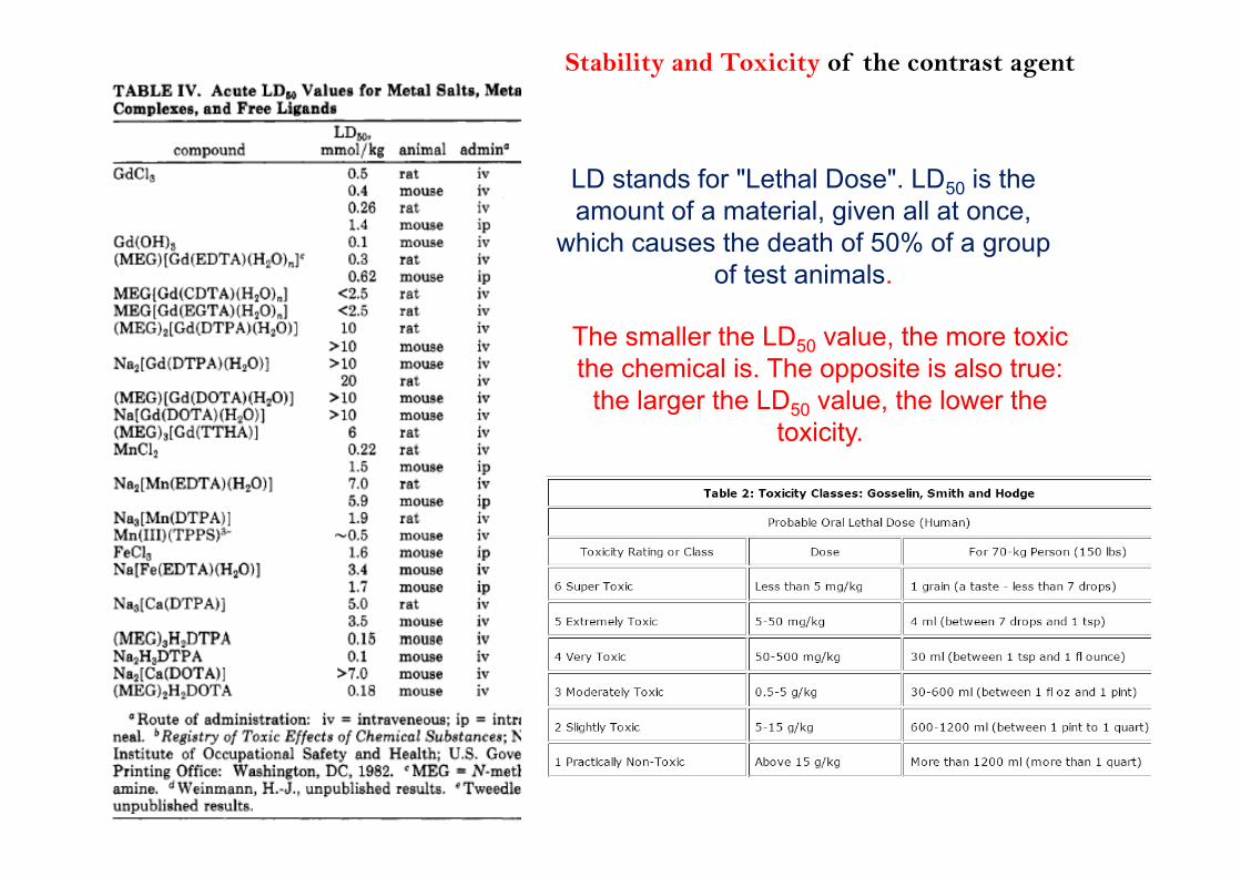
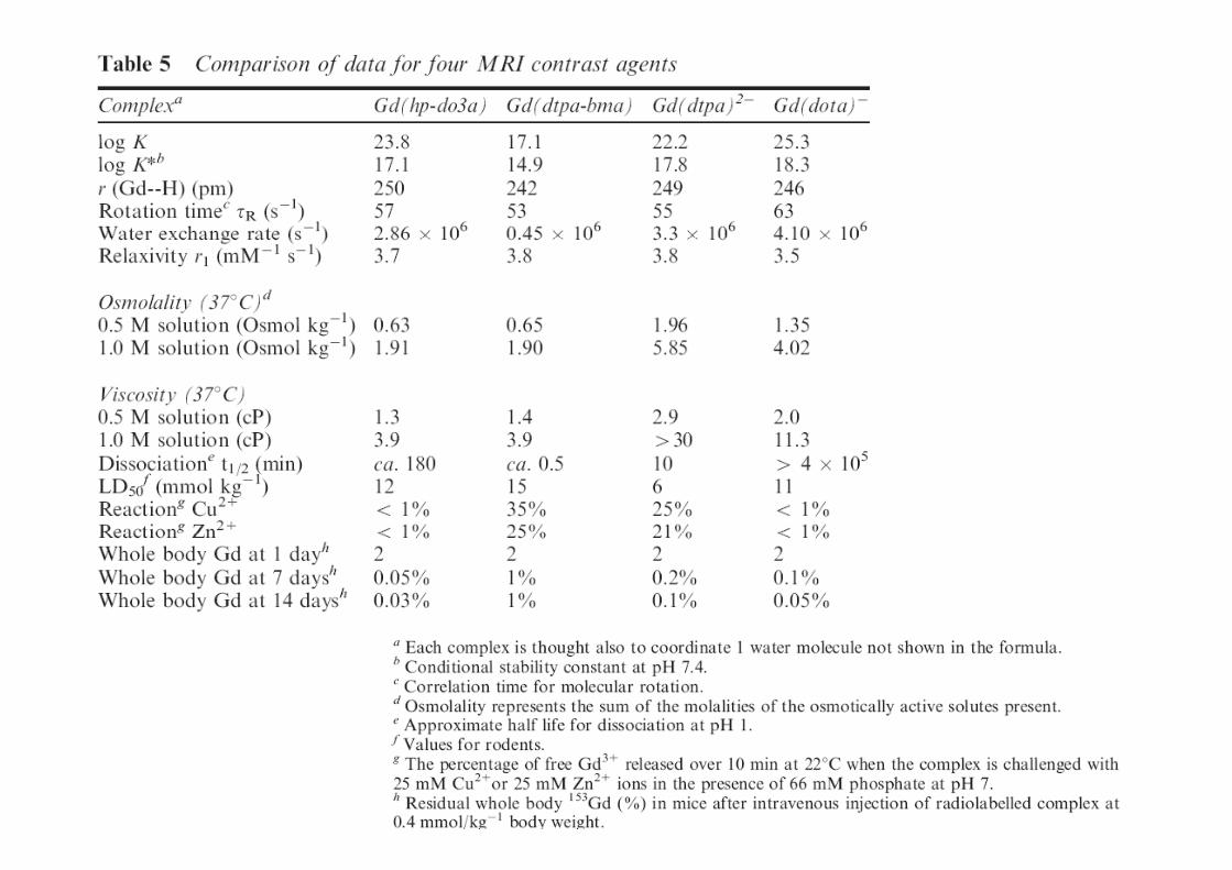
Stability and Toxicity of the contrast agent
LD stands for "Lethal Dose". LD50 is the amount of a material, given all at once,
which causes the death of 50% of a groupwhich causes the death of 50% of a group of test animals.
The smaller the LD value the more toxicThe smaller the LD50 value, the more toxic the chemical is. The opposite is also true:
the larger the LD50 value, the lower the toxicity.toxicity.
Ok…besides the C.A., is everything else safe???
What to rememberWhat to remember…
Literature
1) R. B. Lauffer, Chem.Rev. 87, 901 (1987).
2) V. Alexander, Chem.Rev. 95, 273 (1995).
3) The Basics of MRI, J. P. Hornak, Center for Imaging Science, Rochester Institute of
T h l R h t NY htt // i it d /htb k / i/ (©1996 2008) I t tiTechnology, Rochester, NY, http://www.cis.rit.edu/htbooks/mri/ (©1996-2008), Interactive
Learning Software, Henietta, NY.
4) MRI: From Body to Proton, D. W. McRobbie, E. A. Moore, M. J. Graves and M. R. Prince,4) MRI: From Body to Proton, D. W. McRobbie, E. A. Moore, M. J. Graves and M. R. Prince,
2nd ed., Cambridge University press, 2006.
5) MRI In Practise, C. Westbrook and C. Kaut, 2nd ed., Blackwell publ., 1998.