9 example thesis for sop 2014-02-10
DESCRIPTION
Implants and periodonticsTRANSCRIPT

Joint Degree Master Program of the International Medical College
and the Universities Dresden, Essen, Saarland, Leipzig, Szeged and Bangkok Scientific director: Univ.-Prof. Dr. med. Dr. med. dent. Dr. h.c. U. Joos
Outcome of dental implants in patients with a history of periodontitis
Master thesis
Master of Science in Implantology and Dental Surgery
International Medical College Gartenstraße 21
D-48147 Münster
from:
Dr. S. M. M. Bakush
2013
Dr. surname, street XX, postal code, city
Summary Objectives: the aim of the present study was to perform a systematic review of
prospective and retrospective studies regarding the long term (>5years) outcome of
dental implant placed in patients with previous tooth loss due to the history of
periodontitis and to assess how this impact on implant survival, peri-implantitis risk as
well as soft and hard tissue loss.
Methods: an electronic search of the literature was performed between March and June
2013 using MEDLINE (National Library of Medicine)-PubMed, a literature search for
articles published up to and including year 2012was performed.
Results: the initial database search yielded 1433 studies, 40 studies passed the first
review phase and 15 studies were finally selected with >5years observation period.
Because of considerable discrepancies among these studies, statistical analysis was not
performed.
Conclusions: the survival and success rate of dental implant in periodontally
compromised patients has to be not significant different than periodontally healthy
subjects. However, there was a tendency toward greater amount of marginal bone loss
and higher risk of peri-implant disease in patients with history of periodontitis.
Key words: dental implant, periodontitis, peri-implantitis, survival, bone loss.
2
Contents 1 Introduction ................................................................................................................ 3
2 Material and methods................................................................................................. 5
3 Results ........................................................................................................................ 6
3.1 Periodontitis and peri-implantitis ...................................................................... 7
3.1.1 Periodontal pocket depth (PPD) .................................................................. 7
3.1.2 Bleeding on probing (BoP) ....................................................................... 11
3.1.3 Marginal bone loss .................................................................................... 12
3.1.4 Implant survival ........................................................................................ 13
3.2 The prevalence of peri implantitis................................................................... 14
3.3 Effect of smoking ............................................................................................ 14
4 Discussion ................................................................................................................ 15
5 List of tables............................................................................................................. 22
6 List of abbreviations ................................................................................................ 23
7 Bibiliography ........................................................................................................... 24
3
1 Introduction
During the last decade, the use of dental implants has become an established and widely
used treatment modality for the rehabilitation of patients who have experienced loss of
teeth. Recent systematic reviews (1, 2) have provided evidences that support the suitable
long term prognosis of implant therapy in general population.
Although the general effect of implant therapy provide a high rate of success,
complications do occur which are associated with failure and loss of implant.
Bone quality, surgical trauma, bacterial contamination and overload are factors
associated with early implant failure, while a history of periodontal disease seems to
induce the patient to a higher risk of biological complications (peri-implant disease)
which is considered a late complication of implant therapy (3).
Several risk factors for peri-implant disease have been classified by many studies. It has
been reported that poor oral hygiene can increase the risk of peri-implant disease by 2.5
times as plaque initiated peri-implant mucositis (4) Therefore, patient compliance and
motivation are essential for long term peri-implant health.
A history of periodontal disease with deep pockets is considered another risk factor for
peri-implant disease; this may highlight the important role of undergoing an adequate
periodontal maintenance program (5, 6).
Smoking is associated with high peri-implant marginal bone loss compared with non-
smokers (4, 7). In addition, diabetes represents as a risk factor for periodontitis and
therefore may increase the risk of peri-implant disease (8).
However, there is no enough data available regarding the prognosis of dental implants
in periodontally compromised patients which represent a large proportion of those who
would be seeking implant therapy.
There are two stages of peri implant disease, namely, peri-mucositis and peri-
implantitis. While peri-implant mucositis is a reversible inflammation of the soft tissue
without any bone loss, peri-implantitis affects both the soft and hard tissue resulting in
loss of the supporting peri-implant bone (9).
The pathogenesis of peri-implantitis and periodontitis is very similar, both being
stimulated by a pathogen-containing biofilm. This plaque biofilm that develops at the
implant site has shown to resemble that at the neighboring teeth (10).
4
By placement of dental implant into partially edentulous dentition, the ecological
condition of the oral cavity impacting biofilm formation on implant may vary from that
of fully edentulous patients (11).
Therefore pocket may act as a reservoir for colonization of periodontal pathogens
around implants. This implies the important role of periodontal maintenance programs
which may affect on the prognosis of osseointegrated implants in periodontally
compromised patients which was indicated by several studies (12-15).
In the previously published literature reviews the outcome of dental implant therapy in
individuals with and without history of periodontal disease-associated tooth loss has
been analysed (16,17).In addition the prevalence of peri-implantitis and the amount of
peri-implant marginal bone loss was investigated.
Reviews of short and long terms studies (18,19) evaluated the marginal bone loss
around implants in generalized aggressive and chronic periodontitis patients .The
controversy was whether the previous history of aggressive periodontitis had more
impact on amount of marginal bone loss than a history of chronic periodontitis,and if
there is a difference in the amount of marginal bone loss between short and long term
studies.
In order to achieve suitable results, it is thus important that the included studies in the
present review have long term observation periods, as peri-implantitis disease is
considered to be one of the late complications of implant therapy.
Basically, information regarding the susceptibility of patients with a history of
periodontitis to peri-implantitis is still controversial in the literature, as is the long-term
performance of implants in these patients.
Thesis objective
Therefore, the aim of this review was to evaluate the long term survival rate of dental
implants placed in patients with previous tooth loss due to periodontitis and to assess its
impact on the implant success rate.
In this context the following questions have to be answered:

5
Research questions
1 Do patients susceptible to periodontal disease demonstrate an elevated risk for peri-
implantitis and loss of hard and soft tissue?
2. What are the success and survival rates of implants placed in such patients after a
minimal observation period of 5 years?
2 Material and methods
An electronic search of the literature was performed between March and June 2013 to
identify all articles investigating the above addressed questions. Information about the
survival rate of implants placed in patients with a history of periodontitis was retrieved.
This was evaluated by several parameters such as periodontal pocket depth, bleeding on
probing and marginal bone loss. In the present review, the search was also extended to
evaluate the incidence of peri-implantitis in periodontally-susceptible patients. The
search was conducted using MEDLINE (National Library of Medicine)-PubMed
without restrictions concerning the date of publication. Multiple keywords, including
dental implant, periodontitis, periodontal pocket, bone loss,were used (connecting
different keywords with AND, OR). This was followed by a manual search, and
references were used to identify relevant articles. A second electronic search was
performed using additional keywords such as: bleeding, survival rate and peri-
implantitis. The titles and abstracts of all articles identified from the electronic and
manual search were screened to eliminate articles that clearly failed to meet the
following inclusion and exclusion criteria:
Inclusion criteria:
• Randomized controlled clinical trials, prospective and retrospective clinical
studies.
• The mean follow up time should be at least 5 years.
Exclusion criteria:
• In vitro and animal studies, as well as case reports or case studies.
• Studies in a language other than English or without an English abstract.

6
• When multiple reports of the same study were identified, only the most recent
report was included.
The parameters were investigated based on the following definitions:
Periodontal pocket depth: is measured from the floor of the gingival sulcus to the
gingival margin and measured with the aid of periodontal probe.
Bleeding on probing: the susceptibility of peri-implant tissue to bleed indicate the
presence of inflammation.
Marginal bone loss: was measured as a distance from marginal bone level to the
shoulder of the implant.
Survival rate of implant: preservation of osseointegrated and no need to be removed at
time of examination.
Incidence of peri-implantitis: defined as signs of inflammation and bleeding around
the implant associated with progressive bone loss.
3 Results
The initial database search yielded 1433 studies, 40 studies passed the first review
phase, and 15 studies were finally selected after screening on the basis of the inclusion
and exclusion criteria. The included studies were either prospective or retrospective in
nature and with observation period of at least 5 years.
Two of the included studies reported long-term data about patients with generalized
aggressive periodontitis (20, 21), three studies reported long term data for chronic
periodontitis patients (22-24). In the study of De.Boever 2009, data about both chronic
and aggressive periodontitis was reported (25). A comparison was made in the three of
the included studies between severely, moderately periodontally compromised and
periodontally healthy patients (26-28). By the study of Costa et al .2012, the sample of
the study was divided into two groups: one group with preventive maintenance
(GTPgroup) and other group without preventive maintenance during the study period
(GNTP). In this context, .several procedures during the preventive maintenance visit
were performed, such as assessment of the periodontal and peri-implant status ,oral
hygiene instructions ,the application of disclosing agents and when necessary
mechanical debridement (29). Another study divided the total sample according to the

7
age-related bone loss score (ArB-score) into two groups, subjects with ArB-score <25
were defined as the non-perio group, whereas those with a score >55 was considered as
the perio group (30).
Two studies reported data for periodontally healthy and periodontally-compromised
patients (5, 31). However, one of these studies divided the PHP and PCP patients into 4
groups of 20 patients using different types of implants (31). Therefore, the groups were
further subdivided into PHP-N (patients treated with Nobel Biocare implants) and PHP
–S (patients treated with Straumann dental implants), PCP-N and PCP-S.
The publication date of the studies were no older than 2002. Reasons for exclusion
were: articles written in a language other than English, non-clinical studies (case reports
and reviews) and observation periods less than 5 years.
Due to the heterogeneity of the studies (different observation periods, different sample
size ...etc), it was not possible to perform a statistical analysis of the data.
3.1 Periodontitis and peri-implantitis
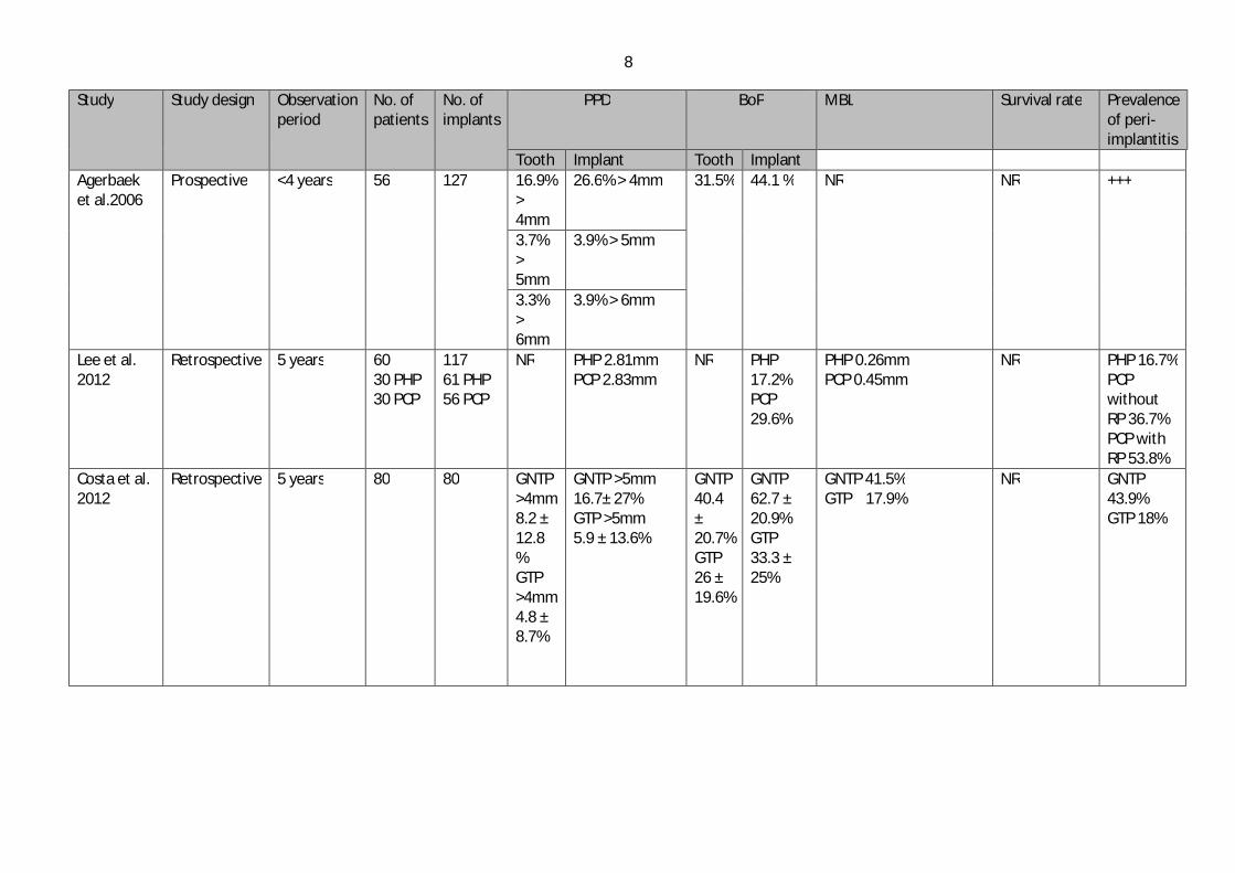
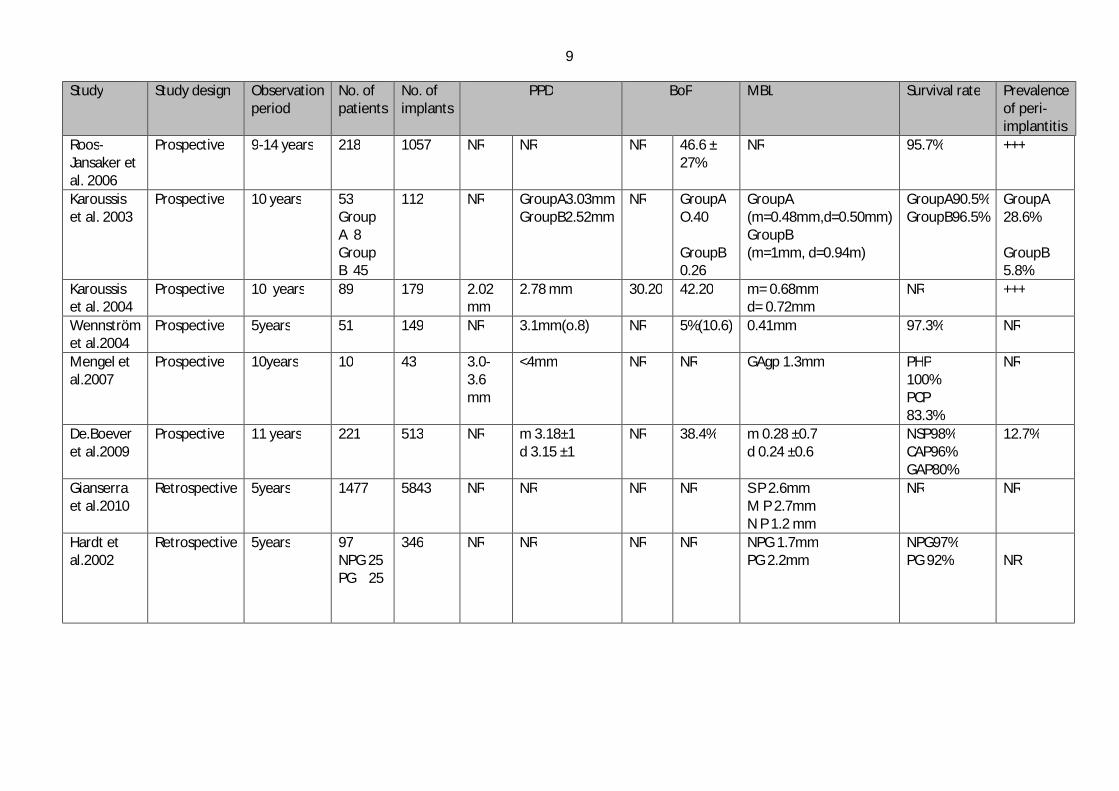
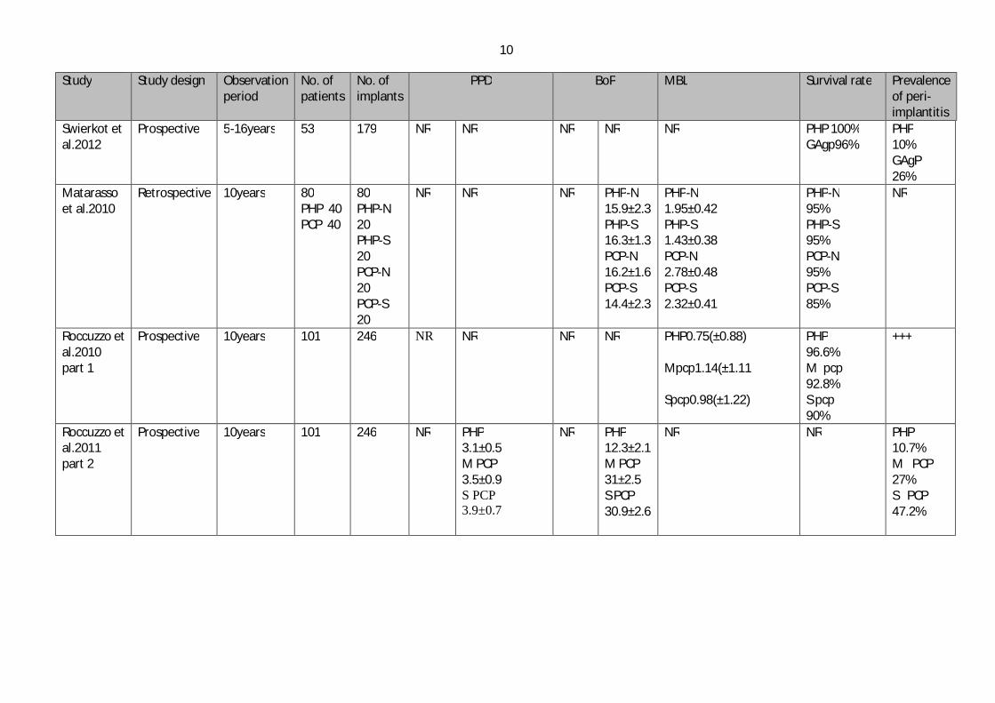
The 15 studies were categorized with respect to the parameters being observed into
periodontal pocket depth (PPD), bleeding on probing (BoP), marginal bone loss (MBL),
survival rate of implants and the prevalence of peri-implantitis (Table 1).
A detailed description of the studies related to each parameter is provided below:
3.1.1 Periodontal pocket depth (PPD)
The periodontal pocket is defined as a pathologically deepened gingival sulcus. The
depth of the periodontal pocket is measured from the floor of the gingival sulcus to the
gingival margin and is measured with the aid of a periodontal probe.
The literature search yielded 9 studies evaluating the periodontal pocket depth (PPD). In
four studies, a comparison was undergone between teeth and implants. Two studies
reported deeper pockets around implants than teeth after an observation period of 5
years. A significant difference was reported especially in case of pockets with a depth of
4 mm, with an incidence of 16.9% and 26.6% around teeth and implants, respectively.
Although patients undergoing a preventive maintenance program showed better results
in terms of periodontal pocket depth, however, the PPD around implants was deeper
also under such maintenance. (29, 32) One long term study (10 years observation
period) showed no significant difference between both teeth and implants, with a PPD
8
Study Study design Observation period
No. of patients
No. of implants
PPD BoP MBL Survival rate Prevalence of peri-implantitis
Tooth Implant Tooth Implant Agerbaek et al.2006
Prospective <4 years 56 127 16.9% > 4mm
26.6% > 4mm 31.5% 44.1 % NR NR +++
3.7% > 5mm
3.9% > 5mm
3.3% > 6mm
3.9% > 6mm
Lee et al. 2012
Retrospective 5 years 60 30 PHP 30 PCP
117 61 PHP 56 PCP
NR PHP 2.81mm PCP 2.83mm
NR PHP 17.2% PCP 29.6%
PHP 0.26mm PCP 0.45mm
NR PHP 16.7% PCP without RP 36.7% PCP with RP 53.8%
Costa et al. 2012
Retrospective 5 years 80 80 GNTP >4mm 8.2 ± 12.8 % GTP >4mm 4.8 ± 8.7%
GNTP >5mm 16.7± 27% GTP >5mm 5.9 ± 13.6%
GNTP 40.4 ± 20.7% GTP 26 ± 19.6%
GNTP 62.7 ± 20.9% GTP 33.3 ± 25%
GNTP 41.5% GTP 17.9%
NR GNTP 43.9% GTP 18%
9
Study Study design Observation period
No. of patients
No. of implants
PPD BoP MBL Survival rate Prevalence of peri-implantitis
Roos-Jansaker et al. 2006
Prospective 9-14 years 218 1057 NR NR NR 46.6 ± 27%
NR 95.7% +++
Karoussis et al. 2003
Prospective 10 years 53 Group A 8 Group B 45
112 NR GroupA3.03mm GroupB2.52mm
NR GroupA O.40 GroupB 0.26
GroupA (m=0.48mm,d=0.50mm) GroupB (m=1mm, d=0.94m)
GroupA90.5% GroupB96.5%
GroupA 28.6% GroupB 5.8%
Karoussis et al. 2004
Prospective 10 years 89 179 2.02 mm
2.78 mm 30.20 42.20 m= 0.68mm d= 0.72mm
NR +++
Wennström et al.2004
Prospective 5years 51 149 NR 3.1mm(o.8) NR 5%(10.6) 0.41mm 97.3% NR
Mengel et al.2007
Prospective 10years 10 43 3.0-3.6 mm
<4mm NR NR GAgp 1.3mm PHP 100% PCP 83.3%
NR
De.Boever et al.2009
Prospective 11 years 221 513 NR m 3.18±1 d 3.15 ±1
NR 38.4% m 0.28 ±0.7 d 0.24 ±0.6
NSP98% CAP96% GAP80%
12.7%
Gianserra et al.2010
Retrospective 5years 1477 5843 NR NR NR NR S P 2.6mm M P 2.7mm N P 1.2 mm
NR NR
Hardt et al.2002
Retrospective 5years 97 NPG 25 PG 25
346 NR NR NR NR NPG 1.7mm PG 2.2mm
NPG97% PG 92%
NR
10
Study Study design Observation period
No. of patients
No. of implants
PPD BoP MBL Survival rate Prevalence of peri-implantitis
Swierkot et al.2012
Prospective 5-16years 53
179 NR NR NR NR NR PHP 100% GAgp96%
PHP 10% GAgP 26%
Matarasso et al.2010
Retrospective 10years 80 PHP 40 PCP 40
80 PHP-N 20 PHP-S 20 PCP-N 20 PCP-S 20
NR NR NR
PHP-N 15.9±2.3 PHP-S 16.3±1.3 PCP-N 16.2±1.6 PCP-S 14.4±2.3
PHP-N 1.95±0.42 PHP-S 1.43±0.38 PCP-N 2.78±0.48 PCP-S 2.32±0.41
PHP-N 95% PHP-S 95% PCP-N 95% PCP-S 85%
NR
Roccuzzo et al.2010 part 1
Prospective
10years
101
246
NR NR NR NR
PHP0.75(±0.88) Mpcp1.14(±1.11 Spcp0.98(±1.22)
PHP 96.6% M pcp 92.8% S pcp 90%
+++
Roccuzzo et al.2011 part 2
Prospective 10years 101 246 NR PHP 3.1±0.5 M PCP 3.5±0.9 S PCP 3.9±0.7
NR PHP 12.3±2.1 M PCP 31±2.5 S PCP 30.9±2.6
NR NR PHP 10.7% M PCP 27% S PCP 47.2%
11
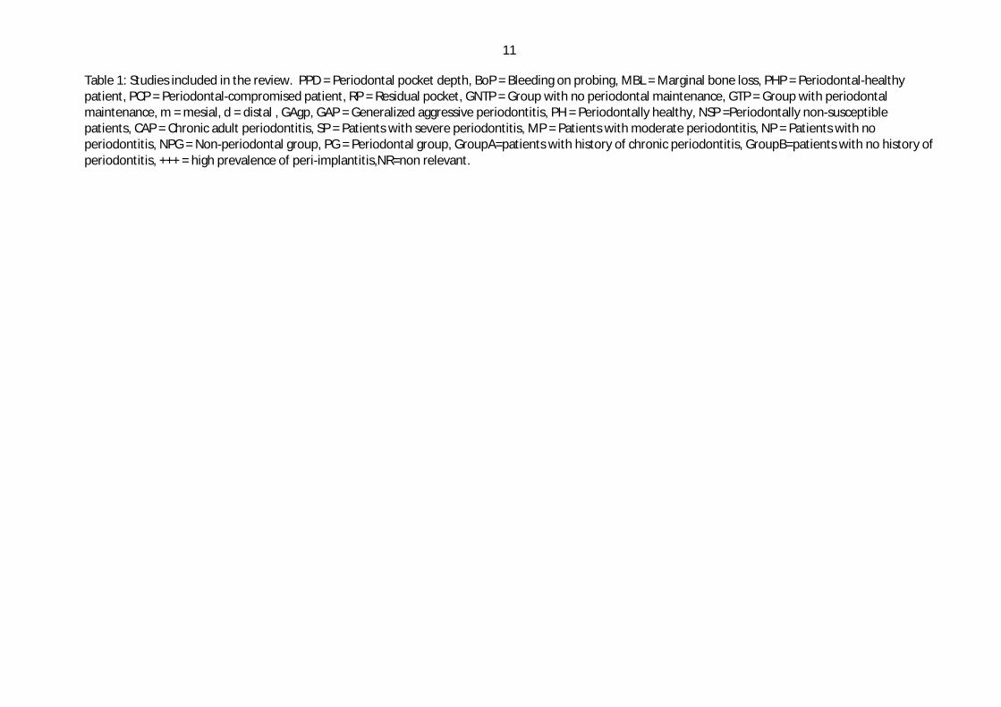
Table 1: Studies included in the review. PPD = Periodontal pocket depth, BoP = Bleeding on probing, MBL = Marginal bone loss, PHP = Periodontal-healthy patient, PCP = Periodontal-compromised patient, RP = Residual pocket, GNTP = Group with no periodontal maintenance, GTP = Group with periodontal maintenance, m = mesial, d = distal , GAgp, GAP = Generalized aggressive periodontitis, PH = Periodontally healthy, NSP =Periodontally non-susceptible patients, CAP = Chronic adult periodontitis, SP = Patients with severe periodontitis, MP = Patients with moderate periodontitis, NP = Patients with no periodontitis, NPG = Non-periodontal group, PG = Periodontal group, GroupA=patients with history of chronic periodontitis, GroupB=patients with no history of periodontitis, +++ = high prevalence of peri-implantitis,NR=non relevant.

11
of 2.02 and 2.78mm respectively (22). In contrast, another study with the same
observation period reported deeper PPD around implants than around teeth (20).
Two studies measured the PPD in periodontally-healthy and periodontally-compromised
patients. Both studies reported statistically no significant differences in PPD (5, 23).
However, in one study significant difference was reported between patients with
residual periodontitis and healthy subjects if the criterion of PPD was ≥ 5 mm. and BoP
were used. In this study, different implant designs and surfaces were used; however,
these factors did not seem to affect the clinical parameters (5).
The two studies evaluated the periodontal pocket depth in periodontitis-susceptible
patients. Here, a PPD of ≤ 3mm in almost 80% of the cases was reported (24, 25).
In the study by Roccuzzo et al.2011, the periodontal pocket depth was compared
between three groups after 10 years observation period. Here, the mean PD was 3.1 ±
0.5 in periodontally healthy individuals (PHP), 3.5 ± 0.9 in moderately periodontally-
compromised patients (PCP) and 3.9 ± 0.7 in severely periodontally-compromised
patients. Here, the difference was considered significant between the PHP and severely
PCP patients (28).
In the same study there was also a periodontal pocket measurement of 6mm or more
around the implants in a percentage of 1.7%, 15.9% and 27.2% in PHP, moderately PCP
and severely PCP respectively (28).
3.1.2 Bleeding on probing (BoP)
Most of the studies showed bleeding on probing with different percentages. In three
studies bleeding on probing was observed around teeth and implants. Implants showed
more susceptibility to bleeding on probing after 5 and 10 years, even if periodontal
maintenance programs were followed (22, 29, 32).
Other studies compared peri-implant bleeding on probing in periodontally-compromised
subjects with periodontally healthy subjects and reported higher percentages in patients
susceptible to periodontitis with a range between 32-40% (5, 23, 25, 33).This was
controversial with another study which reported only 5% of bleeding on probing in
patients susceptible to periodontitis. Here, it is important to mention that all patients
included in this study underwent an individual periodontal maintenance program
throughout the follow-up period (24).

12
There was also a significant difference in BOP between three groups in the study by
Roccuzzo et al 2010 which was 12.3+2.1% in PHP, 31.0+2.5 in moderately PCP and
30.9+2.6 in severely PCP (28).
3.1.3 Marginal bone loss
The marginal bone loss was measured as the distance from the marginal bone level to
the shoulder of the implant.
Marginal bone loss was observed in most of the included studies. In two studies, a
comparison between periodontally healthy and periodontally-compromised subjects
showed higher mean bone loss after observation periods of 10 years (22, 23). In another
study, there was no significant difference in the amount of bone loss between NPG
(1.7mm) and PG (2.2mm) patients (30). However, 64% of the PG patients had a mean
peri-implant loss of >2mm compared with 24% for the NPG patients, and the
percentage of implants showing 2mm of bone loss from baseline (abutment connection)
and after 5years was 62% in the perio group and 44% in the non-perio groups.
Four studies measured bone loss around implants in periodontitis-susceptible patients.
Three of which reported a mean bone loss of less than 1mm (22, 24, 25). One study
showed a mean marginal bone loss of 1.3mm after 10 years in patients with generalized
aggressive periodontitis (20).
In a study, after 10 years observation period, significantly higher amount of bone loss in
periodontally-compromised patients compared to periodontally healthy patients was
reported independent of the implant type used (31). In contrast, the study by Roccuzzo
et al 2010 reported no significant difference between three groups (PHP, moderately
and severely PCP) (28, 31). Both studies reported 3mm or more of bone loss(pathologic
bone resorption due to peri-implantitis) which was four time higher in PCP compared
with PHP (31). .In the other study, the percentage of sites with bone loss 3mm or more
was significant between PHP (4.7%) and severe PCP (15.1%) (28) .
There was a difference in the amount of bone loss between group with preventive
maintenance (GNTP) and group without preventive maintenance (GTP) 41.5% and
17.9% respectively after 5years observation period (29).
In the study by Gianserra et al.2010, the difference was significant in severe
periodontitis (2.6mm) compared to non periodontitis (1.2mm) (26).
13
3.1.4 Implant survival
Survival rate is defined as preservation of osseointegrated implant and not need to be
removed at time of observation. Nine of the included studies was reported the survival
rate of implants in patients with a history of peridontitis. One long term study (more
than 10 years) reported a survival rate of 97.5% in periodontally-susceptible patients
(33).An almost similar high survival rate of implants was reported in another study after
an observation period of 5years (24). In contrast, one 10 year comparative study
reported a lower survival rate of 83.3% in comparison to 100% in the periodontally-
healthy patients. Here, it is noteworthy to mention that the patients included in this
study were treated for generalized aggressive periodontitis (20).This was confirmed by
two studies after more than 10 years observation period which was reported that implant
in generalized aggressive periodontitis had a five time greater risk of failure than
periodontally healthy individuals with 100% survival rate in GAgP and 96% in PHP,
which compared patients with a healthy periodontium, chronic periodontitis and
generalized aggressive periodontitis and reported a survival rate of 98%, 96% and 80%,
respectively (21, 25) .
In two studies, no significant difference was reported after an observation period of 5
years (23, 30). However, one of these studies continued the follow up period to 10 years
and reported a statistically significant difference with a survival rate of 96.5% and
90.5% for the healthy patients and patient with history of chronic periodontitis,
respectively (23) which was in accordance with the survival rate in study by Matarasso
et al.2010 was reported a survival rate between 85%-95% in periodontally compromised
individuals in dependent of the type of implant used (31). However in the same study
there was a trend toward increase loss of implant in periodontally compromised
compared to periodontally healthy subjects.
In the study by Roccuzzo et al.2010, the survival rate for solid- screw implants and all
other types of implants was reported separately (28). These solid screw implants have
not been in use for a long time. However, there was no significant difference
demonstrated between periodontally healthy, moderately-compromised and severely-
compromised subjects (96.6%, 92.8%, and 90%) for all implants and (98%, 94.2%,
90%) for the solid-screw implants.

14
In the same study, there was a high incidence of implant loss in both moderate PCP and
severe PCP who did not adhere to the Supportive periodontal therapy program (SPT)
(28):
3.2 The prevalence of peri implantitis The incidence of peri-implantitis is defined as signs of inflammation and bleeding
around the implants associated with progressive marginal bone loss.
The prevalence of peri implantitis was reported in 10 studies .A correlation between
periodontitis and prevalence of peri implantitis was reported in most of the included
studies. It was reported in several studies with different observation periods, a
significantly greater incidence of peri-implantitis in patients with history of chronic
periodontitis (5, 22, 32, 33).
The use of a preventive maintenance program during the study showed to affect in the
prevalence of peri-implantitis with a percentage of 43.9% and 18% in the GNTP and
GTP, respectively (29). Another study also reported the effect of undergoing a
maintenance program for implant after insertion with 12.7% prevalence of peri-
implantitis (25).
The incidence of peri implantitis was also significantly 14 times higher in patients with
generalized aggressive periodontitis than periodontally healthy patients (21).These
results were in accordance with the study of Karousis et al.2003 which reported 28.6%
of peri-implantitis in chronic periodontitis compared to 5.8% in the non-periodontitis
group (23).
The difference was also significant in the incidence of peri-implantitis between
periodontally healthy (10.7%) and severely periodontally compromised patients (47.2%)
(27).
3.3 Effect of smoking Smoking was reported in five of the included studies. A correlation between peri-
implant marginal bone loss and smoking was reported in two of the included studies
(22, 24)..Marginal bone loss in smokers was approximately 1mm greater compared to
non-smokers (22), and 0.76mm in smokers and 0.22mm in non smokers (24).
In the study by Karoussis et al. 2003, there was no significant difference in incidence
rate of peri-implantitis, success rate, and survival rate between smokers and non-
15
smokers in both groups of patients with and without history of chronic periodontitis
(23). However , there was a tendency for a lower survival rate in smokers (80%) than
non smokers (100%) in patients with a history of periodontitis .This finding suggests
that smokers susceptible to chronic periodontitis yield a higher risk for implant loss than
non-smokers periodontal patients or patients without history of periodontal disease.
In another study, former smokers passed through an extensive period of smoking
cessation (6.3 - 9.2years) which was associated with insignificant occurrence of peri-
implantitis (29).
On the other hand, the study by Agerbaek et al .2006 reported that smoking had no
effect on BOP and on the microflora at implant site (32).
There was also three times higher risk of peri-implantitis in smokers in the study by
Roos-Jansaker et al.2006 (7).
4 Discussion The purpose of this systematic review was to evaluate the long term (>5years) survival
rate of dental implants placed in patients with previous tooth loss due to periodontitis,
and to assess how this impacts on implant survival, peri-implantitis risk as well as soft
and hard tissue loss.
Due to the heterogeneity of the studies (different observation periods, different types of
studies), it was not possible to perform a statistical analysis of the data. The gold
standard for systematic reviews is to study randomized clinical trials (RCT), which are
the studies with the most robust design. Prospective studies were classified in category
B according to the strength of evidence and clearly indicate a higher quality of data
compared to retrospective studies. However, most of the studies included in this review
were prospective in nature.
There is a debate on whether the outcome of dental implants in patients with a history of
periodontitis is as successful as has been observed in patients without periodontitis and
if these patients are prone to an increased incidence of peri-implantitis .
This present review analyzed studies up to 2012 with at least 5 years follow-up period.
One study included a relatively limited number of patients (10 patients) (20), while all
other included studies evaluated larger sample sizes (up to 1000 patients)
16
Periodontally-compromised patients have been defined by patients that have had a
history of periodontitis (chronic or aggressive), but with no active disease at the time of
implant placement. The patients are usually subjected to “successful” periodontal
therapy (nonsurgical and/or surgical) before implant placement. It has been stressed that
neglected or poorly treated periodontitis might increase the risk for peri-implantitis
(34)..However; till now there is no accepted definition for ‘successful’ periodontal
therapy. In this review, the selected studies have not clearly defined the periodontal
status of the patients at the time of implant placement. In addition, certain studies
included a control group comprising periodontally healthy individuals, whereas other
studies included only periodontally compromised patients.
Typical 5-year survival estimates reported in the literature range from 90% to 98% (35-
41), while 10-year survival estimates are in the range of 89% to 95% (34, 42-44).
However, the exact success criteria used vary considerably, but if progressive bone loss
is interpreted as the loss of >1.5 mm bone around the implant, it can be inferred that the
10-year success rate of the implants inserted in the present population of periodontally
compromised patients may be in the range of 60% to 70%. This is considerably lower
than the 10-year success estimates of 90% to 93% that have been reported for implants
inserted in healthier subjects (43). Clearly, the high bone loss rate for implants inserted
in the present periodontal patients may be indicative of a further decrease in the future
of the implant survival. Implant overload was also implicated as one of the possible
factors associated with implant failure (45). No studies considered details on
overloading of implant-supported prostheses, but some studies reported the type of
prostheses provided. Unfortunately, the types of prostheses and potential loading
situations varied widely.
The prevalence of peri-implantitis can vary depending on the criteria used to define it,
and criteria that include both radiographic bone loss and pocket depth result in a more
conservative outcome than the criteria that include pocket depth alone (46). In this
review, the criteria used to identify peri-implantitis included mainly marginal bone loss
and periodontal pocket depth.
Data reported in the literature suggest that PPD around implants placed in patients with
a history of chronic periodontitis tends to increase throughout a long-term period.
Moreover, the proportion of deep pockets seems to be higher in patients with a history
of chronic periodontitis than in periodontally healthy subjects. In one study, 80% of

17
peri-implant sites presented PPD ≤ 3mm, while only 5.3% had a value of ≥ 6mm after a
5 year observation period, resulting in a mean peri-implant PPD of 3.1mm (24). In a 10-
year study (23), demonstrated that implants placed in patients with a history of chronic
periodontitis had statistically significantly greater proportion of PPD > 5mm without
bleeding on probing, as well as of PPD = 5mm with bleeding on probing, compared
with patients without a history of periodontitis. Baelum et al. found a continuous
increase of the percentages of implants exhibiting PPD ≥ 4mm and ≥ 6mm from 1 to 5
years and subsequently from 5 to 10 years (47). Similar results were reported by
Ellegaard et al. (2006) for implants placed in patients with a history of chronic
periodontitis, following a sinus membrane elevation procedure (48). However, these
studies were not included in this review since they were non-clinical studies.
Lee et al 2012 assumed that the maintenance of periodontal health rather than the
previous history of periodontitis is the critical determinant of increased risk of peri-
implant disease (5). In this study, it was reported that patients with at least one residual
periodontal pocket ≥ 6 mm had significantly more implants with PPD ≥ 5 mm with
BOP and radiographic bone loss than both the PHP and the PCP with no residual
periodontal pockets ≥ 6 mm. On the other hand, PCP who did not have any recurrence
of periodontal pockets ≥ 6 mm had similar outcomes to the PHP, Here, the recurrence of
periodontitis, or the presence of new areas of periodontal disease progression was
indicated by the presence of at least one periodontal pocket ≥ 6 mm at the follow-up
examination. In addition, it is important to mention that all patients examined in this
study were suffering from generalized moderate-to-advanced chronic periodontitis.
The results of this study highlight the importance of supportive periodontal therapy
where clinical and radiographic parameters should be re-assessed at every follow-up
visit to detect peri-implant problems as early as possible and to find adequate therapy to
intercept the problems (49). In addition, the SPT program permits monitoring and
maintenance of periodontal stability, as this appears to be a key factor for the success of
implant therapy. This was confirmed by other studies, which also reported a significant
decrease in the incidence of peri-implantitis and implant loss due to the protective effect
of preventive maintenance (16, 29, 50). In the study by Roccuzzo et al.2010 there was a
correlation between implant failure and lack of full adhesion to SPT during the follow-
up in both PCP groups (28). On the other hand,, correctly performed SPT can maintain
high chance of implant survival even in PCP. However, it remains unclear whether
more aggressive treatment, including more surgical intervention, systemic antibiotic
18
therapy would decrease the incidence of peri-implantitis, or whether presence of
residual pocketing at the follow up visit is a marker of inherent patient susceptibility
that would not be affected by additional periodontal therapy. This was seen in the study
by Roccuzzo et al.2011 where antibiotic and /or surgical therapy was performed in 10%
of the cases in the PHP group, 27% of cases in moderate PCP and in 47.2% of cases in
the severe PCP and showed a significant differences between the PHP and severe PCP
group (27).
The second parameter evaluated in this review was the marginal bone loss. Included
studies that had a control group comprising periodontally healthy individuals indicated
that the long-term mean peri-implant marginal bone loss for patients with a history of
chronic periodontitis may be considered comparable to what has been presented for the
general population (5, 26, 30): On the other hand, one controlled study has found a
statistically significant difference in mean peri-implant marginal bone loss between
patients with a history of chronic periodontitis and periodontally healthy subjects (23).
Similar results were reported by De Boever et al. 2009, here PH patients and patients
with CP showed no difference in peri-implant variables, but patients with GAP had
more marginal bone loss, and more peri-implantitis (25)..These long term studies
suggest increased susceptibility to progressive marginal bone loss around implants in
patients with GAP. Therefore, marginal bone loss at implants in patients with GAP as
compared with implants in PH patients or CP patients was not significantly greater.
However, peri-implantitis does not seem to be system-dependent, but rather a condition
associated with several risk factors and host susceptibility. These factors include:
smoking, medical conditions such as diabetes, and the edentulous situation of the patient
(partially or completely edentulous)
It has been suggested that implants placed in partially edentulous patients are more at
risk for bacterial colonization with a perio-pathogenic micro-flora emerging from the
periodontal pockets around diseased teeth in the same mouth (13, 51, 52). They
proposed that partially edentulous patients with titanium implants will easily be
colonized by putative periodontal pathogens in contrast to fully edentulous patients.
However, if a destruction of the marginal bone around the implants occurs, this does not
seem to be solely related to the presence of a perio-pathogenic microflora. It is rather
the result of a complex interaction between the microorganisms and host factors, similar
to what has been seen around natural teeth affected with destructive periodontitis.
19
Accordingly a past history of periodontitis may represent a significant risk factor for
complications around implants in patients that have been treated for advanced
periodontitis. Untreated periodontal disease and refractory periodontitis patients are at
risk for complications and a regular maintenance program is essential to keep the
periodontal and peri-implant tissues healthy
One study demonstrated that bacterial colonization of the implant surface may cause
peri-implant mucositis (13). Consequently, it has been suggested that patients should
not be subjected to dental implant therapy if they present with inflammation or
inadequate oral hygiene (53). Therefore, infection control including extraction of non-
retainable teeth, oral hygiene instruction, scaling, root planning, and periodontal
surgery, if indicated, was performed before implant treatment. The importance of pre-
implant infection control is supported by experimental studies in humans (54-58).
Smoking is considered another factor, as it has been identified as a strong risk factor
associated with periimplant diseases (59-61). Several studies found an increased risk for
implant failure by a factor of almost 2.5 among smokers (62, 63). Smoking status was
however not always reported in the selected studies. Karoussis et al. divided both
periodontitis and non-periodontitis patients in a smoker and non-smoker group. In
patients in the periodontitis group, 47.6% of the implants were installed in smokers.
This was 19.8% of the implants in the patients without a history of periodontitis.
However, owing to the limited number of subjects followed over 10 years, the
differences in survival, incidence rates of peri-implantitis or success rates between
smokers and nonsmokers in both groups of patients, with and without a history of
chronic periodontitis, did not reach statistical significance (23). Nevertheless, there was
a trend for a lower survival rate of implants in smokers vs. non-smokers (80% vs.
100%) in patients with a history of chronic periodontitis. This finding indicates that
smokers susceptible to chronic periodontitis yield a higher risk for implant loss than
non-smoking periodontal patients (23).
Moreover, one study with a 5-year follow-up period reported that smokers exhibited
statistically significantly higher mean peri-implant marginal bone loss than non-smokers
(0.76mm vs. 0.22mm, respectively) (24).. In addition a long period of smoking
cessation reduces the harmful effects of previous tobacco history on periodontal clinical
parameters (64-67). It should be emphasized that, in the present study (29), former
20
smokers passed through an extensive period of smoking cessation (9.2 ± 6.3 years) and
were not significantly associated with the occurrence of peri-implantitis.
Conflicting results have been published about the influence of diabetes associated with a
higher risk of peri-implant disease. Some studies report a positive association (68, 69),
whereas others found no association (64, 70).Thus, diabetes was not associated with the
occurrence of peri-implant disease in subjects with pre-existing peri-implant mucositis.
This finding may well be related to the small number of diabetics in the initial sample.
Some studies mentioned other factors that may affect the survival rate of implants, such
as the type of implant placed and the type of surgery for implant placement, whether it
was a one stage or a two stage surgery. One study reported a significant drop in one
stage implant survival after 10 years, with a survival rate of 78%, although higher
results were observed after 5 years follow up (47). Here, it was assumed that the
relatively decreased survival rate of these one-stage implants could be related to the
type of implant, since all implants placed using this approach were hollow-screwed,
which are impossible to treat once peri-implantitis has developed. These results were
confirmed by another study which observed that hollow screw implants had a lower
survival rate than solid screw implants (37). However, in this study no indication was
found that the difference between the two types of implants should be attributed to the
implant type. Rather, it was indicated that other factors such as implant length and the
type of implant placement (one-stage or two-stage) might be the explanatory factor.
Implant surface could also affect the outcome of dental implants. A high incidence of
implant loss was reported for implants with very rough surfaces (23, 30). This was
confirmed by two other studies, which reported a positive relationship between implants
with minimal rough surfaces with the presence of SPT and the incidence of peri-
implantitis (50, 71). Here, the incidences of peri-implantitis did not vary between
patients with or without a history of periodontitis .An experimental study on dogs with
different surface roughness of implants reported that rougher surface had a high rate of
progression of an already established peri-implantitis (72, 73).
In the study by Rosenberg et al.2004 reported increase the implant survival rate for
periodontally compromised patients (from 81% to 90.6%) and for periodontally healthy
individuals (from 92.6% to 93.7%) in the exclusion of hydroxyapatite-coated implant
from the overall number of implants (74).
21
However, there was no significant difference in peri-implant bone loss between
machined and rough surface designs was reported in the study by Wennström et al.2004
(24).
Microbiology:
In one study, a comparison between the the microbiota presented in teeth and implants
was performed, according to the periodontal pocket depth and bleeding on probing.
Periodontal pocket depths of >4mm and >5mm at tooth site harbored more bacteria than
implant sites, although all subjects were incorporated into a designed maintenance care
program (32),
It was reported by another study that the higher proportion of Gram-negative species
colonizing implants in the subjects with history of periodontal disease compared with
non periodontally subjects. This indicated the transmission of species from previously
extracted periodontally involved teeth (49) .
Therefore, it was concluded that a history of periodontitis had a greater influence on the
peri-implant microbiota than implant loading time (75)
Conclusion
The survival and success rate of dental implant in periodontally compromised patients
has shown to be not significantly different from periodontally healthy subjects.
However, there is a tendency toward a greater amount of marginal bone loss and higher
risk of peri-implant disease in patients with a history of periodontitis and especially
generalized aggressive periodontitis.
Furthermore, patients with a history of periodontitis should be strongly motivated to
adhere to adequate and correct periodontal maintenance program as it has proven to be a
key factor in enhancing the long term outcome of implant therapy by controlling re-
infection.

22
5 List of tables
Table 1 Studies included in the review 8-11
23
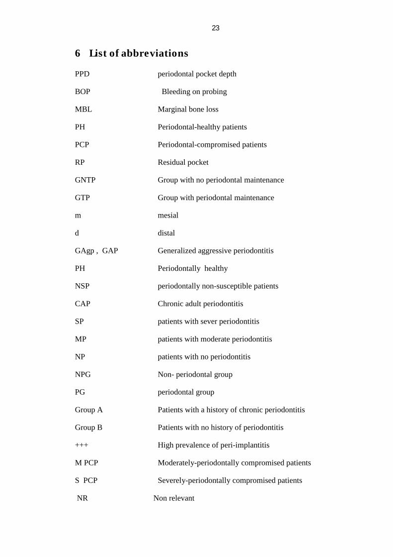
6 List of abbreviations
PPD periodontal pocket depth
BOP Bleeding on probing
MBL Marginal bone loss
PH Periodontal-healthy patients
PCP Periodontal-compromised patients
RP Residual pocket
GNTP Group with no periodontal maintenance
GTP Group with periodontal maintenance
m mesial
d distal
GAgp , GAP Generalized aggressive periodontitis
PH Periodontally healthy
NSP periodontally non-susceptible patients
CAP Chronic adult periodontitis
SP patients with sever periodontitis
MP patients with moderate periodontitis
NP patients with no periodontitis
NPG Non- periodontal group
PG periodontal group
Group A Patients with a history of chronic periodontitis
Group B Patients with no history of periodontitis
+++ High prevalence of peri-implantitis
M PCP Moderately-periodontally compromised patients
S PCP Severely-periodontally compromised patients
NR Non relevant
24
7 Bibiliography
1. Berglundh T, Persson L, Klinge B. A systematic review of the incidence of biological and technical complications in implant dentistry reported in prospective longitudinal studies of at least 5 years. J Clin Periodontol 2002;29 Suppl 3:197-212; discussion 232-193.
2. Pjetursson BE, Tan K, Lang NP, Bragger U, Egger M, Zwahlen M. A systematic review of the survival and complication rates of fixed partial dentures (FPDs) after an observation period of at least 5 years. Clin Oral Implants Res 2004;15:667-676.
3. Esposito M, Hirsch JM, Lekholm U, Thomsen P. Biological factors contributing to failures of osseointegrated oral implants. (II). Etiopathogenesis. Eur J Oral Sci 1998;106:721-764.
4. Heitz-Mayfield LJ. Peri-implant diseases: diagnosis and risk indicators. J Clin Periodontol 2008;35:292-304.
5. Cho-Yan Lee J, Mattheos N, Nixon KC, Ivanovski S. Residual periodontal pockets are a risk indicator for peri-implantitis in patients treated for periodontitis. Clin Oral Implants Res 2012;23:325-333.
6. Renvert S, Persson GR. Periodontitis as a potential risk factor for peri-implantitis. J Clin Periodontol 2009;36 Suppl 10:9-14.
7. Roos-Jansaker AM, Lindahl C, Renvert H, Renvert S. Nine- to fourteen-year follow-up of implant treatment. Part II: presence of peri-implant lesions. J Clin Periodontol 2006;33:290-295.
8. Ferreira SD, Silva GL, Cortelli JR, Costa JE, Costa FO. Prevalence and risk variables for peri-implant disease in Brazilian subjects. J Clin Periodontol 2006;33:929-935.
9. Albrektsson T IF. Consensus report of session IV. In: Proceedings of the First European Workshop on Periodontology. Quintessence 1994:365-369.
10. De Boever AL, De Boever JA. Early colonization of non-submerged dental implants in patients with a history of advanced aggressive periodontitis. Clin Oral Implants Res 2006;17:8-17.
11. Mombelli A, van Oosten MA, Schurch E, Jr., Land NP. The microbiota associated with successful or failing osseointegrated titanium implants. Oral Microbiol Immunol 1987;2:145-151.
12. Koka S, Razzoog ME, Bloem TJ, Syed S. Microbial colonization of dental implants in partially edentulous subjects. J Prosthet Dent 1993;70:141-144.
13. Leonhardt A, Adolfsson B, Lekholm U, Wikstrom M, Dahlen G. A longitudinal microbiological study on osseointegrated titanium implants in partially edentulous patients. Clin Oral Implants Res 1993;4:113-120.
14. Leonhardt A, Berglundh T, Ericsson I, Dahlen G. Putative periodontal pathogens on titanium implants and teeth in experimental gingivitis and periodontitis in beagle dogs. Clin Oral Implants Res 1992;3:112-119.
15. Mombelli A, Marxer M, Gaberthuel T, Grunder U, Lang NP. The microbiota of osseointegrated implants in patients with a history of periodontal disease. J Clin Periodontol 1995;22:124-130.

25
16. Schou S, Holmstrup P, Worthington HV, Esposito M. Outcome of implant therapy in patients with previous tooth loss due to periodontitis. Clin Oral Implants Res 2006;17 Suppl 2:104-123.
17. Van der Weijden GA, van Bemmel KM, Renvert S. Implant therapy in partially edentulous, periodontally compromised patients: a review. J Clin Periodontol 2005;32:506-511.
18. Karoussis IK, Kotsovilis S, Fourmousis I. A comprehensive and critical review of dental implant prognosis in periodontally compromised partially edentulous patients. Clin Oral Implants Res 2007;18:669-679.
19. Kim KK, Sung HM. Outcomes of dental implant treatment in patients with generalized aggressive periodontitis: a systematic review. J Adv Prosthodont 2012;4:210-217.
20. Mengel R, Behle M, Flores-de-Jacoby L. Osseointegrated implants in subjects treated for generalized aggressive periodontitis: 10-year results of a prospective, long-term cohort study. J Periodontol 2007;78:2229-2237.
21. Swierkot K, Lottholz P, Flores-de-Jacoby L, Mengel R. Mucositis, peri-implantitis, implant success, and survival of implants in patients with treated generalized aggressive periodontitis: 3- to 16-year results of a prospective long-term cohort study. J Periodontol 2012;83:1213-1225.
22. Karoussis IK, Muller S, Salvi GE, Heitz-Mayfield LJ, Bragger U, Lang NP. Association between periodontal and peri-implant conditions: a 10-year prospective study. Clin Oral Implants Res 2004;15:1-7.
23. Karoussis IK, Salvi GE, Heitz-Mayfield LJ, Bragger U, Hammerle CH, Lang NP. Long-term implant prognosis in patients with and without a history of chronic periodontitis: a 10-year prospective cohort study of the ITI Dental Implant System. Clin Oral Implants Res 2003;14:329-339.
24. Wennstrom JL, Ekestubbe A, Grondahl K, Karlsson S, Lindhe J. Oral rehabilitation with implant-supported fixed partial dentures in periodontitis-susceptible subjects. A 5-year prospective study. J Clin Periodontol 2004;31:713-724.
25. De Boever AL, Quirynen M, Coucke W, Theuniers G, De Boever JA. Clinical and radiographic study of implant treatment outcome in periodontally susceptible and non-susceptible patients: a prospective long-term study. Clin Oral Implants Res 2009;20:1341-1350.
26. Gianserra R, Cavalcanti R, Oreglia F, Manfredonia MF, Esposito M. Outcome of dental implants in patients with and without a history of periodontitis: a 5-year pragmatic multicentre retrospective cohort study of 1727 patients. Eur J Oral Implantol 2010;3:307-314.
27. Roccuzzo M, Bonino F, Aglietta M, Dalmasso P. Ten-year results of a three arms prospective cohort study on implants in periodontally compromised patients. Part 2: clinical results. Clin Oral Implants Res 2012;23:389-395.
28. Roccuzzo M, De Angelis N, Bonino L, Aglietta M. Ten-year results of a three-arm prospective cohort study on implants in periodontally compromised patients. Part 1: implant loss and radiographic bone loss. Clin Oral Implants Res 2010;21:490-496.
29. Costa FO, Takenaka-Martinez S, Cota LO, Ferreira SD, Silva GL, Costa JE. Peri-implant disease in subjects with and without preventive maintenance: a 5-year follow-up. J Clin Periodontol 2012;39:173-181.
26
30. Hardt CR, Grondahl K, Lekholm U, Wennstrom JL. Outcome of implant therapy in relation to experienced loss of periodontal bone support: a retrospective 5- year study. Clin Oral Implants Res 2002;13:488-494.
31. Matarasso S, Rasperini G, Iorio Siciliano V, Salvi GE, Lang NP, Aglietta M. A 10-year retrospective analysis of radiographic bone-level changes of implants supporting single-unit crowns in periodontally compromised vs. periodontally healthy patients. Clin Oral Implants Res 2010;21:898-903.
32. Agerbaek MR, Lang NP, Persson GR. Comparisons of bacterial patterns present at implant and tooth sites in subjects on supportive periodontal therapy. I. Impact of clinical variables, gender and smoking. Clin Oral Implants Res 2006;17:18-24.
33. Roos-Jansaker AM, Lindahl C, Renvert H, Renvert S. Nine- to fourteen-year follow-up of implant treatment. Part I: implant loss and associations to various factors. J Clin Periodontol 2006;33:283-289.
34. Leonhardt A, Grondahl K, Bergstrom C, Lekholm U. Long-term follow-up of osseointegrated titanium implants using clinical, radiographic and microbiological parameters. Clin Oral Implants Res 2002;13:127-132.
35. Behneke A, Behneke N, d'Hoedt B. The longitudinal clinical effectiveness of ITI solid-screw implants in partially edentulous patients: a 5-year follow-up report. Int J Oral Maxillofac Implants 2000;15:633-645.
36. Brocard D, Barthet P, Baysse E, Duffort JF, Eller P, Justumus P, et al. A multicenter report on 1,022 consecutively placed ITI implants: a 7-year longitudinal study. Int J Oral Maxillofac Implants 2000;15:691-700.
37. Buser D, Mericske-Stern R, Bernard JP, Behneke A, Behneke N, Hirt HP, et al. Long-term evaluation of non-submerged ITI implants. Part 1: 8-year life table analysis of a prospective multi-center study with 2359 implants. Clin Oral Implants Res 1997;8:161-172.
38. Chuang SK, Tian L, Wei LJ, Dodson TB. Kaplan-Meier analysis of dental implant survival: a strategy for estimating survival with clustered observations. J Dent Res 2001;80:2016-2020.
39. Lindh T, Gunne J, Tillberg A, Molin M. A meta-analysis of implants in partial edentulism. Clin Oral Implants Res 1998;9:80-90.
40. Naert I, Koutsikakis G, Duyck J, Quirynen M, Jacobs R, van Steenberghe D. Biologic outcome of implant-supported restorations in the treatment of partial edentulism. part I: a longitudinal clinical evaluation. Clin Oral Implants Res 2002;13:381-389. 41. Vehemente VA, Chuang SK, Daher S, Muftu A, Dodson TB. Risk factors affecting dental implant survival. J Oral Implantol 2002;28:74-81.
42. Boioli LT, Penaud J, Miller N. A meta-analytic, quantitative assessment of osseointegration establishment and evolution of submerged and non-submerged endosseous titanium oral implants. Clin Oral Implants Res 2001;12:579-588.
43. Ferrigno N, Laureti M, Fanali S, Grippaudo G. A long-term follow-up study of non-submerged ITI implants in the treatment of totally edentulous jaws. Part I: Ten-year life table analysis of a prospective multicenter study with 1286 implants. Clin Oral Implants Res 2002;13:260-273.

27
44. Lekholm U, Gunne J, Henry P, Higuchi K, Linden U, Bergstrom C, et al. Survival of the Branemark implant in partially edentulous jaws: a 10-year prospective multicenter study. Int J Oral Maxillofac Implants 1999;14:639-645.
45. Tonetti MS, Schmid J. Pathogenesis of implant failures. Periodontol 2000 1994;4:127-138.
46. Ong CT, Ivanovski S, Needleman IG, Retzepi M, Moles DR, Tonetti MS, et al. Systematic review of implant outcomes in treated periodontitis subjects. J Clin Periodontol 2008;35:438-462.
47. Baelum V, Ellegaard B. Implant survival in periodontally compromised patients. J Periodontol 2004;75:1404-1412.
48. Ellegaard B, Baelum V, Kolsen-Petersen J. Non-grafted sinus implants in periodontally compromised patients: a time-to-event analysis. Clin Oral Implants Res 2006;17:156-164.
49. Mombelli A, Lang NP. The diagnosis and treatment of peri-implantitis. Periodontol 2000 1998;17:63-76.
50. Quirynen M, Abarca M, Van Assche N, Nevins M, van Steenberghe D. Impact of supportive periodontal therapy and implant surface roughness on implant outcome in patients with a history of periodontitis. J Clin Periodontol 2007;34:805-815.
51. Meffert RM. Periodontitis and periimplantitis: one and the same? Pract Periodontics Aesthet Dent 1993;5:79-80, 82.
52. Nevins M. Will implants survive well in patients with a history of inflammatory periodontal disease? J Periodontol 2001;72:113-117.
53. Buser D, Weber HP, Bragger U. The treatment of partially edentulous patients with ITI hollow-screw implants: presurgical evaluation and surgical procedures. Int J Oral Maxillofac Implants 1990;5:165-175.
54. Abrahamsson I, Berglundh T, Lindhe J. Soft tissue response to plaque formation at different implant systems. A comparative study in the dog. Clin Oral Implants Res 1998;9:73-79.
55. Berglundh T, Lindhe J, Marinello C, Ericsson I, Liljenberg B. Soft tissue reaction to de novo plaque formation on implants and teeth. An experimental study in the dog. Clin Oral Implants Res 1992;3:1-8.
56. Ericsson I, Berglundh T, Marinello C, Liljenberg B, Lindhe J. Long-standing plaque and gingivitis at implants and teeth in the dog. Clin Oral Implants Res 1992;3:99-103.
57. Pontoriero R, Tonelli MP, Carnevale G, Mombelli A, Nyman SR, Lang NP. Experimentally induced peri-implant mucositis. A clinical study in humans. Clin Oral Implants Res 1994;5:254-259.
58. Zitzmann NU, Berglundh T, Marinello CP, Lindhe J. Experimental peri-implant mucositis in man. J Clin Periodontol 2001;28:517-523.
59. Aglietta M, Siciliano VI, Rasperini G, Cafiero C, Lang NP, Salvi GE. A 10-year retrospective analysis of marginal bone-level changes around implants in periodontally healthy and periodontally compromised tobacco smokers. Clin Oral Implants Res 2011;22:47-53.
28
60. Heitz-Mayfield LJ, Huynh-Ba G. History of treated periodontitis and smoking as risks for implant therapy. Int J Oral Maxillofac Implants 2009;24 Suppl:39-68.
61. Karbach J, Callaway A, Kwon YD, d'Hoedt B, Al-Nawas B. Comparison of five parameters as risk factors for peri-mucositis. Int J Oral Maxillofac Implants 2009;24:491-496.
62. Leonhardt A, Dahlen G, Renvert S. Five-year clinical, microbiological, and radiological outcome following treatment of peri-implantitis in man. J Periodontol 2003;74:1415-1422.
63. Wilson TG, Jr., Nunn M. The relationship between the interleukin-1 periodontal genotype and implant loss. Initial data. J Periodontol 1999;70:724-729.
64. Anner R, Grossmann Y, Anner Y, Levin L. Smoking, diabetes mellitus, periodontitis, and supportive periodontal treatment as factors associated with dental implant survival: a long-term retrospective evaluation of patients followed for up to 10 years. Implant Dent 2010;19:57-64.
65. Bain CA. Smoking and implant failure--benefits of a smoking cessation protocol. Int J Oral Maxillofac Implants 1996;11:756-759.
66. Lambert PM, Morris HF, Ochi S. The influence of smoking on 3-year clinical success of osseointegrated dental implants. Ann Periodontol 2000;5:79-89.
67. Rosa EF, Corraini P, de Carvalho VF, Inoue G, Gomes EF, Lotufo JP, et al. A prospective 12-month study of the effect of smoking cessation on periodontal clinical parameters. J Clin Periodontol 2011;38:562-571.
68. Moy PK, Medina D, Shetty V, Aghaloo TL. Dental implant failure rates and associated risk factors. Int J Oral Maxillofac Implants 2005;20:569-577.
69. Zupnik J, Kim SW, Ravens D, Karimbux N, Guze K. Factors associated with dental implant survival: a 4-year retrospective analysis. J Periodontol 2011;82:1390-1395.
70. Abdulwassie H, Dhanrajani PJ. Diabetes mellitus and dental implants: a clinical study. Implant Dent 2002;11:83-86.
71. Evian CI, Emling R, Rosenberg ES, Waasdorp JA, Halpern W, Shah S, et al. Retrospective analysis of implant survival and the influence of periodontal disease and immediate placement on long-term results. Int J Oral Maxillofac Implants 2004;19:393-398.
72. Albouy JP, Abrahamsson I, Persson LG, Berglundh T. Spontaneous progression of peri-implantitis at different types of implants. An experimental study in dogs. I: clinical and radiographic observations. Clin Oral Implants Res 2008;19:997-1002. 73. Berglundh T, Gotfredsen K, Zitzmann NU, Lang NP, Lindhe J. Spontaneous progression of ligature induced peri-implantitis at implants with different surface roughness: an experimental study in dogs. Clin Oral Implants Res 2007;18:655-661.
74. Rosenberg ES, Cho SC, Elian N, Jalbout ZN, Froum S, Evian CI. A comparison of characteristics of implant failure and survival in periodontally compromised and periodontally healthy patients: a clinical report. Int J Oral Maxillofac Implants 2004;19:873-879.
75. Lee KH, Maiden MF, Tanner AC, Weber HP. Microbiota of successful osseointegrated dental implants. J Periodontol 1999;70:131-138.

29
Declaration of academic integrity
I declare that I independently completed this thesis and this thesis was not previously
submitted to another academic institution. I also confirm that no other sources have
been used than those indicated in this thesis and the thoughts taken directly or indirectly
from external sources are properly marked as such.
Oldenburg, and 25/07/2013
Dr. S. M. M. Bakush