62 hfhs rev1 - shimadzu · on the presentation by dr. flynn of henry ford health system at rsna...
TRANSCRIPT
MEDICAL NOW No.62 (2007.7)
Digital R/F
Evaluation of Tomosynthesis Using SONIALVISION safire Digital Table − Based on Presentation by Dr. Flynn of Henry Ford Health System at RSNA 2006−
Medical Systems Division, Shimadzu Corporation
Tetsuo Imanishi
1. Introduction
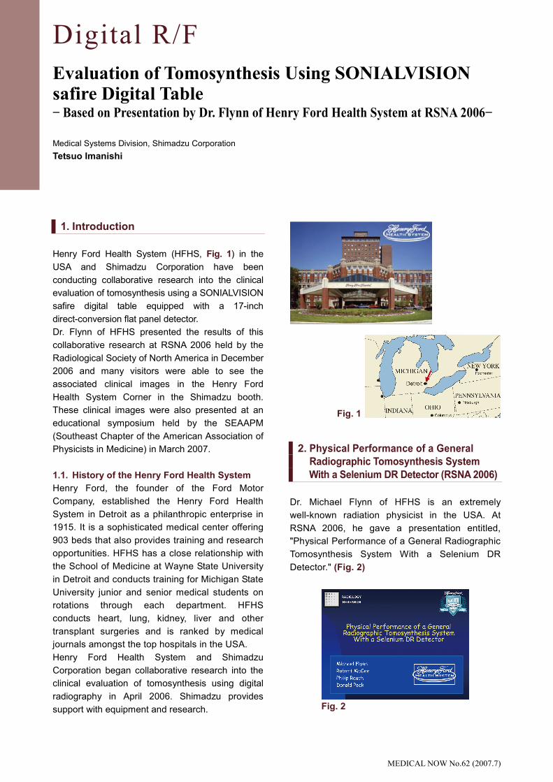
Henry Ford Health System (HFHS, Fig. 1) in the
USA and Shimadzu Corporation have been
conducting collaborative research into the clinical
evaluation of tomosynthesis using a SONIALVISION
safire digital table equipped with a 17-inch
direct-conversion flat panel detector.
Dr. Flynn of HFHS presented the results of this
collaborative research at RSNA 2006 held by the
Radiological Society of North America in December
2006 and many visitors were able to see the
associated clinical images in the Henry Ford
Health System Corner in the Shimadzu booth.
These clinical images were also presented at an
educational symposium held by the SEAAPM
(Southeast Chapter of the American Association of
Physicists in Medicine) in March 2007.
1.1. History of the Henry Ford Health System
Henry Ford, the founder of the Ford Motor
Company, established the Henry Ford Health
System in Detroit as a philanthropic enterprise in
1915. It is a sophisticated medical center offering
903 beds that also provides training and research
opportunities. HFHS has a close relationship with
the School of Medicine at Wayne State University
in Detroit and conducts training for Michigan State
University junior and senior medical students on
rotations through each department. HFHS
conducts heart, lung, kidney, liver and other
transplant surgeries and is ranked by medical
journals amongst the top hospitals in the USA.
Henry Ford Health System and Shimadzu
Corporation began collaborative research into the
clinical evaluation of tomosynthesis using digital
radiography in April 2006. Shimadzu provides
support with equipment and research.
2. Physical Performance of a General
Radiographic Tomosynthesis System
With a Selenium DR Detector (RSNA 2006)
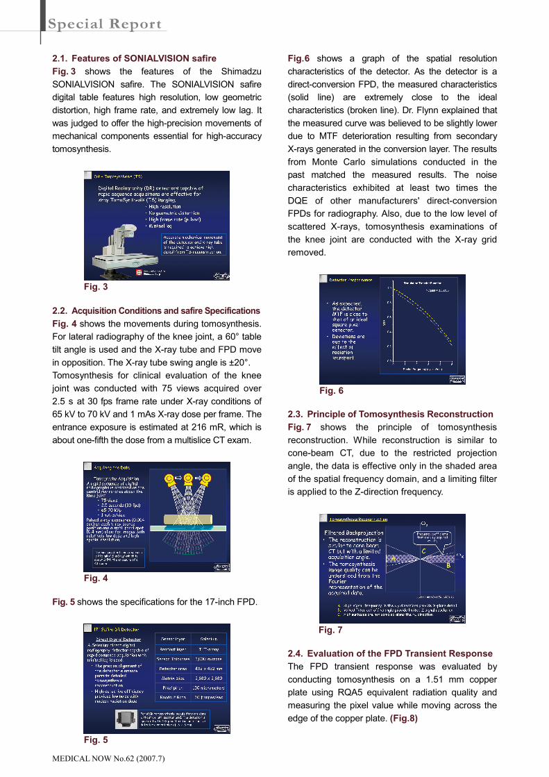
Dr. Michael Flynn of HFHS is an extremely
well-known radiation physicist in the USA. At
RSNA 2006, he gave a presentation entitled,
"Physical Performance of a General Radiographic
Tomosynthesis System With a Selenium DR
Detector." (Fig. 2)
Fig. 2
Fig. 1
MEDICAL NOW No.62 (2007.7)
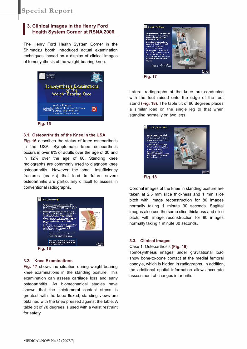
2.1. Features of SONIALVISION safire
Fig. 3 shows the features of the Shimadzu
SONIALVISION safire. The SONIALVISION safire
digital table features high resolution, low geometric
distortion, high frame rate, and extremely low lag. It
was judged to offer the high-precision movements of
mechanical components essential for high-accuracy
tomosynthesis.
2.2. Acquisition Conditions and safire Specifications
Fig. 4 shows the movements during tomosynthesis.
For lateral radiography of the knee joint, a 60° table
tilt angle is used and the X-ray tube and FPD move
in opposition. The X-ray tube swing angle is ±20°.
Tomosynthesis for clinical evaluation of the knee
joint was conducted with 75 views acquired over
2.5 s at 30 fps frame rate under X-ray conditions of
65 kV to 70 kV and 1 mAs X-ray dose per frame. The
entrance exposure is estimated at 216 mR, which is
about one-fifth the dose from a multislice CT exam.
Fig. 5 shows the specifications for the 17-inch FPD.
Fig. 6 shows a graph of the spatial resolution
characteristics of the detector. As the detector is a
direct-conversion FPD, the measured characteristics
(solid line) are extremely close to the ideal
characteristics (broken line). Dr. Flynn explained that
the measured curve was believed to be slightly lower
due to MTF deterioration resulting from secondary
X-rays generated in the conversion layer. The results
from Monte Carlo simulations conducted in the
past matched the measured results. The noise
characteristics exhibited at least two times the
DQE of other manufacturers' direct-conversion
FPDs for radiography. Also, due to the low level of
scattered X-rays, tomosynthesis examinations of
the knee joint are conducted with the X-ray grid
removed.
2.3. Principle of Tomosynthesis Reconstruction
Fig. 7 shows the principle of tomosynthesis
reconstruction. While reconstruction is similar to
cone-beam CT, due to the restricted projection
angle, the data is effective only in the shaded area
of the spatial frequency domain, and a limiting filter
is applied to the Z-direction frequency.
2.4. Evaluation of the FPD Transient Response
The FPD transient response was evaluated by
conducting tomosynthesis on a 1.51 mm copper
plate using RQA5 equivalent radiation quality and
measuring the pixel value while moving across the
edge of the copper plate. (Fig.8)
Fig. 4
Fig. 3
Fig. 5
Fig. 6
Fig. 7

MEDICAL NOW No.62 (2007.7)
Graphs of the pixel value in each frame plotted
while moving from light to dark and from dark to
light indicate the excellent lag characteristics of the
Shimadzu FPD. (Fig. 9)
2.5. Measuring Slice Thickness and Spatial
Resolution
The slice thickness and spatial resolution were
measured using a test phantom (Fig. 10). The
phantom comprises an 80 µm tungsten wire inclined
at 1:10 pitch, which is an angle of about 6 degrees.
The tungsten wire is 80 µm thick and the FPD pixel
size is 300 × 300 µm max. with 2 × 2 binning. A
magnified image is shown in Fig. 11.
Fig. 12 shows the tomosynthesis image of the
tungsten wire. The image was reconstructed with a
1 mm slice interval. The tomographic slice thickness
was measured as approximately 2.5 mm from the
FWHM of the profile along the tungsten wire.
In addition, the profile perpendicular to the
tungsten wire in the tomosynthesis image of the
tungsten wire (Fig. 13) yields FWHM = 0.238 mm.
Normally, the Modulation Transfer Function (MTF)
curve is the Fourier transform of the line spread
function. However, a feature of this method is the
loss of DC components due to effects of filtered
backprojection (Fig. 14).
Fig. 10
Fig. 9
Fig. 11 Fig. 8
Fig. 12
Fig. 14
Fig. 13
MEDICAL NOW No.62 (2007.7)
3. Clinical Images in the Henry Ford
Health System Corner at RSNA 2006
The Henry Ford Health System Corner in the
Shimadzu booth introduced actual examination
techniques, based on a display of clinical images
of tomosynthesis of the weight-bearing knee.
3.1. Osteoarthritis of the Knee in the USA
Fig. 16 describes the status of knee osteoarthritis
in the USA. Symptomatic knee osteoarthritis
occurs in over 6% of adults over the age of 30 and
in 12% over the age of 60. Standing knee
radiographs are commonly used to diagnose knee
osteoarthritis. However the small insufficiency
fractures (cracks) that lead to future severe
osteoarthritis are particularly difficult to assess in
conventional radiographs.
3.2. Knee Examinations
Fig. 17 shows the situation during weight-bearing
knee examinations in the standing posture. This
examination can assess cartilage loss and early
osteoarthritis. As biomechanical studies have
shown that the tibiofemoral contact stress is
greatest with the knee flexed, standing views are
obtained with the knee pressed against the table. A
table tilt of 70 degrees is used with a waist restraint
for safety.
Lateral radiographs of the knee are conducted
with the foot raised onto the edge of the foot
stand (Fig. 18). The table tilt of 60 degrees places
a similar load on the single leg to that when
standing normally on two legs.
Coronal images of the knee in standing posture are
taken at 2.5 mm slice thickness and 1 mm slice
pitch with image reconstruction for 80 images
normally taking 1 minute 30 seconds. Sagittal
images also use the same slice thickness and slice
pitch, with image reconstruction for 80 images
normally taking 1 minute 30 seconds.
3.3. Clinical Images
Case 1: Osteoarthosis (Fig. 19)
Tomosynthesis images under gravitational load
show bone-to-bone contact at the medial femoral
condyle, which is hidden in radiographs. In addition,
the additional spatial information allows accurate
assessment of changes in arthritis.
Fig. 15
Fig. 16
Fig. 17
Fig. 18
MEDICAL NOW No.62 (2007.7)
Case 2: Sudden Onset Left Knee Pain (Fig. 20)
In this case, initially no abnormality could be seen
in the frontal and lateral radiographs but MRI
follow-up images reveal insufficiency fractures in
the medial femoral condyle and lateral femoral
condyle. Similarly, tomosynthesis discovers the
cracks but offers much higher spatial resolution
and greater utility than MRI.
Fig. 19
Fig. 20

MEDICAL NOW No.62 (2007.7)
Case 3: Tibial Plateau Fracture (Fig. 21)
After an initial CT evaluation, the healed fracture
remained painful. Follow up by tomosynthesis after
one year revealed a residual step-off deformity that
was believed to be the cause of the pain.
4. Clinical Images at a SEAAPM
Educational Symposium
The tomosynthesis cases presented by Dr. Flynn
were introduced at an educational symposium held
by the SEAAPM (Southeast Chapter of the
American Association of Physicists in Medicine) in
March 2007.
Case 4: Suspected Trochanter Fracture (Fig. 22)
While not identifiable in radiographs, the edema
apparent in the MR image suggests the need for
surgery. However, tomosynthesis showed that the
fracture was restricted to the non-weight-bearing
head of the trochanter, so that surgery was not
required.
5. Conclusions
This paper introduced the evaluation of
tomosynthesis using SONIALVISION safire based
on the presentation by Dr. Flynn of Henry Ford
Health System at RSNA 2006 and using the
clinical images displayed in the Henry Ford Health
System Corner in the Shimadzu booth at RSNA
2006 and presented by Dr. Flynn at the SEAAPM
educational symposium.
In the future, Shimadzu intends to further expand
the utility and possibilities of tomosynthesis.
Fig. 21
Fig. 22