41 urethral disease
TRANSCRIPT

41 Urethral Disease

CLINICAL IMAGAGINGAN ATLAS OF DIFFERENTIAL DAIGNOSIS
EISENBERG
DR. Muhammad Bin Zulfiqar PGR-FCPS III SIMS/SHL
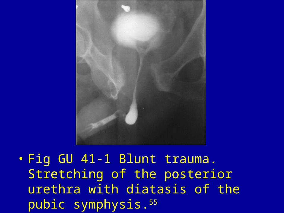
• Fig GU 41-1 Blunt trauma. Stretching of the posterior urethra with diatasis of the pubic symphysis.55

• Fig GU 41-2 Blunt trauma. Extensive extravasation of contrast material without flow into the prostatic urethra or bladder due to complete rupture of the posterior urethra. A fracture of the right pubic ramus was diagnosed.55
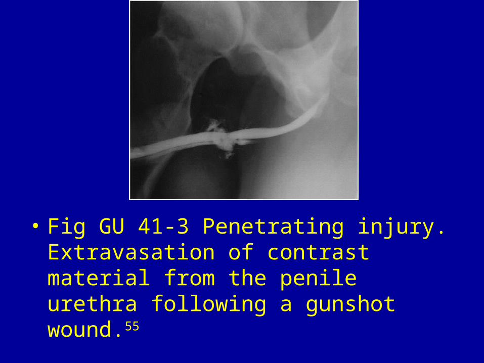
• Fig GU 41-3 Penetrating injury. Extravasation of contrast material from the penile urethra following a gunshot wound.55

• Fig GU 41-4 Penile fracture. Extravasation of contrast material in the penile urethra adjacent to the site of a corpus cavernosal injury.55

• Fig GU 41-5 Gonococcal urethral stricture. Irregular, beaded narrowing of a segment of the distal bulbous urethra with opacification of the left Cowper duct (arrow).

• Fig GU 41-6 Gonococcal urethral stricture with periurethral abscess. Long segment of irregular, beaded narrowing in the bulbous urethra with opacification of the Littré glands (arrow). Note the irregular periurethral cavity originating from the ventral aspect of the bulbous urethra.55

• Fig GU 41-7 Condyloma acuminata. Multiple small filling defects in the anterior urethra.55

• Fig GU 41-8 Urethral diverticulum. (A) Postvoiding image obtained during excretory urography demonstrates a contrast-filled urethral diverticulum (arrow). (B) T2-weighted MR image shows a large diverticulum surrounding the urethra (arrow), with a septum that results in an impression at the bladder base (female prostate sign). B, bladder; S, pubic symphysis.55

• Fig GU 41-9 Calculi associated with urethral stricture. The stones (arrowhead) lie in a segment of an anterior urethral stricture.55

• Fig GU 41-10 Squamous cell carcinoma. Irregular stricture of the bulbous urethra.55

• Fig GU 41-11 Adenocarcinoma of the urethra. Contrast CT scan in a woman with previous hysterectomy shows an inhomogeneous soft-tissue mass of the urethra (m) representing a high-grade tumor.55

