3d culture brochure 2015 axil - apical scientific · 3500-096-01 3445-096-cp 3550-096-k 4892-010-k...
TRANSCRIPT

3-D CU
LTURE
REGENERATIVEMEDICINE
DRU
GD
ISCOVERY
CANCERRESEARCH
GEN
ETIC
TOXI
COLO
GY
3-D CULTURE
3-D ASSAY KITS AND 3-D MATRICES OrganoidsSpheroids
Embryoid BodiesAcinar Structures
In Vitro Tumor Co-Culture

Recent studies indicate that the composition of the extracellular environment influences cellular responses to apoptosis inducing agents implicating a role for extracellular proteins in influencing both toxicity and drug resistance. As a result, this environment must be mimicked during the course of cell-based studies to provide the most accurate transla-tion to animal models.
Formation of tri-culture structure on a mixture of Cultrex® ECM matrices, over 10 days. Red: breast cancer cell line (MDA-MB-231), Green: human umbilical vein endothelial cells (HUVECs); Blue: human adipose-derived mesenchymal stem cells (hMSCs).
Nuclear morphology of MCF-10A, mammary epithelial acinar structures, as depicted using 1X SYBR Green reagent. 3-D structures were imaged using a Nikon Eclipse E400 microscope (100X magnification) using epifluorescence with a FITC filter and images were captured using a Q Imaging Micropublisher 3.3 camera.
Detection of luminal hollowing in MCF-10A, mammary epithelial acinar structures. Cells were labeled with 2 μM Calcein AM and 1 μM EtBr for 15 min. prior to imaging. Green cells indicate conversion of calcein AM to calcein by living cells, and red cells indicate compromised plasma membrane of dead cells.
3-D CANCER CELL ASSAYS
CATALOG #PRODUCT NAME
3445-001-013445-005-013445-010-013532-001-023532-005-023532-010-023533-001-023533-005-023533-010-023446-005-013447-020-013448-020-K3500-096-K3500-096-033510-096-K3511-096-K3500-096-013445-096-CP3550-096-K4892-010-K 3575-096-K
SIZE
1 ml5 ml2 X 5 ml1 ml5 ml2 x 5 ml1 ml5 ml2 x 5 ml30 mg20 ml20 tests96 samples6 ml96 samples96 samples600 µl96 samples96 samples1000 wells96 Tests
Cultrex® 3-D Culture Matrix™ Reduced Growth Factor Basement Membrane Extract, PathClear®
(Type 2), PathClear®
Membrane Extract (Type 2), PathClear®
Cultrex® 3-D Culture Matrix Laminin ICultrex® 3-D Culture Matrix Rat Collagen ICultrex® 3-D Culture Cell Harvesting KitCultrex® 3-D Spheroid Cell Invasion AssayCultrex® 3-D Spheroid Invasion MatrixCultrex® 3-D Spheroid Fluorometric Proliferation/Viability AssayCultrex® 3-D Spheroid Colorimetric Proliferation/Viability AssayCultrex® 3-D Spheroid Formation MatrixCultrex® 3-D Culture Matrix BME Coated 96 Well PlateCultrex® Embryoid Body Formation KitCalcein-AM Cell Viability Assay KitCultrex® 3-D Microtumor Co-Culture Kit*
The behavior of cancer cells has both intrigued and plagued scientists for years. As a provider of tools for cancer research, Trevigen has developed and optimized assay formats and characterized the factors affecting basement membrane protein-dependent in vitro assays for cancer and endothelial cells. Keep-ing the researcher in mind, we emphasized sensitivity, accuracy, and ease of use. The culmination of this development work is a series of products and methods designed to study cancer progression at the cellular level. These include assays that measure the critical cellular functions of adhesion, proliferation, migration, and invasion, as well as 3-D assays that may be used to assess cellular differentiation, mor-phology, angiogenic potential, and molecular composition of cells within their physiological microenvi-ronment.
Background photo: Primary embryonic submandibular epithelium cultured in Cultrex® 3-D Laminin I. Image courtesy of Matthew P. Hoffman.
*Coming Soon
3-D culture proliferation of MDA-MB-231 breast cancer spheroids (using Cultrex 3-D Spheroid Proliferation/Viability Assay) time lapse expansion of MDA-MB-231 spheroids over a 96 hour period.
Spheroid invasion by MDA-MB-231 breast cancer spheroids over a 96 hour period.
Inhibition of spheroid invasion by MDA-MB-231 breast cancer spheroids by Sulforaphane over a 96 hour period.
Morphology of MCF-10A normal mamma-ry epithelial cells in traditional 2-D (A) and 3-D BME (B) cell culture and MCF-7 mammary adenocarcinoma cells in traditional 2-D (C) and 3-D BME (D) cell culture, scale = 250 μm.
SUPPORTING DATA
3-D Spheroid Assays
3-D Morphology
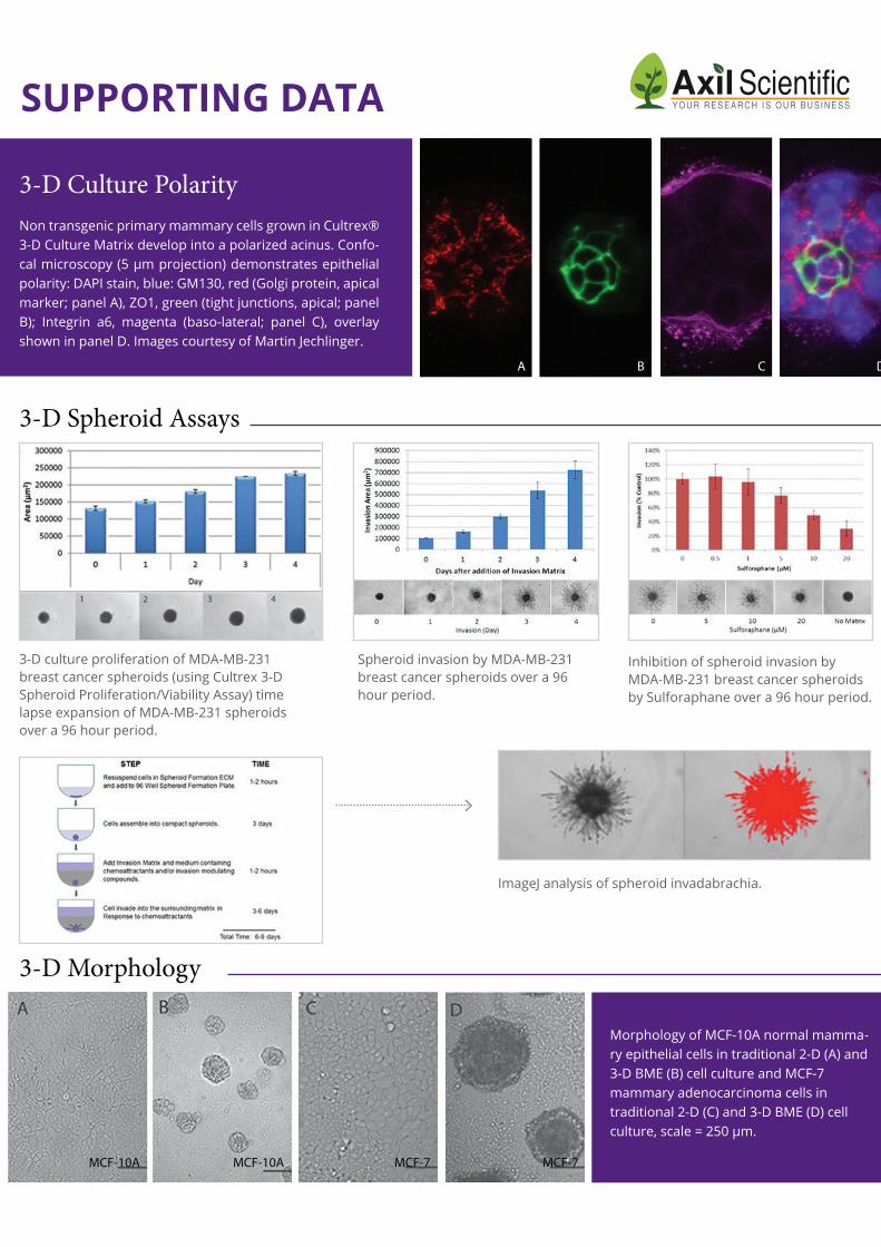
Non transgenic primary mammary cells grown in Cultrex® 3-D Culture Matrix develop into a polarized acinus. Confo-cal microscopy (5 μm projection) demonstrates epithelial polarity: DAPI stain, blue: GM130, red (Golgi protein, apical marker; panel A), ZO1, green (tight junctions, apical; panel B); Integrin a6, magenta (baso-lateral; panel C), overlay shown in panel D. Images courtesy of Martin Jechlinger.
3-D Culture Polarity
A B C D
MCF-10A MCF-10A MCF-7 MCF-7
ImageJ analysis of spheroid invadabrachia.
CITATIONS
Long-term culture of genome-stable bipotent stem cells from adult human liver.Huch M, Gehart H, van Boxtel R, Hamer K, Blokzijl F, Verstegen MM, Ellis E, van Wenum M, Fuchs SA, de Ligt J, van de Wetering M, Sasaki N, Boers SJ, Kemperman H, de Jonge J, Ijzermans JN, Nieuwenhuis EE, Hoekstra R, Strom S, Vries RR, van der Laan LJ, Cuppen E, Clevers H.Cell. 2015 Jan 15;160(1-2):299-312. doi: 10.1016/j.cell.2014.11.050. Epub 2014 Dec 18.
In vitro microtumors provide a physiologically predictive tool for breast cancer therapeutic screening.Benton G., DeGray G., Kleinman HK., George J., Arnaoutova I.PLoS One. 2015 Apr 9;10(4):e0123312. doi: 10.1371/journal.pone.0123312
Prospective derivation of a 'Living Organoid Biobank' of colorectal cancer patients.van de Wetering, M., Francies, H.E., Francis, J.M., Bounova, G., Iorio, F., Pronk, A., van Houdt, W., van Gorp, J., Taylor-Weiner, A., Kester, L., McLaren-Douglas, A., Blokker, J., Jaksani, S., Bartfeld, S., Volckman, R., van Sluis, P., Li, V.S.W., Seepo, S., Sekhar Pedamallu, C., Cibulskis, C., Carter, S.L., McKenna, A., Lawrence, M.S., Lichtenstein, L., Stewart, C., Koster, J., Versteeg, R., van Oudenaarden, A., Saez-Rodriguez, J., Vries, R.G.J., Getz, G., Wessels, L., Stratton, M.R., McDermott, U., Meyerson, M., Garnett, M.J., Clevers, H. Cell. 2015 May 7;161(4):933-45. doi: 10.1016/j.cell.2015.03.053.
In vitro differentiation of human amniotic epithelial cells into insulin producing 3D spheroids.Okere B, Alviano F, Costa R, Quaglino D, Ricci F, Dominici M, Paolucci P, Bonsi L, Iughetti L.Int J Immunopathol Pharmacol. 2015 Jul 27. pii: 0394632015588439
Endophilin A2 Promotes TNBC Cell Invasion and Tumor Metastasis.Baldassarre T, Watt K, Truesdell P, Meens J, Schneider MM, Sengupta SK, Craig AW.Mol Cancer Res. 2015 Jun;13(6):1044-55. doi: 10.1158/1541-7786.MCR-14-0573. Epub 2015 Mar 17.
Silencing β3 Integrin by Targeted ECO/siRNA Nanoparticles Inhibits EMT and Metastasis of Triple-Negative Breast Cancer.Parvani JG, Gujrati MD, Mack MA, Schiemann WP, Lu ZR.Cancer Res. 2015 Jun 1;75(11):2316-25. doi: 10.1158/0008-5472.CAN-14-3485. Epub 2015 Apr 9.
Organotypic Culture of Untransformed and Tumorigenic Primary Mammary Epithelial Cells.Martin JechlingerCold Spring Harb Protoc. 2015 May 1;2015(5):457-61. doi: 10.1101/pdb.prot078295.
Matrigel: From discovery and ECM mimicry to assays and models for cancer research.Benton G, Arnaoutova I, George J, Kleinman HK, Koblinski JAdv Drug Deliv Rev. 2014 Dec 15;79-80:3-18. doi: 10.1016/j.addr.2014.06.005. Epub 2014 Jul 2.
1 800-TREVIGEN

FAQSWHAT ARE 3-D CULTURES?
3-D cultures are in vitro cultures where immortalized cell lines, primary cell lines, stem cells, or explants are placed within hydrogel matrices that mimic in vivo cell environments.
WHAT IS THE ADVANTAGE OF 3-D CULTURE OVER TRADITIONAL 2-D CULTURE?
While 2-D culture has been used for studying many aspects of cell function and behavior, the tissue-culture treated plastic environment is unlike anything found within living organ-isms. As result, cells in 2-D culture exhibit altered morphology, function, proliferation and gene expression when compared to their emanating tissues. By placing these cells in a 3-D environment, they assume biological and biochemical characteristics similar to what is observed in vivo.
WHAT ARE THE VARIABLES ASSOCIATED WITH 3-D CULTURE?
The major variables associated with 3-D culture are cell type, cell seeding density, com-position of hydrogel, thickness of hydrogel, stiffness of hydrogel, composition of cell culture medium, and time of culture.
WHAT ARE THE DIFFERENT TYPES OF 3-D CULTURE?
The two principal methods for conducting 3-D culture are the top assay and embedded assay. For the top assay, cells are seeded on a thick gel and a thin overlay is applied with the cell culture medium. For the embedded assay, cells are resuspended within a thick gel and the culture media is applied on top. The top assay is easier to setup, to control seeding densities, and to keep cells within one focal plane for analysis.
WHICH MATRIX SHOULD I USE FOR 3-D CULTURE?
Choice of matrix should correspond to the environment that you wish to recapitulate. A basement membrane extract (BME) will reca-pitulate the basal lamina, which underlie most cells of epithelial or endothelial origin. Colla-gen I is the major constituent of connective tissue, and it is commonly inhabited by stationary cells, such as fibrocytes and adipose
cells, as well as migrating cells, such as mast cells, macrophages, monocytes, lymphocytes, plasma cells, and eosinophils.
HOW SHOULD CELLS BE CULTURED PRIOR TO SETTING UP THE 3-D CULTURE?
Cells need to be healthy and actively dividing in 2-D culture. Cells should be passaged two or three times after resuspension from cryopreservation, and they should never surpass 80% confluency during each passage. Cells should also be assessed using trypan blue, and they should exhibit less than 5% staining.
WHAT TYPE OF ANALYSIS IS TYPICALLY APPLIED TO 3-D CULTURES?
Within the cultures, cells may be assessed for mor-phology, apical/basal polarity, protein localization, and relative proliferation. In addition, cells may be isolated from the 3-D culture and evaluated for levels of RNA and protein expression, as well as modifica
-
tions to DNA.
HOW CAN I HARVEST MY CELLS FOR SUBSEQUENT ANALYSIS?
Cells may be harvested from 3-D culture using the Cultrex® 3-D Culture Cell Harvesting Kit.
CAN I TRANSFER ORGANOIDS FROM ONE MATRIX TO ANOTHER?
We strongly recommend, as is the standard practice in cell culture, that you maintain the same matrix throughout your process of deriving and expanding your organoids. We’ve learned that intestinal and liver organoid culture is most successful using Cultrex® Basement Membrane Extract, Type 2 from start to finish rather than switching between matrices.
WHAT IS A RECOMMENDED PROTOCOL FOR ORGANOID CULTURE?
· Air Liquid Interface (ALI) organoid cultures: an insert containing an acellular layer of ECM is used to suspend organoids above the culture medium level to create the air-liquid interface. ALI organoids include tissue stroma, and they usually utilize a collagen-1 ECM.
· Submerged organoid cultures: isolated epithelial stem cells are embedded in a BME Type 2 hydrogel that is positioned in the middle of a culture vessel. These stem cells routinely require medium containing Wnt, EGF, Noggin, and R-spondin-1 (WENR).

RELATED PRODUCTS
ABOUT US
SIZEDESCRIPTION
24 Samples 24 Samples 24 Samples 24 Samples 24 Samples 96 Samples 96 Samples96 Samples96 Samples96 Samples
CATALOG NUMBER
3480-024-K3455-024-K3457-024-K3458-024-K3456-024-K3455-096-K3457-096-K3458-096-K3456-096-K3471-096-K
CultreCoat® 24 Well BME Cell Invasion Assay Cultrex® 24 Well BME Cell Invasion Assay Cultrex® 24 Well Collagen I Cell Invasion Assay Cultrex® 24 Well Collagen IV Cell Invasion Assay Cultrex® 24 Well Laminin I Cell Invasion Assay Cultrex® 96 Well BME Cell Invasion Assay Cultrex® 96 Well Collagen I Cell Invasion Assay Cultrex® 96 Well Collagen IV Cell Invasion Assay Cultrex® 96 Well Laminin I Cell Invasion Assay Cultrex® Endothelial Cell Invasion Kit
Trevigen, Inc is an innovative biotechnology company focused on the development of products and technologies for cell behavior, including stem cell/ regenerative medicine and cancer research. Trevgien has core technologies in protein purification, cell biology and DNA damage and repair. The company is the recipient of several NIH SBIR grants from the National Institute of Health, concerning technology development for the analysis of DNA Damage, and an SBIR Contract for the development of an in vitro co-culture breast cancer model. The Trevigen Research and Development team has a number of thought leaders in the fast growing 3-D Cell Culture research field, who have helped the company become a pioneer in the industry by developing some of the first commercially availa-
ble products for 3-D Culture research.
WHAT CUSTOMERS SAYI have been using the kit "Cultrex® 3-D Spheroid Invasion Assay" in order to test the effect of chemopreventive drugs on tumor cells (both cell lines and primary cells directly otained from patients) invasion. This is an easy-to-use kit which worked very well for my cells. As expected, spheroids were formed at 72h after plating. Then, I was able to easily follow the invasive properties of tumor cells at the different timepoints indicated by the manufacturer instructions. In addition, the kit's datasheet provides a step by step guid-ance for image analysis (by using a a free software such as image-j) and data interpreta-tion. They were very helpful to finally obtain the figures I will include in my next research paper. Overall, I am fully satisfied by this product and I would recommend it for invasion studies by other researchers.
“We have used Trevgen's Cultrex® Reduced Growth Factor BME in our studies on DCIS. When grown in 3D, MCF10.DCIS forms dysplastic structures that recapitulate DCIS in the mammary gland of the patient. This matrix is also a reliable BME, which helps us study signaling in DCIS when grown in 3D.
Seema Shah Wayne State
University
Katiuscia Dallaglio Azienda Ospedaliera S.Maria Nuova-Reggio Emilia