2. necrosis severe damage metabolism stop structure destroy function lose classification: necrosis...
TRANSCRIPT
2. Necrosis2. Necrosis
Severe damage Metabolism stop Structure destroy Function lose
Classification: necrosis & apoptosis
(1) Definition: Localized death of cell or tissue occurring in the living body.
① Ultrastructural changes
Margination or progressive loss of nuclear chromatin
Focal rupture of the nuclear membrane
Breakdown of the plasmalemma.
Development of flocculent densities in mitochondria.
(2) Cell death is recognized by:
② Changes in the nucleus.
Pyknosis: condensation of chromatin of chromatin and shrinkage of the nucleus.
Karyorrhexis: fragmentation of the nucleus.
Karyolysis: dissolution of the nucleus.
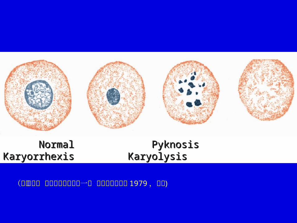
Normal Pyknosis Karyorrhexis Normal Pyknosis Karyorrhexis KaryolysisKaryolysis
(参照武忠弼 病理学规划教材第一版 人民卫生出版社 1979 ,修改 )
③ Changes in cytoplasm staining
Positive staining with vital dyes such as Trepan blue which reflects abnormal membrane permeability.
Opacification:Opacification: denaturation of proteins lead to aggregation with resultant opacification of the cytoplasm.
Eosino0.philia:Eosino0.philia: exposure of basic amino groups results in increased affinity for acidic dyes such as eosin.
④ Biochemical changes
Release of K+ by dead cells.
Release of enzymes into the blood. e. g. increased plasma levels of creatine kinases, lactic dehydrogenase and aspartate aminotransferase.
Release of protein or protein breakdown products into the blood.
⑤Postmortem change: General of normal tissues occurring dead body, generally distinguished from necrosis by being diffuse and not associated with inflammatory response.
⑥Autolysis: Digestion of cell by enzymes released from lysosome; occurs after cell dies.
(3) Types:
①① Coagulative necrosis:Coagulative necrosis:
Gross features:Gross features: The necrosis area is swollen, firm and pale.
LM:LM: cell detail is lost, but architecture preserved. The dead cells retain their outline but only indistinctly.
This type of necrosis is frequently caused by lack of blood supply and is exemplified well in infarcts of solid organs, e. g. heart, spleen, kidney.
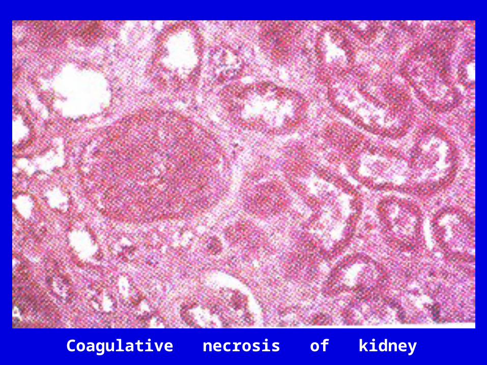
Coagulative necrosis of kidney
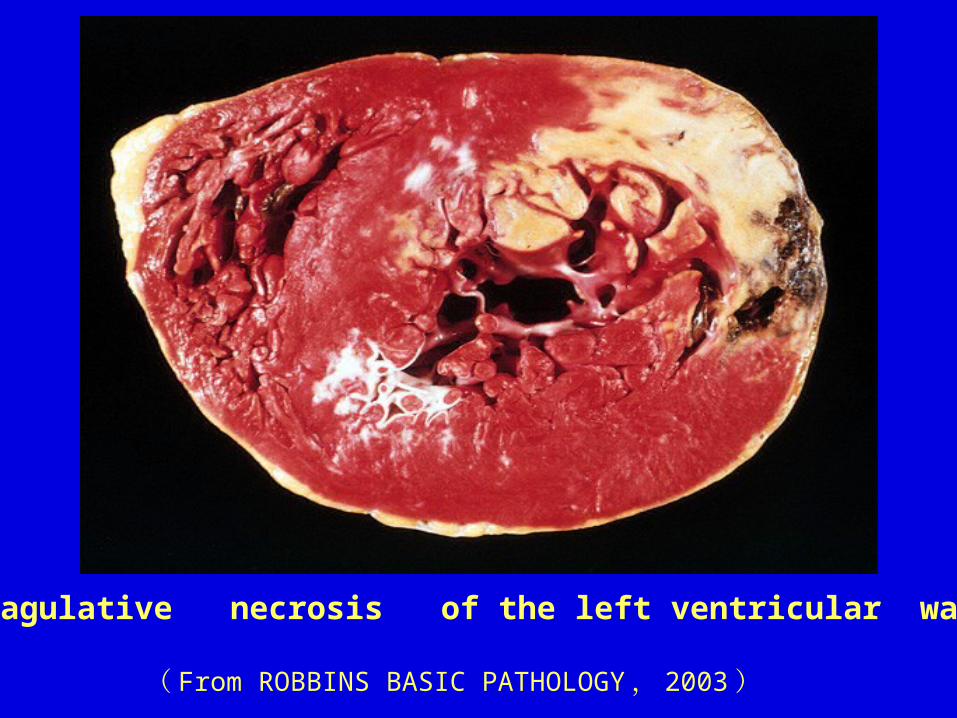
Coagulative necrosis of the left ventricular wall
( From ROBBINS BASIC PATHOLOGY , 2003 )
A. Caseous necrosis:A. Caseous necrosis: Gross features:Gross features: soft, granular, and
friable a cream-cheesy appearance. granular, eosinophilic.
LM:LM: architecture completely destroyed.
i. e. Tuberculosis, syphilis, some sarcoma.
Special types of coagulative necrosis
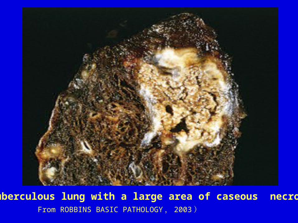
From ROBBINS BASIC PATHOLOGY , 2003 )A tuberculous lung with a large area of caseous necrosis
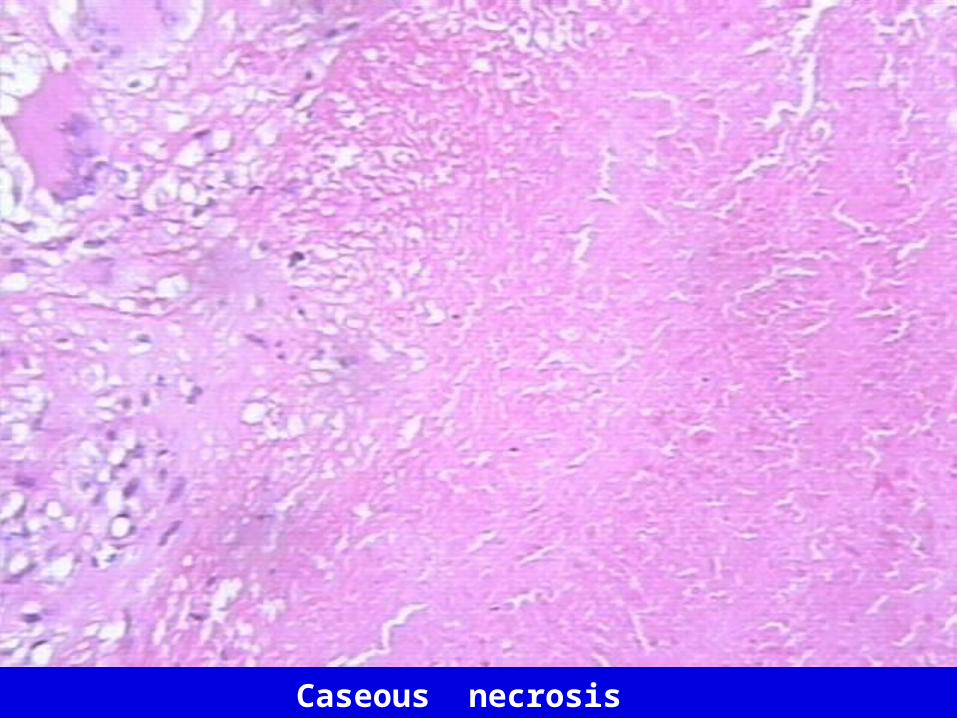
Caseous necrosis
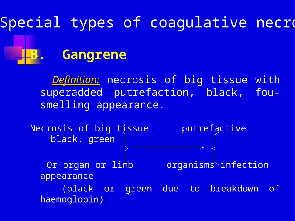
B. Gangrene
Definition:Definition: necrosis of big tissue with superadded putrefaction, black, fou-smelling appearance.
Necrosis of big tissue putrefactive black, green
Or organ or limb organisms infection appearance
(black or green due to breakdown of haemoglobin)
Special types of coagulative necrosis
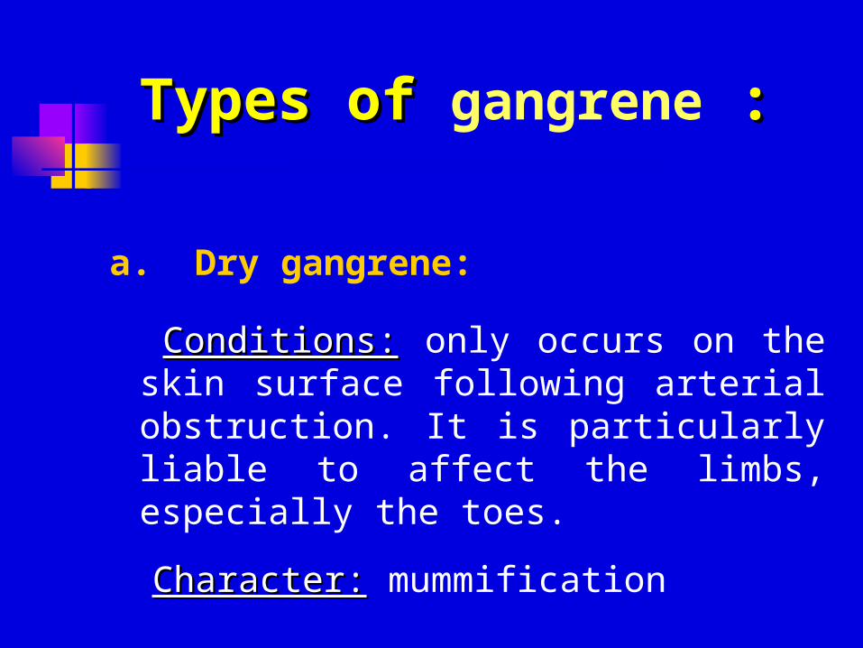
a. Dry gangrene:
Conditions:Conditions: only occurs on the skin surface following arterial obstruction. It is particularly liable to affect the limbs, especially the toes.
Character:Character: mummification
Types of Types of gangrene ::
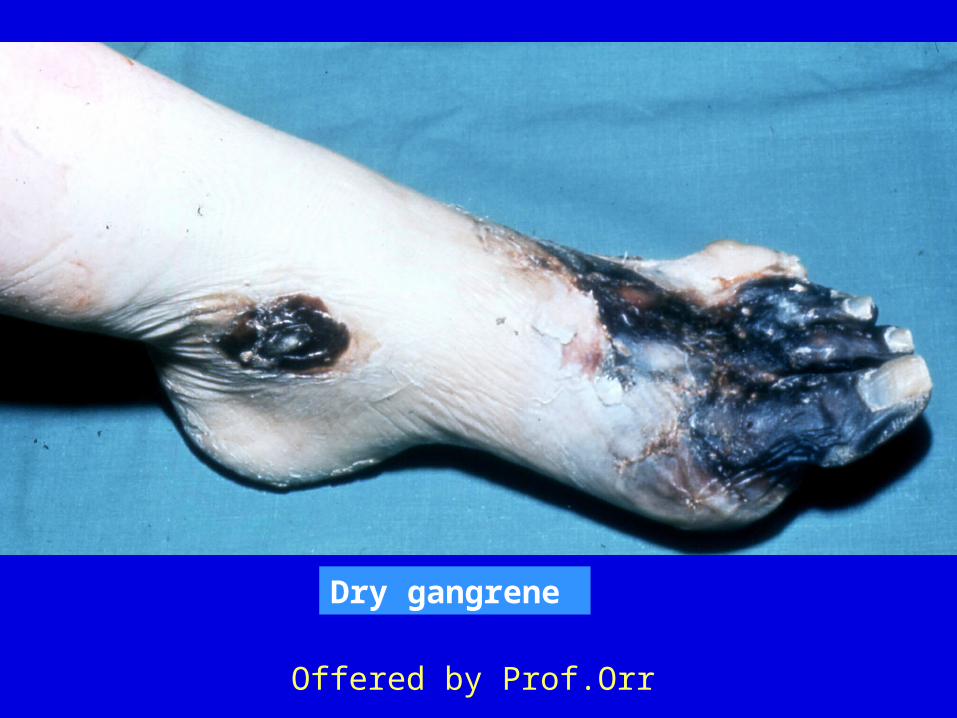
Dry gangrene
Offered by Prof.Orr
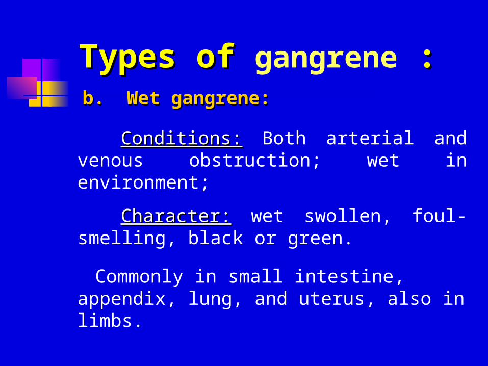
b. Wet gangrene:b. Wet gangrene:
Conditions:Conditions: Both arterial and venous obstruction; wet in environment;
Character:Character: wet swollen, foul-smelling, black or green.
Commonly in small intestine, appendix, lung, and uterus, also in limbs.
Types of Types of gangrene ::
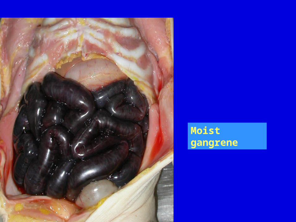
Moist gangrene
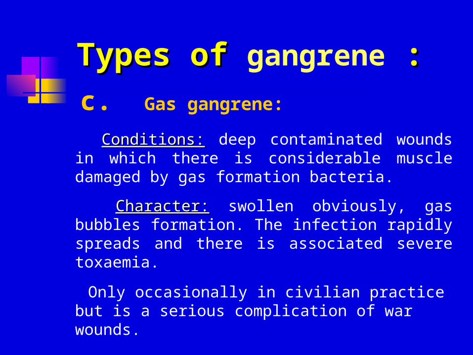
c. Gas gangrene:
Conditions:Conditions: deep contaminated wounds in which there is considerable muscle damaged by gas formation bacteria.
Character:Character: swollen obviously, gas bubbles formation. The infection rapidly spreads and there is associated severe toxaemia.
Only occasionally in civilian practice but is a serious complication of war wounds.
Types of Types of gangrene ::
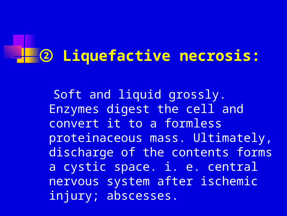
② Liquefactive necrosis:
Soft and liquid grossly. Enzymes digest the cell and convert it to a formless proteinaceous mass. Ultimately, discharge of the contents forms a cystic space. i. e. central nervous system after ischemic injury; abscesses.
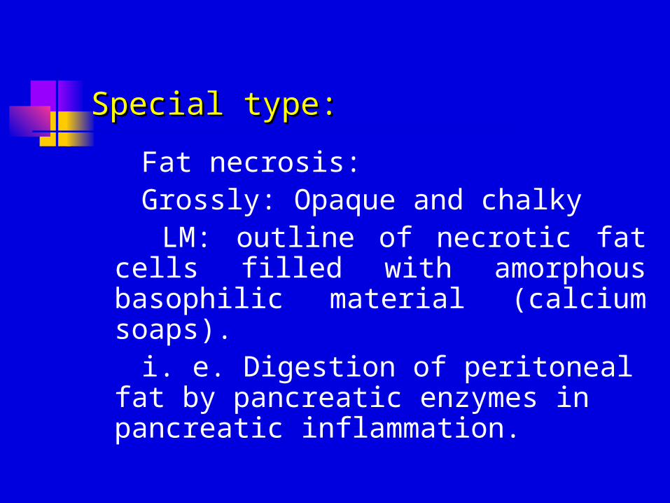
Special type:Special type:
Fat necrosis: Grossly: Opaque and chalky LM: outline of necrotic fat cells
filled with amorphous basophilic material (calcium soaps).
i. e. Digestion of peritoneal fat by pancreatic enzymes in pancreatic inflammation.
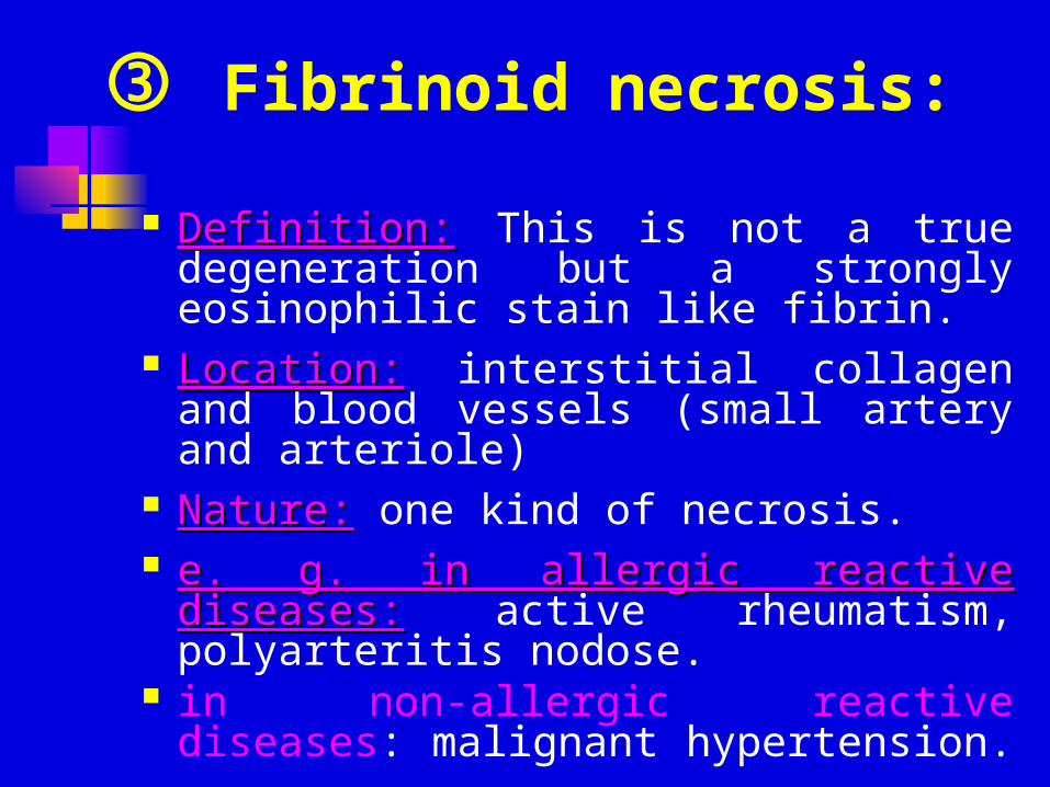
Definition:Definition: This is not a true degeneration but a strongly eosinophilic stain like fibrin.
Location:Location: interstitial collagen and blood vessels (small artery and arteriole)
Nature:Nature: one kind of necrosis. e. g. in allergic reactive diseases:e. g. in allergic reactive diseases:
active rheumatism, polyarteritis nodose.
in non-allergic reactive diseases: malignant hypertension.
Fibrinoid necrosis:
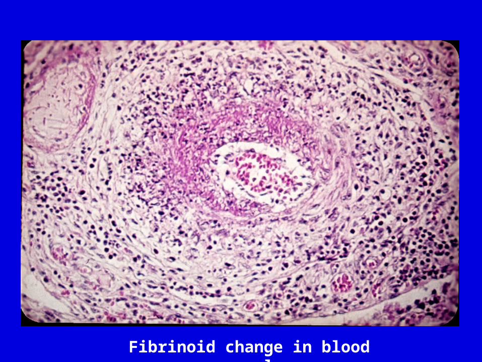
Fibrinoid change in blood vessel
(4) Consequences of necrosis
① Acute or chronic inflammation
② Immunological reactions to sub cellular components released by dead tissue or self-antigens altered by denaturation.
③ lysis and absorption
④ Isolation and discharge: ulceration and cavity formation
⑤ organization
⑥ encapsulation, calcification.