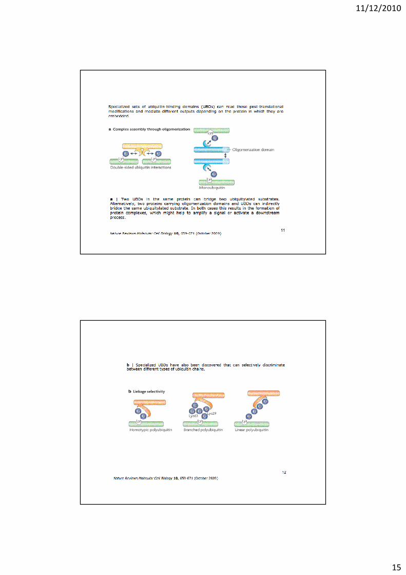
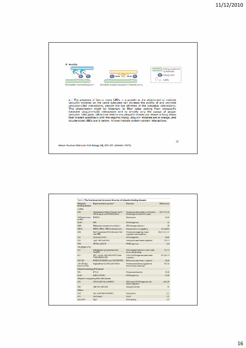
17 ubiquitination 12 11 10 - biologia.i-learn.unito.it
TRANSCRIPT
11/12/2010
1
11/12/2010
2
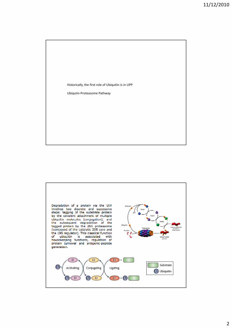
Historically, the first role of Ubiquitin is in UPP
Ubiqutin-Proteasome Pathway
11/12/2010
3
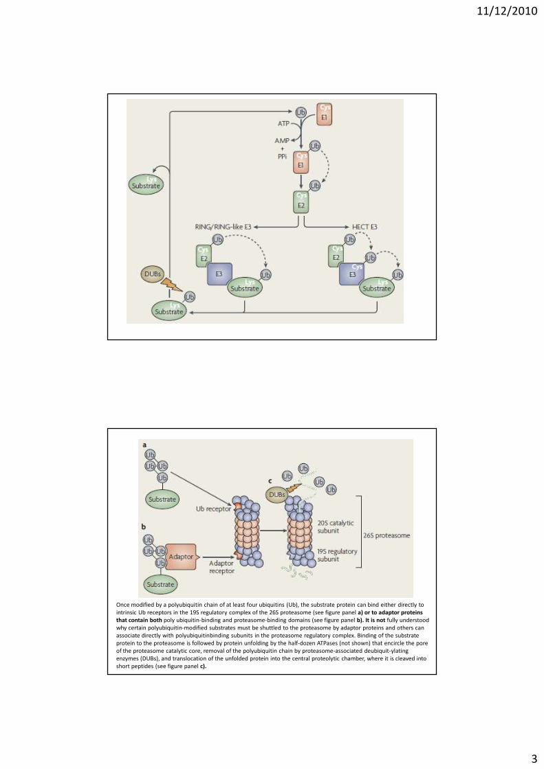
Once modified by a polyubiquitin chain of at least four ubiquitins (Ub), the substrate protein can bind either directly to
intrinsic Ub receptors in the 19S regulatory complex of the 26S proteasome (see figure panel a) or to adaptor proteins
that contain both poly ubiquitin-binding and proteasome-binding domains (see figure panel b). It is not fully understood
why certain polyubiquitin-modified substrates must be shuttled to the proteasome by adaptor proteins and others can
associate directly with polyubiquitinbinding subunits in the proteasome regulatory complex. Binding of the substrate
protein to the proteasome is followed by protein unfolding by the half-dozen ATPases (not shown) that encircle the pore
of the proteasome catalytic core, removal of the polyubiquitin chain by proteasome-associated deubiquit-ylating
enzymes (DUBs), and translocation of the unfolded protein into the central proteolytic chamber, where it is cleaved into
short peptides (see figure panel c).
11/12/2010
4
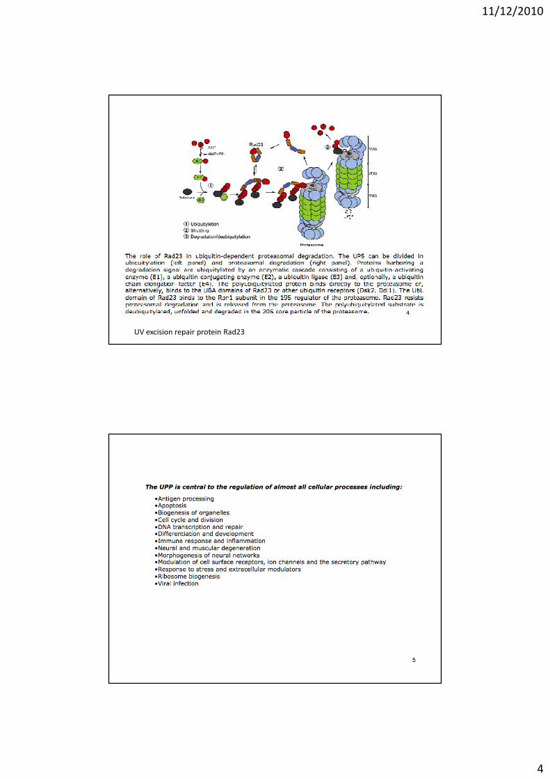
UV excision repair protein Rad23
11/12/2010
5
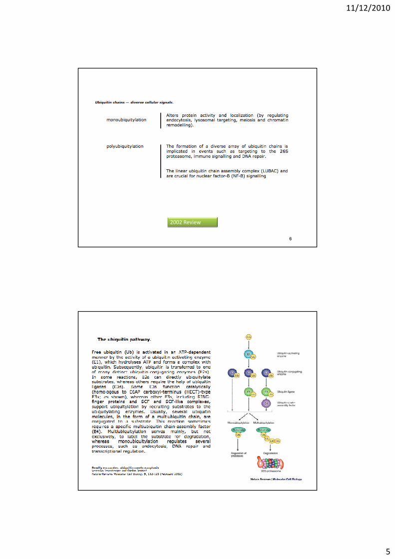
2002 Review
11/12/2010
6
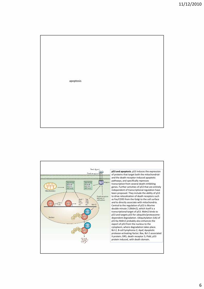
apoptosis
p53 and apoptosis. p53 induces the expression
of proteins that target both the mitochondrial-
and the death-receptor-induced apoptotic
pathways, and specifically represses
transcription from several death-inhibiting
genes. Further activities of p53 that are entirely
independent of transcriptional regulation have
been proposed. They include the ability of p53
to drive relocalization of death receptors such
as Fas/CD95 from the Golgi to the cell surface
and to directly associate with mitochondria.
Central to the regulation of p53 is Murine
double minute 2 (Mdm2), which itself is a
transcriptional target of p53. Mdm2 binds to
p53 and targets p53 for ubiquitin/proteasome-
dependent degradation. Ubiquitylation (Ub) of
p53 by Mdm2 probably also enhances the
export of p53 from the nucleus to the
cytoplasm, where degradation takes place.
Bcl-2, B-cell lymphoma 2; Apaf, Apoptotic
protease-activating-factor; Bax, Bcl-2 associated
X protein; DR5, death receptor 5; Pidd, p53
protein induced, with death domain.
11/12/2010
7
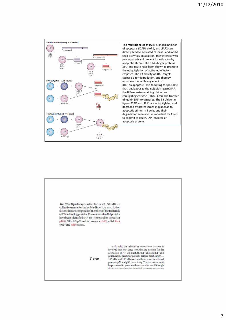
The multiple roles of IAPs. X-linked inhibitor
of apoptosis (XIAP), cIAP1, and cIAP2 can
directly bind to activated caspases and inhibit
their activities. In addition, they interact with
procaspase-9 and prevent its activation by
apoptotic stimuli. The RING-finger proteins
XIAP and cIAP2 have been shown to promote
the ubiquitylation of activated effector
caspases. The E3 activity of XIAP targets
caspase-3 for degradation, and thereby
enhances the inhibitory effect of
XIAP on apoptosis. It is tempting to speculate
that, analagous to the ubiquitin ligase XIAP,
the BIR-repeat-containing ubiquitin-
conjugating enzyme (BRUCE) can also transfer
ubiquitin (Ub) to caspases. The E3 ubiquitin
ligases XIAP and cIAP1 are ubiquitylated and
degraded by proteasomes in response to
apoptotic stimuli in T cells, and their
degradation seems to be important for T cells
to commit to death. IAP, inhibitor of
apoptosis protein.
1° step
11/12/2010
8
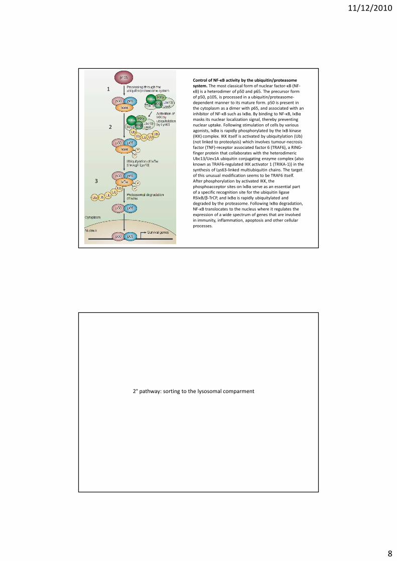
Control of NF-κB activity by the ubiquitin/proteasome
system. The most classical form of nuclear factor-κB (NF-
κB) is a heterodimer of p50 and p65. The precursor form
of p50, p105, is processed in a ubiquitin/proteasome-
dependent manner to its mature form. p50 is present in
the cytoplasm as a dimer with p65, and associated with an
inhibitor of NF-κB such as IκBα. By binding to NF-κB, IκBα
masks its nuclear localization signal, thereby preventing
nuclear uptake. Following stimulation of cells by various
agonists, IκBα is rapidly phosphorylated by the IκB kinase
(IKK) complex. IKK itself is activated by ubiquitylation (Ub)
(not linked to proteolysis) which involves tumour-necrosis
factor (TNF)-receptor associated factor 6 (TRAF6), a RING-
finger protein that collaborates with the heterodimeric
Ubc13/Uev1A ubiquitin conjugating enzyme complex (also
known as TRAF6-regulated IKK activator 1 (TRIKA-1)) in the
synthesis of Lys63-linked multiubiquitin chains. The target
of this unusual modification seems to be TRAF6 itself.
After phosphorylation by activated IKK, the
phosphoacceptor sites on IκBα serve as an essential part
of a specific recognition site for the ubiquitin ligase
RSIκB/β-TrCP, and IκBα is rapidly ubiquitylated and
degraded by the proteasome. Following IκBα degradation,
NF-κB translocates to the nucleus where it regulates the
expression of a wide spectrum of genes that are involved
in immunity, inflammation, apoptosis and other cellular
processes.
1
2
3
2° pathway: sorting to the lysosomal comparment
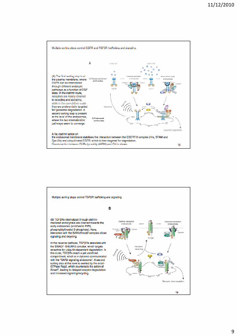
11/12/2010
9
11/12/2010
10
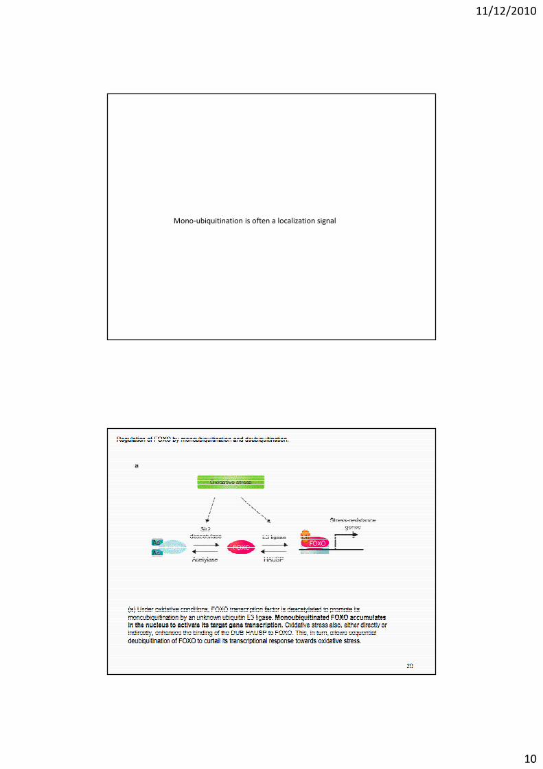
Mono-ubiquitination is often a localization signal
11/12/2010
11
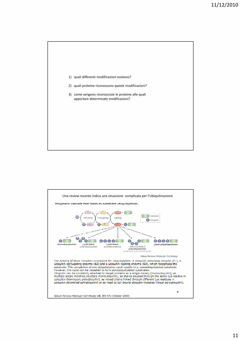
1) quali differenti modificazioni esistono?
2) quali proteine riconoscono queste modificazioni?
3) come vengono riconosciute le proteine alle quali
apportare determinate modificazioni?
Una review recente indica una situazione complicata per l’Ubiquitinazione
11/12/2010
12
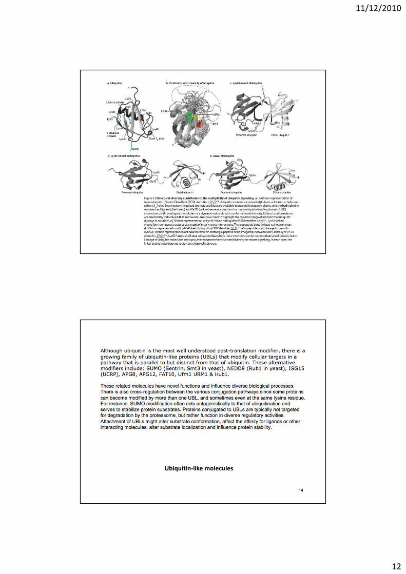
Ubiquitin-like molecules
11/12/2010
13
11/12/2010
14
Ubiquitin binding domains
NRMCB 2009
11/12/2010
15
11/12/2010
16
11/12/2010
17
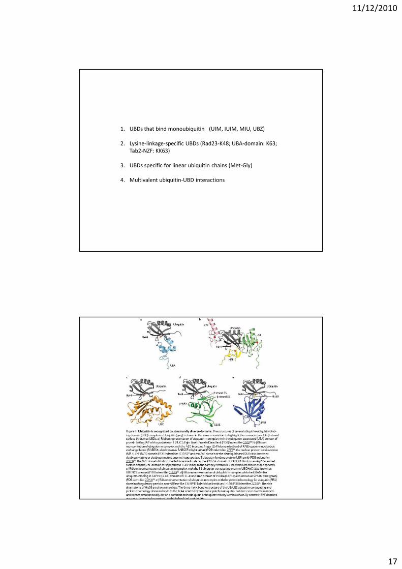
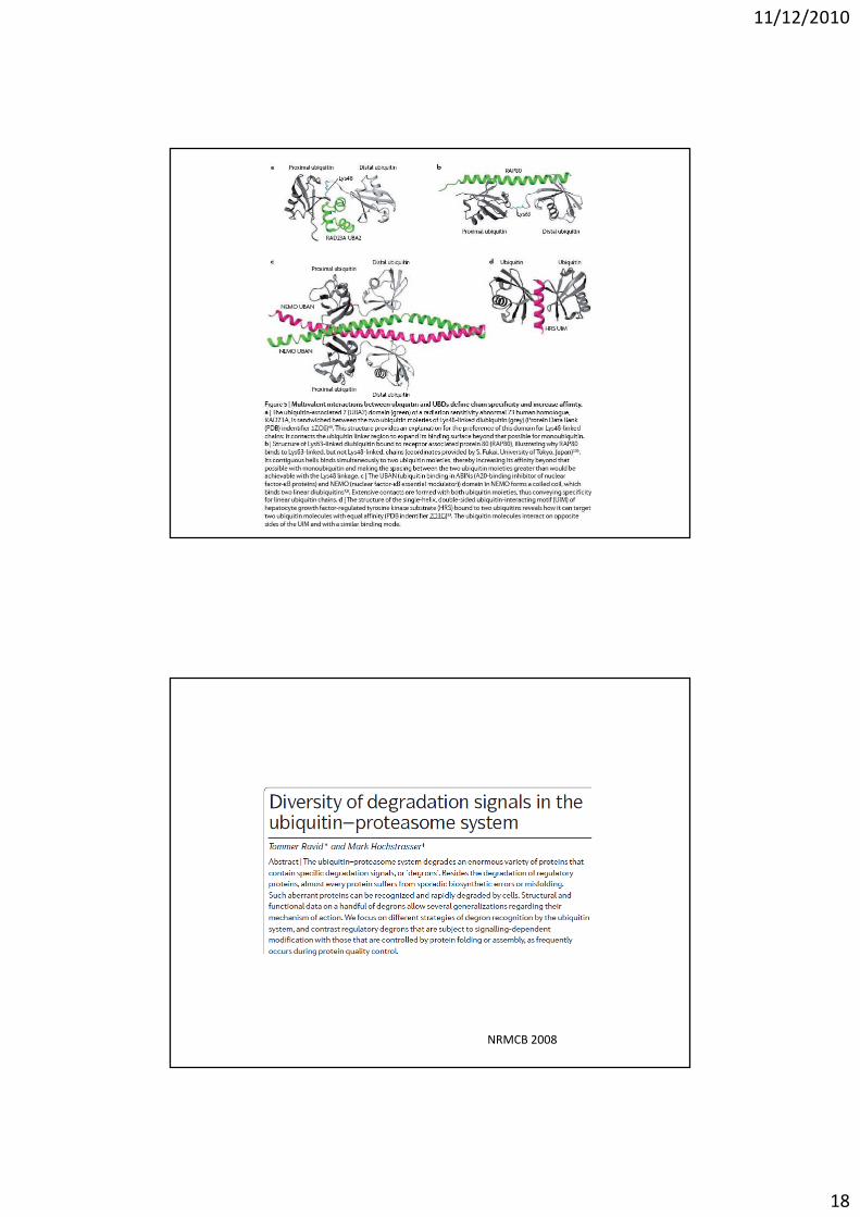
1. UBDs that bind monoubiquitin (UIM, IUIM, MIU, UBZ)
2. Lysine-linkage-specific UBDs (Rad23-K48; UBA-domain: K63;
Tab2-NZF: KK63)
3. UBDs specific for linear ubiquitin chains (Met-Gly)
4. Multivalent ubiquitin-UBD interactions
11/12/2010
18
NRMCB 2008
11/12/2010
19
Segnale nella proteina da degradare: “degron”
11/12/2010
20
Unfolded protein response:
unfolded, damaged proteins � expose degrons that otherwise
are inside
1. The N-end rule pathway
2. E3α is the enzyme recognizin the N-terminus (RING-domain)
11/12/2010
21
Phosphodegrons
11/12/2010
22
11/12/2010
23
11/12/2010
24