153a winter 2011 review session for final examrebecca/153a/w11/153a_final_review.pdf · 153a winter...
TRANSCRIPT
153A Winter 2011
Review session for Final Exam
Thursday, March 10
5-7 pm in Dodd 147
Theresa and Megan
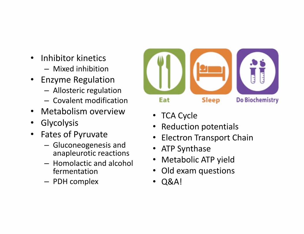
• Inhibitor kinetics– Mixed inhibition
• Enzyme Regulation– Allosteric regulation
– Covalent modification
• Metabolism overview
• Glycolysis
• Fates of Pyruvate– Gluconeogenesis and
anapleurotic reactions
– Homolactic and alcohol fermentation
– PDH complex
• TCA Cycle
• Reduction potentials
• Electron Transport Chain
• ATP Synthase
• Metabolic ATP yield
• Old exam questions
• Q&A!
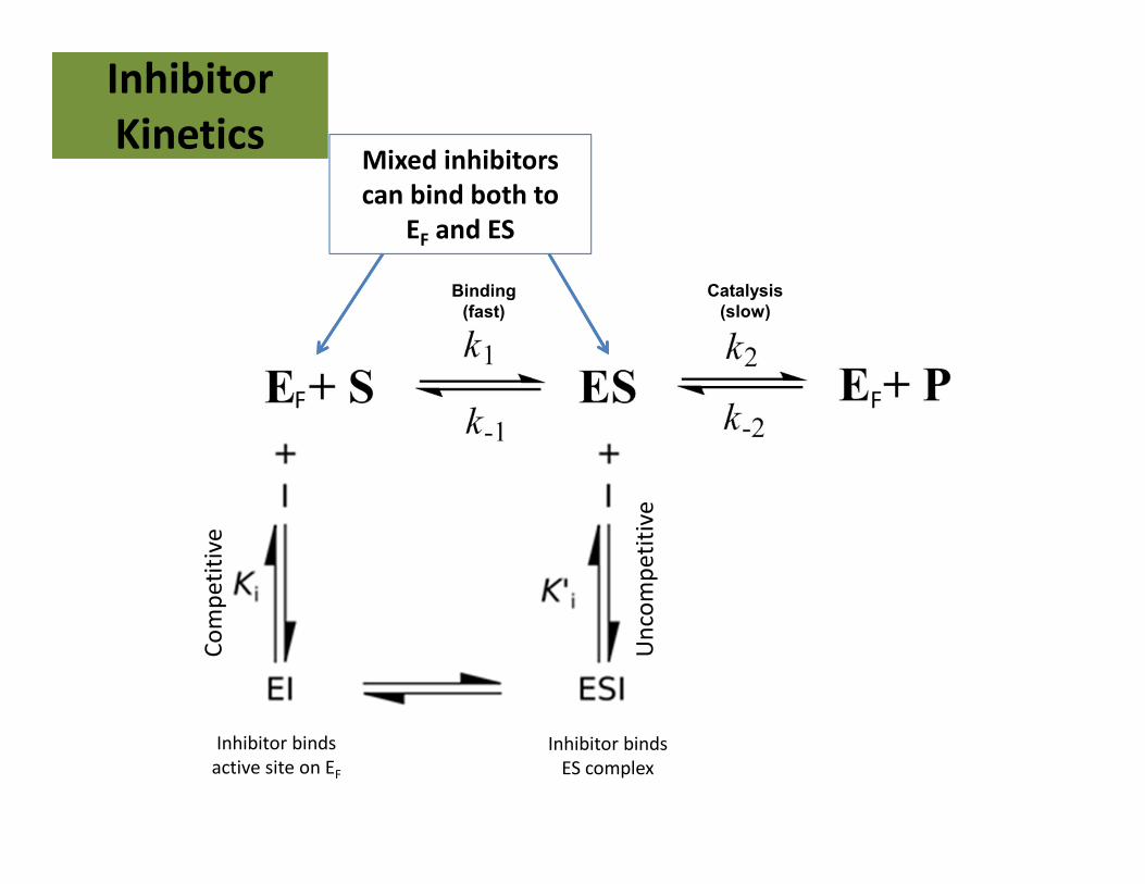
Inhibitor
Kinetics
Inhibitor
Kinetics
Binding
(fast)
Catalysis
(slow)
F F
Inhibitor binds
active site on EF
Inhibitor binds
ES complex
Mixed inhibitors
can bind both to
EF and ESC
om
pe
titi
ve
Un
com
pe
titi
ve
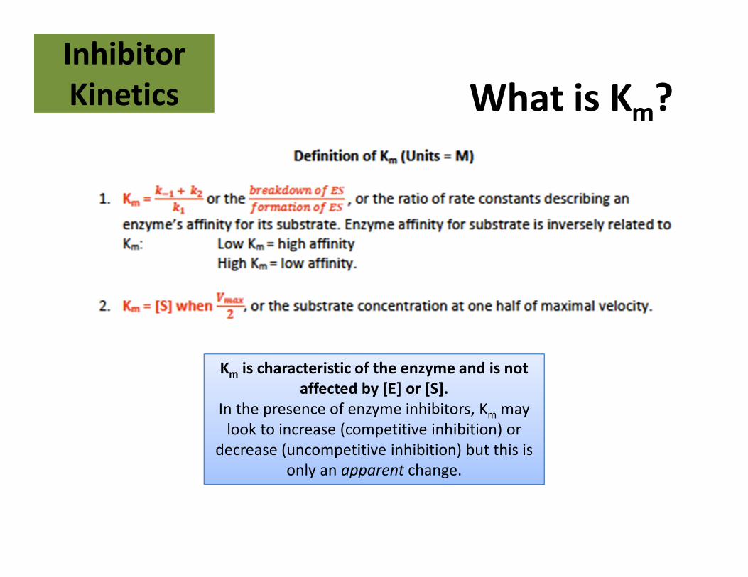
What is Km?Inhibitor
Kinetics
Inhibitor
Kinetics
Km is characteristic of the enzyme and is not
affected by [E] or [S].
In the presence of enzyme inhibitors, Km may
look to increase (competitive inhibition) or
decrease (uncompetitive inhibition) but this is
only an apparent change.
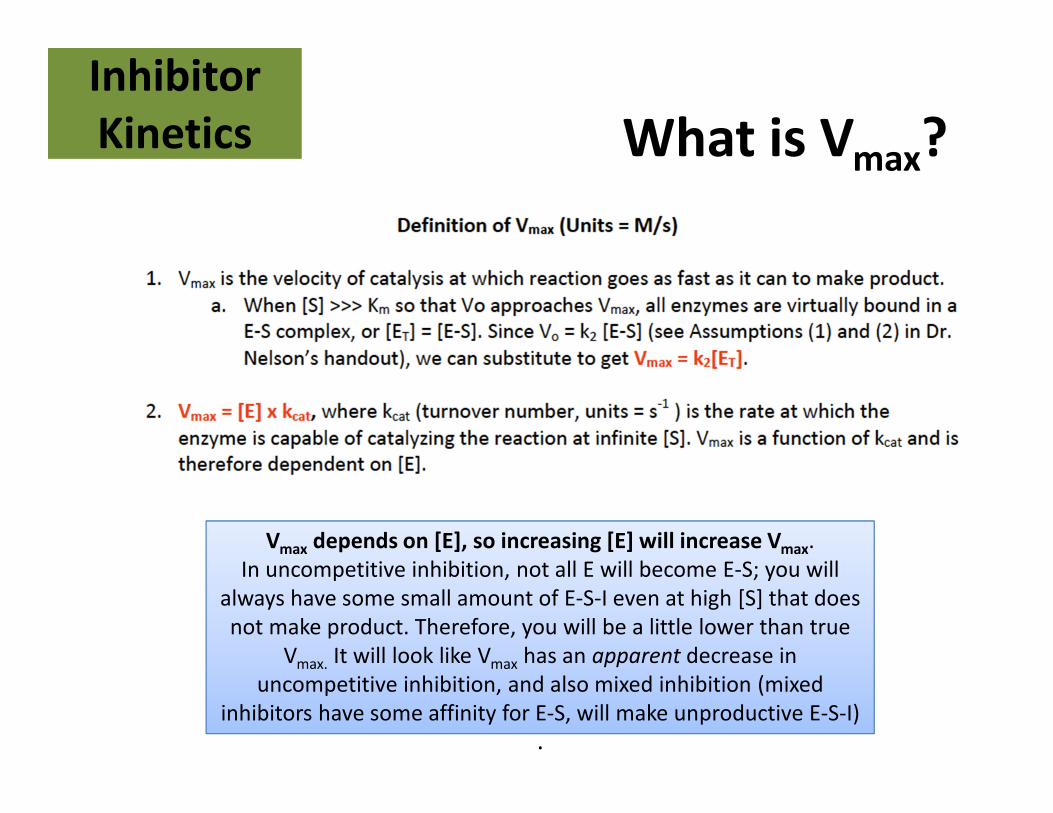
What is Vmax?Inhibitor
Kinetics
Inhibitor
Kinetics
.
Vmax depends on [E], so increasing [E] will increase Vmax.
In uncompetitive inhibition, not all E will become E-S; you will
always have some small amount of E-S-I even at high [S] that does
not make product. Therefore, you will be a little lower than true
Vmax. It will look like Vmax has an apparent decrease in
uncompetitive inhibition, and also mixed inhibition (mixed
inhibitors have some affinity for E-S, will make unproductive E-S-I)
.
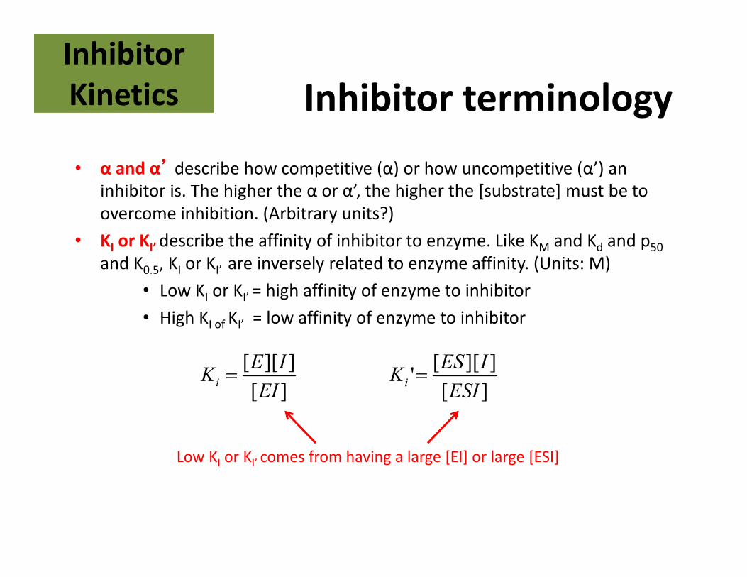
Inhibitor terminology
• α and α’’’’ describe how competitive (α) or how uncompetitive (α’) an
inhibitor is. The higher the α or α’, the higher the [substrate] must be to
overcome inhibition. (Arbitrary units?)
• KI or KI’ describe the affinity of inhibitor to enzyme. Like KM and Kd and p50
and K0.5, KI or KI’ are inversely related to enzyme affinity. (Units: M)
• Low KI or KI’ = high affinity of enzyme to inhibitor
• High KI of KI’ = low affinity of enzyme to inhibitor
Inhibitor
Kinetics
Inhibitor
Kinetics
][
]][[
EI
IEKi=
][
]][['
ESI
IESKi=
Low KI or KI’ comes from having a large [EI] or large [ESI]
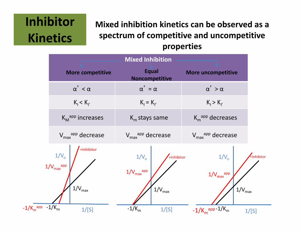
Mixed inhibition kinetics can be observed as a
spectrum of competitive and uncompetitive
properties
Inhibitor
Kinetics
Inhibitor
Kinetics
Mixed Inhibition
α’ < α α’ = α α’ > α
KI < KI’ KI = KI’ KI > KI’
KMapp increases Km stays same Km
app decreases
Vmaxapp decrease Vmax
app decrease Vmaxapp decrease
More competitive More uncompetitiveEqual
Noncompetitive
1/Vo
1/[S]-1/Km-1/Kmapp
1/Vmax
1/Vmaxapp
+Inhibitor
1/Vo
1/[S]-1/Km-1/Kmapp
1/Vmax
1/Vmaxapp
1/Vo
1/[S]-1/Km
1/Vmax
1/Vmaxapp
+Inhibitor +Inhibitor
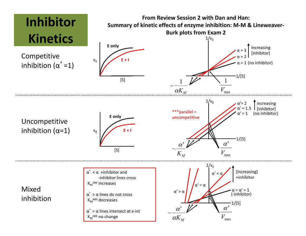
From Review Session 2 with Dan and Han:
Summary of kinetic effects of enzyme inhibition: M-M & Lineweaver-
Burk plots from Exam 2
Competitive
inhibition (α’=1)
Mixed
inhibition
α' = 1α' = 1.5α'= 2 increasing
[inhibitor](no inhibitor)
1/v0
1/[S]
MK
'α−
max
'
V
α
α = 1
α = 2
α = 3increasing
[inhibitor]
(no inhibitor)
1/v0
1/[S]
MKα
1−
max
1
V
α’ < α +inhibitor and
-inhibitor lines cross
KMapp increases
α’ > α lines do not cross
KMapp decreases
α’ = α lines intersect at x-int
KMapp no change
***parallel =
uncompetitive
α = α' = 1
[increasing]
+inhibitor
(-inhibitor)
1/v0
1/[S]
max
'
V
α
MKα
α '−
α' > α
α' = α
α' < α
Uncompetitive
inhibition (α=1) v0
[S]
E only
E + I
v0
[S]
E only
E + I
Inhibitor
Kinetics
Inhibitor
Kinetics
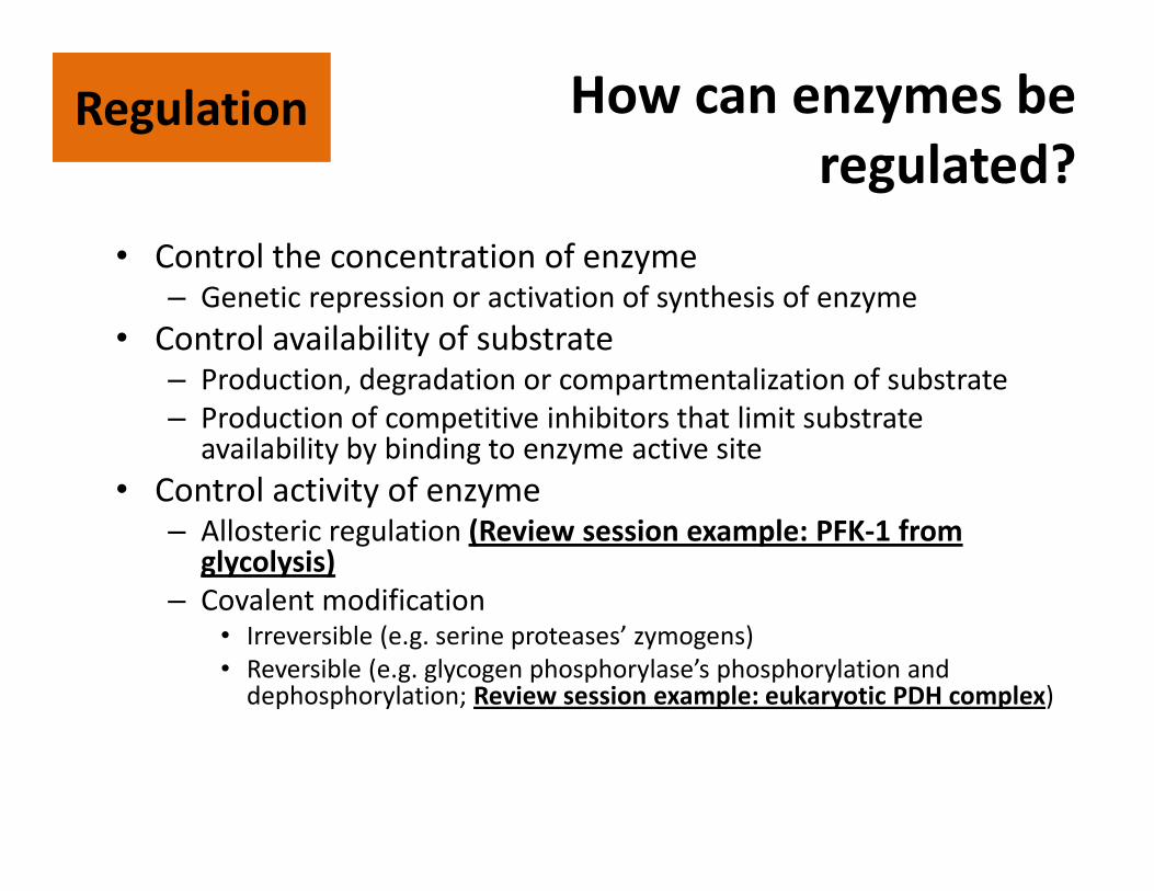
How can enzymes be
regulated?
• Control the concentration of enzyme– Genetic repression or activation of synthesis of enzyme
• Control availability of substrate– Production, degradation or compartmentalization of substrate
– Production of competitive inhibitors that limit substrate availability by binding to enzyme active site
• Control activity of enzyme– Allosteric regulation (Review session example: PFK-1 from
glycolysis)
– Covalent modification• Irreversible (e.g. serine proteases’ zymogens)
• Reversible (e.g. glycogen phosphorylase’s phosphorylation and dephosphorylation; Review session example: eukaryotic PDH complex)
RegulationRegulation
RegulationRegulation
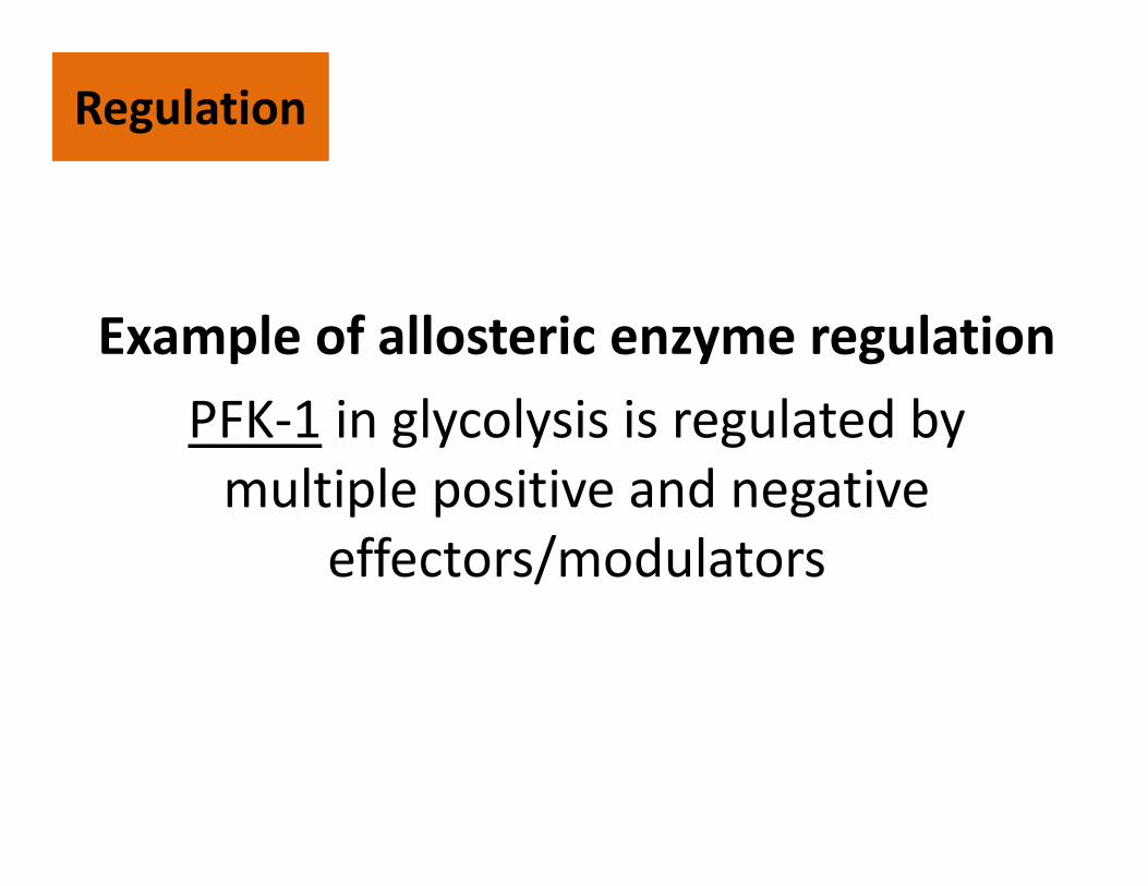
Example of allosteric enzyme regulation
PFK-1 in glycolysis is regulated by
multiple positive and negative
effectors/modulators
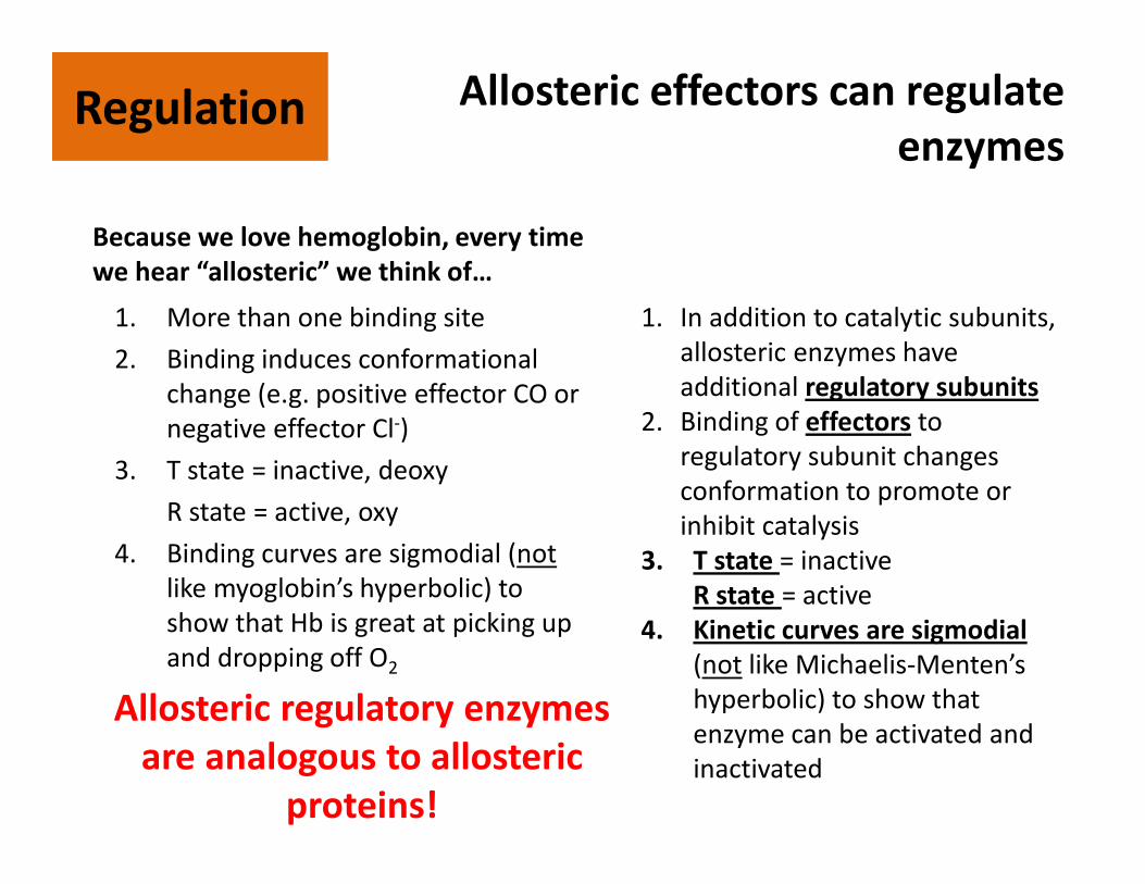
Allosteric effectors can regulate
enzymes
1. More than one binding site
2. Binding induces conformational
change (e.g. positive effector CO or
negative effector Cl-)
3. T state = inactive, deoxy
R state = active, oxy
4. Binding curves are sigmodial (not
like myoglobin’s hyperbolic) to
show that Hb is great at picking up
and dropping off O2
RegulationRegulation
Allosteric regulatory enzymes
are analogous to allosteric
proteins!
1. In addition to catalytic subunits,
allosteric enzymes have
additional regulatory subunits
2. Binding of effectors to
regulatory subunit changes
conformation to promote or
inhibit catalysis
3. T state = inactive
R state = active
4. Kinetic curves are sigmodial
(not like Michaelis-Menten’s
hyperbolic) to show that
enzyme can be activated and
inactivated
Because we love hemoglobin, every time
we hear “allosteric” we think of…
Allosteric effectors can regulate
enzymesRegulationRegulation
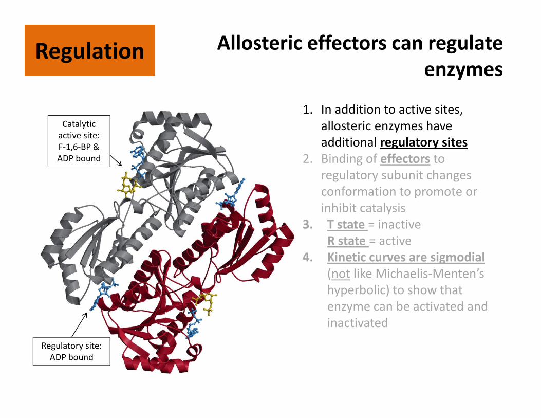
1. In addition to active sites,
allosteric enzymes have
additional regulatory sites
2. Binding of effectors to
regulatory subunit changes
conformation to promote or
inhibit catalysis
3. T state = inactive
R state = active
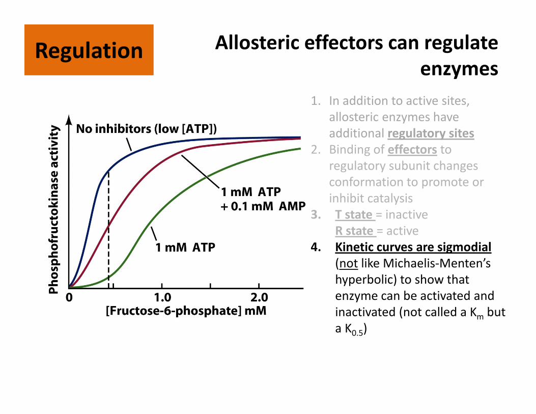
4. Kinetic curves are sigmodial
(not like Michaelis-Menten’s
hyperbolic) to show that
enzyme can be activated and
inactivated
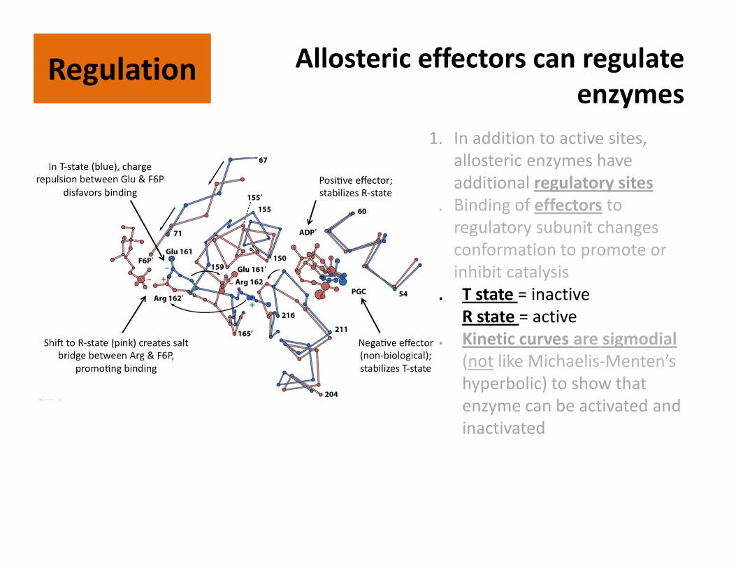
Catalytic
active site:
F-1,6-BP &
ADP bound
Regulatory site:
ADP bound
Allosteric effectors can regulate
enzymesRegulationRegulation
1. In addition to active sites,
allosteric enzymes have
additional regulatory sites
2. Binding of effectors to
regulatory subunit changes
conformation to promote or
inhibit catalysis
3. T state = inactive
R state = active
4. Kinetic curves are sigmodial
(not like Michaelis-Menten’s
hyperbolic) to show that
enzyme can be activated and
inactivated
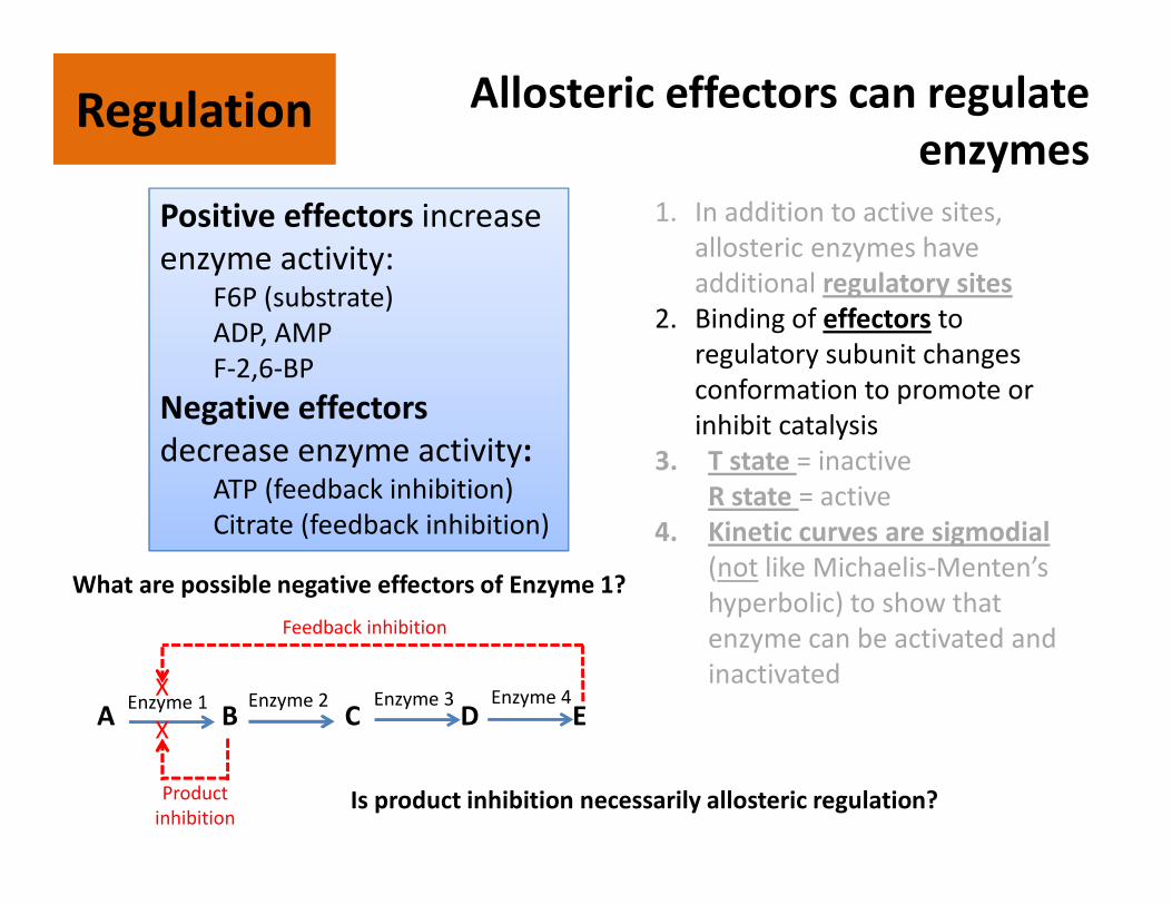
Positive effectors increase
enzyme activity:F6P (substrate)
ADP, AMP
F-2,6-BP
Negative effectors
decrease enzyme activity:ATP (feedback inhibition)
Citrate (feedback inhibition)
A B C D EEnzyme 1 Enzyme 2 Enzyme 3 Enzyme 4
What are possible negative effectors of Enzyme 1?
Product
inhibition
X
X
Feedback inhibition
Is product inhibition necessarily allosteric regulation?
Allosteric effectors can regulate
enzymesRegulationRegulation
1. In addition to active sites,
allosteric enzymes have
additional regulatory sites
2. Binding of effectors to
regulatory subunit changes
conformation to promote or
inhibit catalysis
3. T state = inactive
R state = active
4. Kinetic curves are sigmodial
(not like Michaelis-Menten’s
hyperbolic) to show that
enzyme can be activated and
inactivated
Allosteric effectors can regulate
enzymesRegulationRegulation
1. In addition to active sites,
allosteric enzymes have
additional regulatory sites
2. Binding of effectors to
regulatory subunit changes
conformation to promote or
inhibit catalysis
3. T state = inactive
R state = active
4. Kinetic curves are sigmodial
(not like Michaelis-Menten’s
hyperbolic) to show that
enzyme can be activated and
inactivated (not called a Km but
a K0.5)
RegulationRegulation
Example of covalent modification for
enzyme regulation
Pyruvate dehydrogenase complex is
regulated by phosphorylation and
dephosphorylation
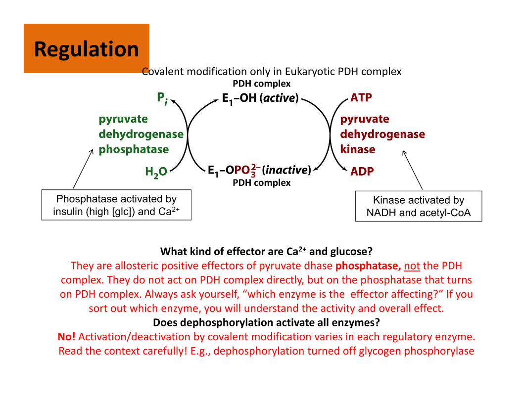
RegulationRegulation
Kinase activated by
NADH and acetyl-CoA
Phosphatase activated by
insulin (high [glc]) and Ca2+
What kind of effector are Ca2+ and glucose?
They are allosteric positive effectors of pyruvate dhase phosphatase, not the PDH
complex. They do not act on PDH complex directly, but on the phosphatase that turns
on PDH complex. Always ask yourself, “which enzyme is the effector affecting?” If you
sort out which enzyme, you will understand the activity and overall effect.
Does dephosphorylation activate all enzymes?
No! Activation/deactivation by covalent modification varies in each regulatory enzyme.
Read the context carefully! E.g., dephosphorylation turned off glycogen phosphorylase
PDH complex
PDH complex
Covalent modification only in Eukaryotic PDH complex
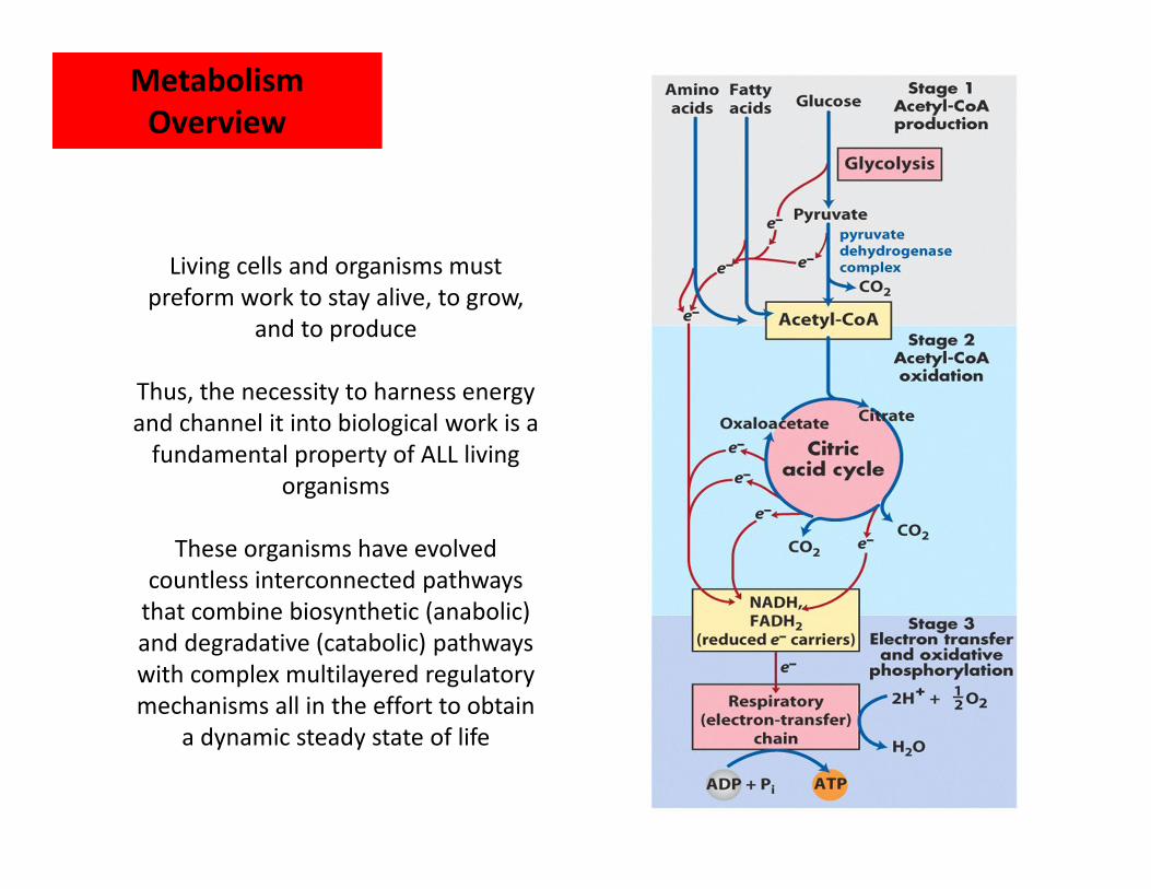
Living cells and organisms must
preform work to stay alive, to grow,
and to produce
Thus, the necessity to harness energy
and channel it into biological work is a
fundamental property of ALL living
organisms
These organisms have evolved
countless interconnected pathways
that combine biosynthetic (anabolic)
and degradative (catabolic) pathways
with complex multilayered regulatory
mechanisms all in the effort to obtain
a dynamic steady state of life
Metabolism
Overview
Metabolism
Overview
Metabolism
Overview
Metabolism
Overview
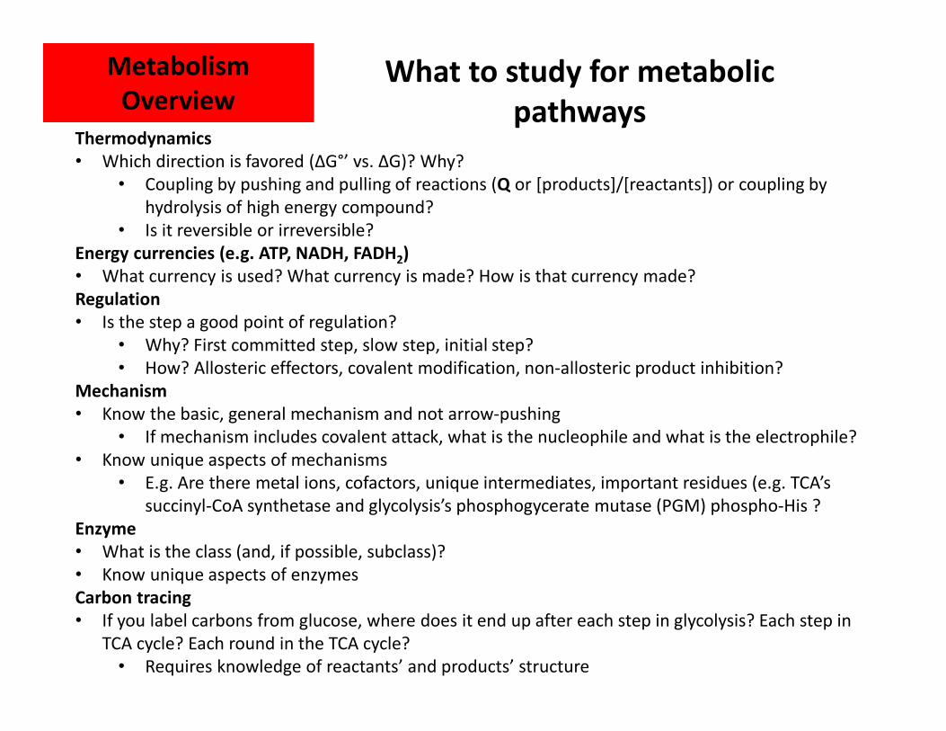
Thermodynamics
• Which direction is favored (ΔG°’ vs. ΔG)? Why?
• Coupling by pushing and pulling of reactions (Q or [products]/[reactants]) or coupling by
hydrolysis of high energy compound?
• Is it reversible or irreversible?
Energy currencies (e.g. ATP, NADH, FADH2)
• What currency is used? What currency is made? How is that currency made?
Regulation
• Is the step a good point of regulation?
• Why? First committed step, slow step, initial step?
• How? Allosteric effectors, covalent modification, non-allosteric product inhibition?
Mechanism
• Know the basic, general mechanism and not arrow-pushing
• If mechanism includes covalent attack, what is the nucleophile and what is the electrophile?
• Know unique aspects of mechanisms
• E.g. Are there metal ions, cofactors, unique intermediates, important residues (e.g. TCA’s
succinyl-CoA synthetase and glycolysis’s phosphogycerate mutase (PGM) phospho-His ?
Enzyme
• What is the class (and, if possible, subclass)?
• Know unique aspects of enzymes
Carbon tracing
• If you label carbons from glucose, where does it end up after each step in glycolysis? Each step in
TCA cycle? Each round in the TCA cycle?
• Requires knowledge of reactants’ and products’ structure
What to study for metabolic
pathways
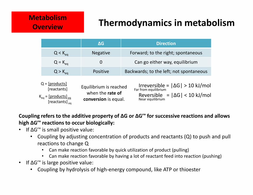
Thermodynamics in metabolism
ΔG Direction
Q < Keq Negative Forward; to the right; spontaneous
Q = Keq 0 Can go either way, equilibrium
Q > Keq Positive Backwards; to the left; not spontaneous
Q = [products]
[reactants]
Keq = [products] eq
[reactants] eq
Irreversible = |ΔG| > 10 kJ/mol
Reversible = |ΔG| < 10 kJ/mol
Equilibrium is reached
when the rate of
conversion is equal. Near equilibrium
Far from equilibrium
Coupling refers to the additive property of ΔG or ΔG’° for successive reactions and allows
high ΔG’° reactions to occur biologically:
• If ΔG’° is small positive value:
• Coupling by adjusting concentration of products and reactants (Q) to push and pull
reactions to change Q • Can make reaction favorable by quick utilization of product (pulling)
• Can make reaction favorable by having a lot of reactant feed into reaction (pushing)
• If ΔG’° is large positive value:
• Coupling by hydrolysis of high-energy compound, like ATP or thioester
Metabolism
Overview
Metabolism
Overview
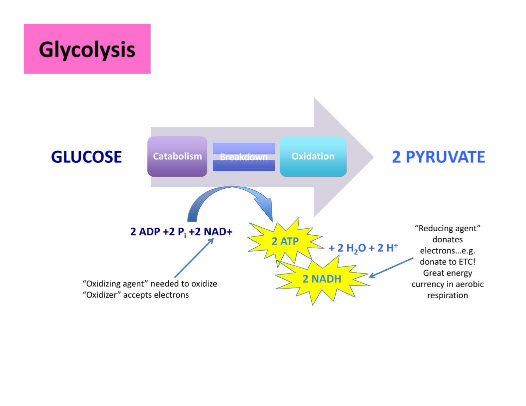
GlycolysisGlycolysis
CatabolismCatabolism BreakdownBreakdown OxidationOxidationGLUCOSE 2 PYRUVATE
2 ADP +2 Pi +2 NAD+2 ATP
2 NADH
+ 2 H2O + 2 H+
“Oxidizing agent” needed to oxidize
“Oxidizer” accepts electrons
“Reducing agent”
donates
electrons…e.g.
donate to ETC!
Great energy
currency in aerobic
respiration

isomerization
GlycolysisGlycolysis
PREPARATORY PHASEPAYOFF PHASE
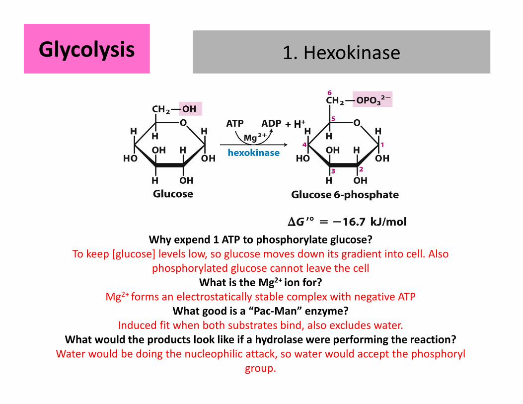
1. Hexokinase1. HexokinaseGlycolysisGlycolysis
+ H+
Why expend 1 ATP to phosphorylate glucose?
To keep [glucose] levels low, so glucose moves down its gradient into cell. Also
phosphorylated glucose cannot leave the cell
What is the Mg2+ ion for?
Mg2+ forms an electrostatically stable complex with negative ATP
What good is a “Pac-Man” enzyme?
Induced fit when both substrates bind, also excludes water.
What would the products look like if a hydrolase were performing the reaction?
Water would be doing the nucleophilic attack, so water would accept the phosphoryl
group.
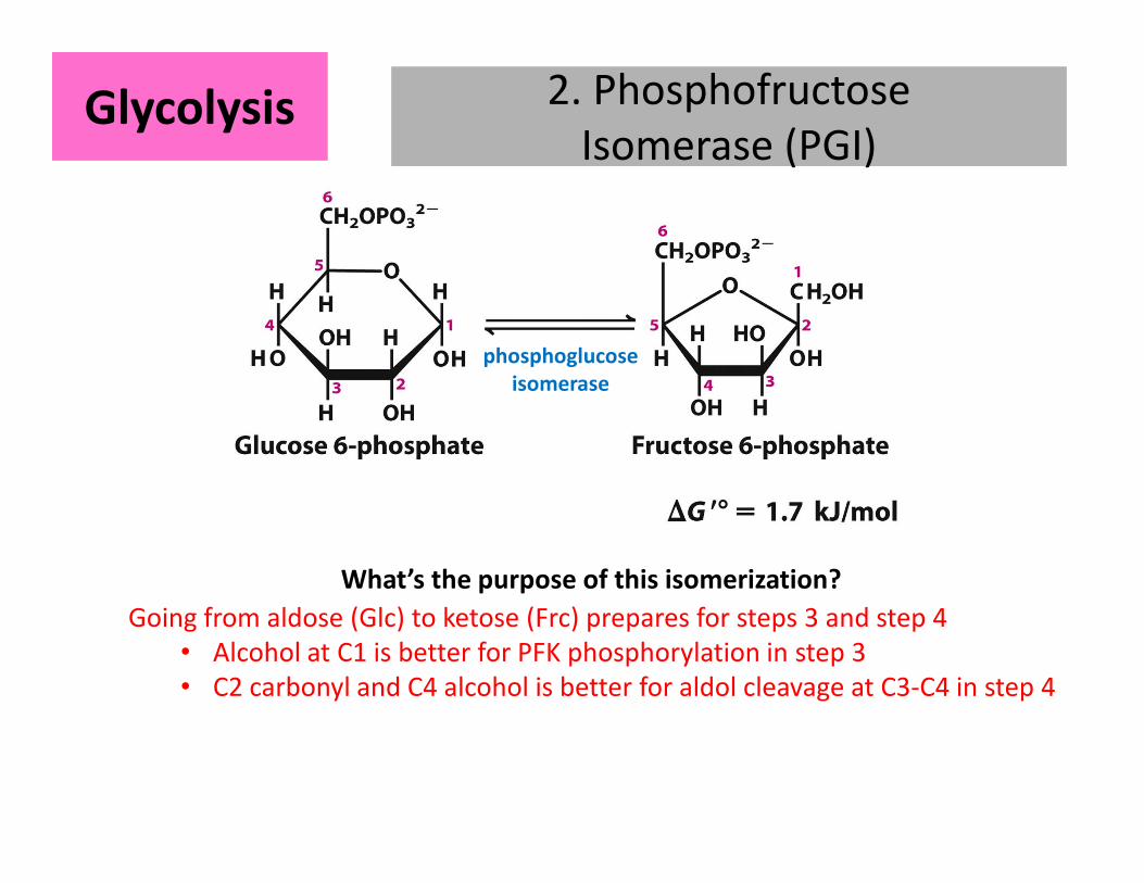
2. Phosphofructose
Isomerase (PGI)
2. Phosphofructose
Isomerase (PGI)
phosphoglucose
isomerase
GlycolysisGlycolysis
Going from aldose (Glc) to ketose (Frc) prepares for steps 3 and step 4
• Alcohol at C1 is better for PFK phosphorylation in step 3
• C2 carbonyl and C4 alcohol is better for aldol cleavage at C3-C4 in step 4
What’s the purpose of this isomerization?
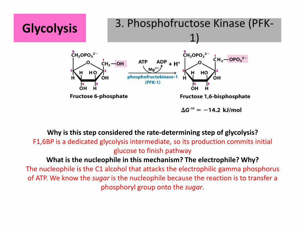
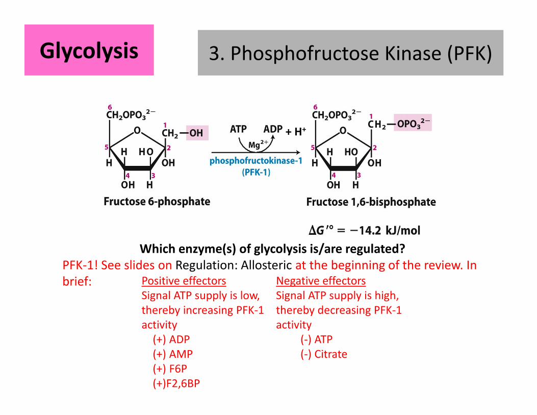
3. Phosphofructose Kinase (PFK-
1)
3. Phosphofructose Kinase (PFK-
1)
+ H+
GlycolysisGlycolysis
Why is this step considered the rate-determining step of glycolysis?
F1,6BP is a dedicated glycolysis intermediate, so its production commits initial
glucose to finish pathway
What is the nucleophile in this mechanism? The electrophile? Why?
The nucleophile is the C1 alcohol that attacks the electrophilic gamma phosphorus
of ATP. We know the sugar is the nucleophile because the reaction is to transfer a
phosphoryl group onto the sugar.
3. Phosphofructose Kinase (PFK)3. Phosphofructose Kinase (PFK)
+ H+
GlycolysisGlycolysis
Which enzyme(s) of glycolysis is/are regulated?
PFK-1! See slides on Regulation: Allosteric at the beginning of the review. In
brief: Positive effectors
Signal ATP supply is low,
thereby increasing PFK-1
activity
(+) ADP
(+) AMP
(+) F6P
(+)F2,6BP
Negative effectors
Signal ATP supply is high,
thereby decreasing PFK-1
activity
(-) ATP
(-) Citrate
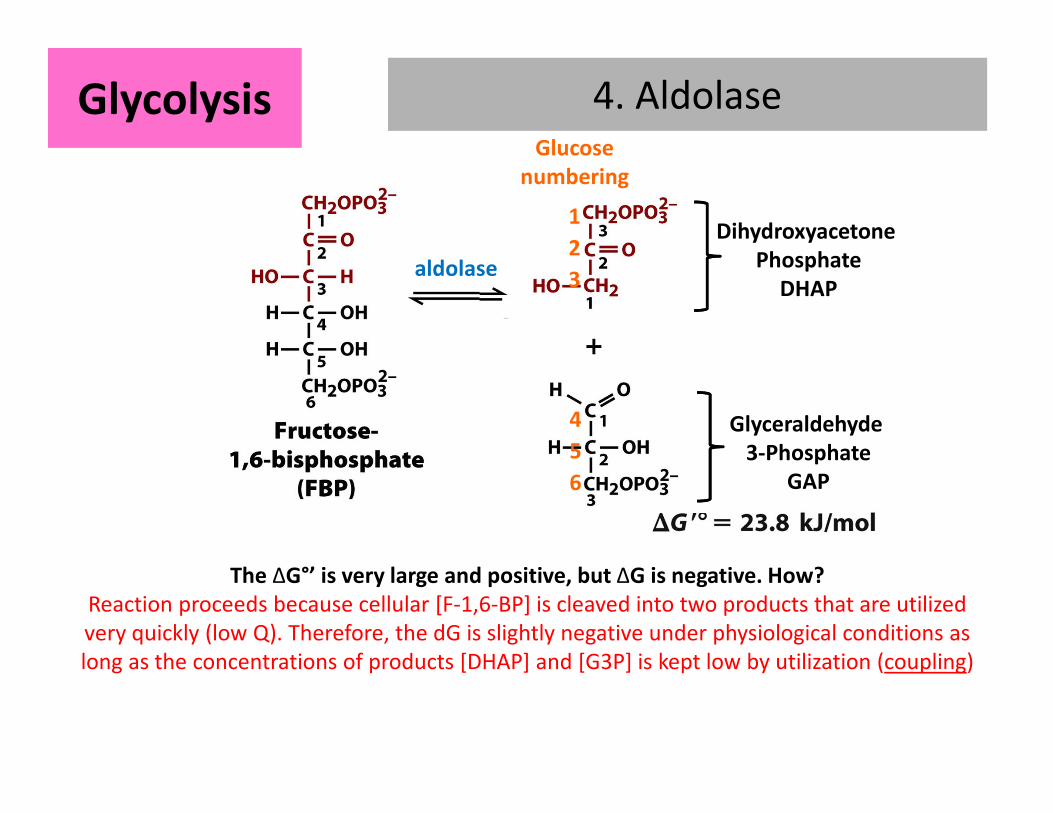
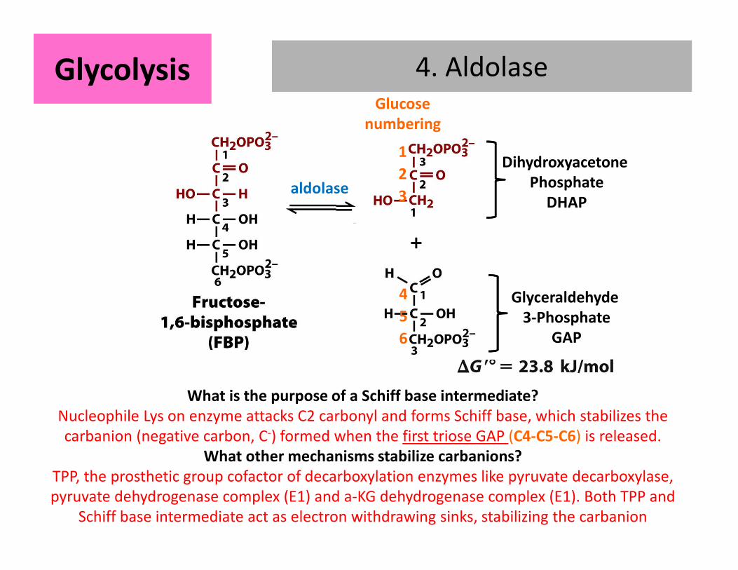
4. AldolaseGlycolysisGlycolysisGlucose
numberinga
1
2
3
4
5
6
Dihydroxyacetone
Phosphate
DHAP
Glyceraldehyde
3-Phosphate
GAP
aldolase
The ΔG°’ is very large and positive, but ΔG is negative. How?
Reaction proceeds because cellular [F-1,6-BP] is cleaved into two products that are utilized
very quickly (low Q). Therefore, the dG is slightly negative under physiological conditions as
long as the concentrations of products [DHAP] and [G3P] is kept low by utilization (coupling)
4. Aldolase4. AldolaseGlycolysisGlycolysisGlucose
numberinga
1
2
3
4
5
6
Dihydroxyacetone
Phosphate
DHAP
Glyceraldehyde
3-Phosphate
GAP
aldolase
What is the purpose of a Schiff base intermediate?
Nucleophile Lys on enzyme attacks C2 carbonyl and forms Schiff base, which stabilizes the
carbanion (negative carbon, C-) formed when the first triose GAP (C4-C5-C6) is released.
What other mechanisms stabilize carbanions?
TPP, the prosthetic group cofactor of decarboxylation enzymes like pyruvate decarboxylase,
pyruvate dehydrogenase complex (E1) and a-KG dehydrogenase complex (E1). Both TPP and
Schiff base intermediate act as electron withdrawing sinks, stabilizing the carbanion
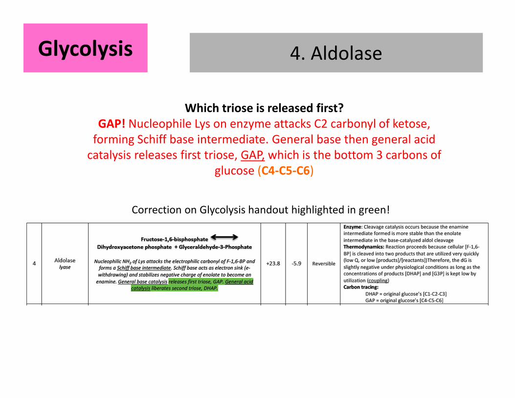
4. Aldolase4. AldolaseGlycolysisGlycolysis
Correction on Glycolysis handout highlighted in green!
Which triose is released first?
GAP! Nucleophile Lys on enzyme attacks C2 carbonyl of ketose,
forming Schiff base intermediate. General base then general acid
catalysis releases first triose, GAP, which is the bottom 3 carbons of
glucose (C4-C5-C6)
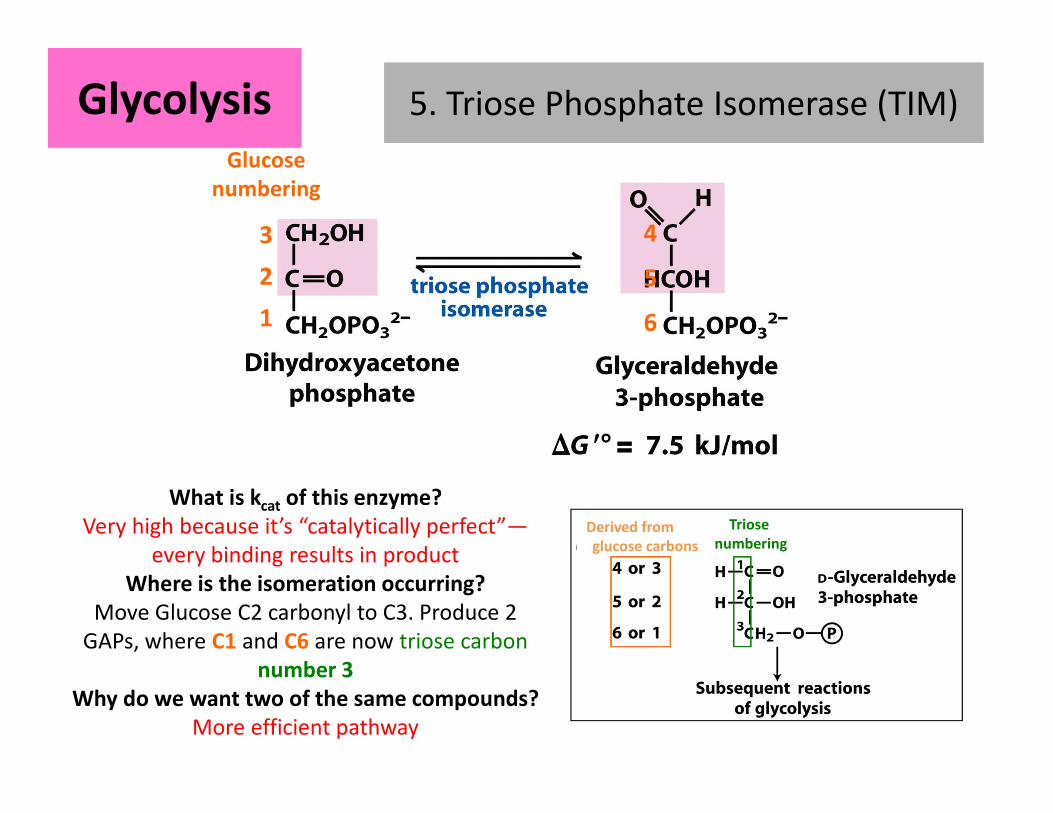
5. Triose Phosphate Isomerase (TIM)5. Triose Phosphate Isomerase (TIM)GlycolysisGlycolysis
What is kcat of this enzyme?
Very high because it’s “catalytically perfect”—
every binding results in product
Where is the isomeration occurring?
Move Glucose C2 carbonyl to C3. Produce 2
GAPs, where C1 and C6 are now triose carbon
number 3
Why do we want two of the same compounds?
More efficient pathway
Glucose
numberingA
3
2
1
4
5
6
Derived from
glucose carbons
Triose
numbering
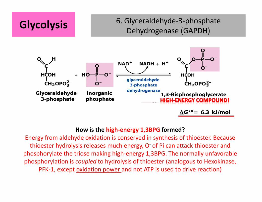
6. Glyceraldehyde-3-phosphate
Dehydrogenase (GAPDH) GlycolysisGlycolysis
How is the high-energy 1,3BPG formed?
Energy from aldehyde oxidation is conserved in synthesis of thioester. Because
thioester hydrolysis releases much energy, O- of Pi can attack thioester and
phosphorylate the triose making high-energy 1,3BPG. The normally unfavorable
phosphorylation is coupled to hydrolysis of thioester (analogous to Hexokinase,
PFK-1, except oxidation power and not ATP is used to drive reaction)
1 ATP
per GAP
1 ATP
made per GAP
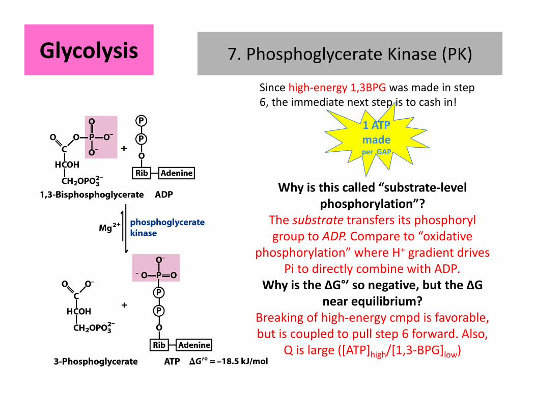
7. Phosphoglycerate Kinase (PK)GlycolysisGlycolysis
Since high-energy 1,3BPG was made in step
6, the immediate next step is to cash in!
Why is this called “substrate-level
phosphorylation”?
The substrate transfers its phosphoryl
group to ADP. Compare to “oxidative
phosphorylation” where H+ gradient drives
Pi to directly combine with ADP.
Why is the ΔG°’ so negative, but the ΔG
near equilibrium?
Breaking of high-energy cmpd is favorable,
but is coupled to pull step 6 forward. Also,
Q is large ([ATP]high/[1,3-BPG]low)
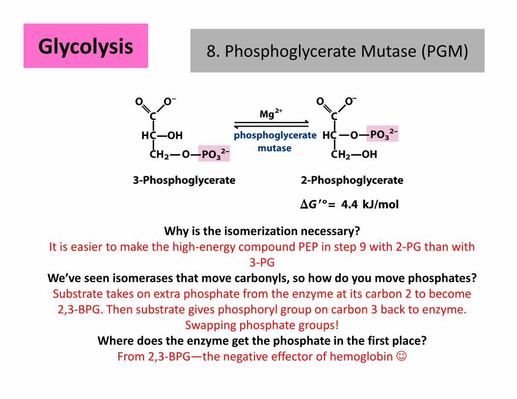
8. Phosphoglycerate Mutase (PGM)GlycolysisGlycolysis
Why is the isomerization necessary?
It is easier to make the high-energy compound PEP in step 9 with 2-PG than with
3-PG
We’ve seen isomerases that move carbonyls, so how do you move phosphates?
Substrate takes on extra phosphate from the enzyme at its carbon 2 to become
2,3-BPG. Then substrate gives phosphoryl group on carbon 3 back to enzyme.
Swapping phosphate groups!
Where does the enzyme get the phosphate in the first place?
From 2,3-BPG—the negative effector of hemoglobin ☺
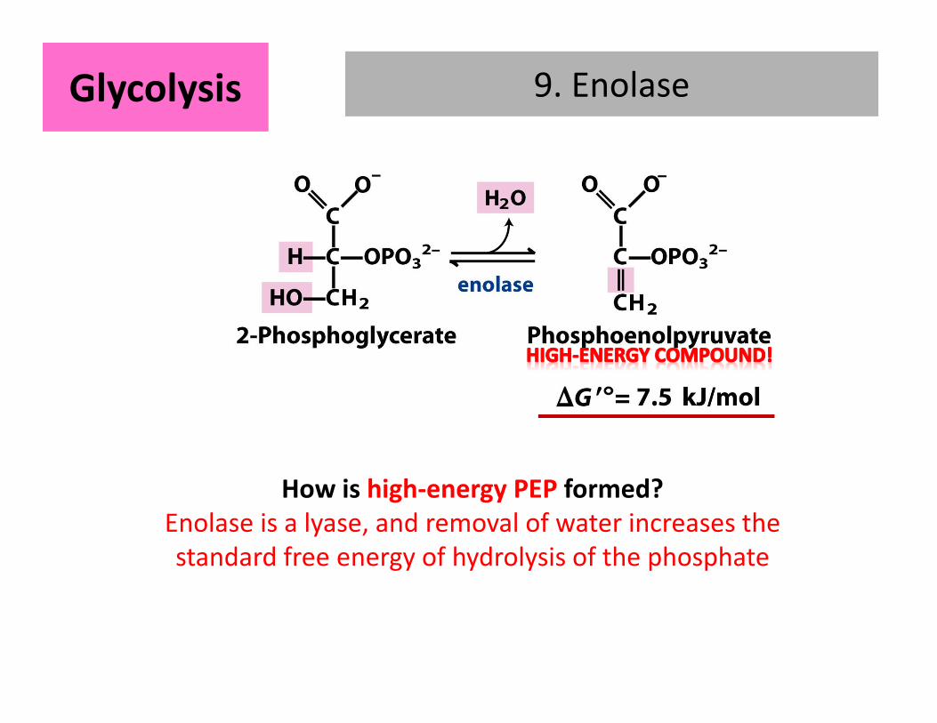
9. EnolaseGlycolysisGlycolysis
How is high-energy PEP formed?
Enolase is a lyase, and removal of water increases the
standard free energy of hydrolysis of the phosphate
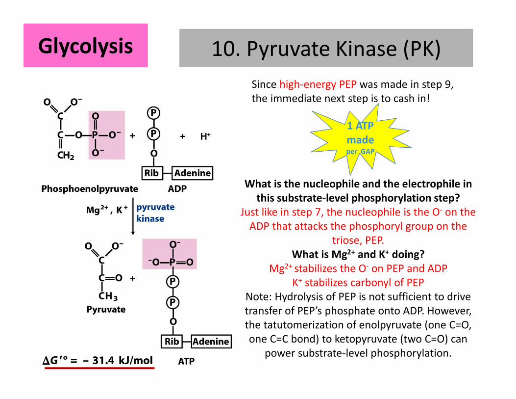
10. Pyruvate Kinase (PK)
+ H+ H+ H+ H++++
GlycolysisGlycolysis
1 ATP
per GAP
1 ATP
made per GAP
Since high-energy PEP was made in step 9,
the immediate next step is to cash in!
What is the nucleophile and the electrophile in
this substrate-level phosphorylation step?
Just like in step 7, the nucleophile is the O- on the
ADP that attacks the phosphoryl group on the
triose, PEP.
What is Mg2+ and K+ doing?
Mg2+ stabilizes the O- on PEP and ADP
K+ stabilizes carbonyl of PEP
Note: Hydrolysis of PEP is not sufficient to drive
transfer of PEP’s phosphate onto ADP. However,
the tatutomerization of enolpyruvate (one C=O,
one C=C bond) to ketopyruvate (two C=O) can
power substrate-level phosphorylation.

Immediate fates of pyruvate
1. Gluconeogenesis and
Anapleuorotic reaction: Pyruvate
carboxylase
– first step in remaking glucose via
gluconeogenesis
– Anapleurotic reaction to replenish OAA
for TCA
2. Homolactic Fermentation (e.g. in
mammals): Lactate dehydrogenase
– Reduce and Regenerate NAD+
3. Alcohol Fermentation (e.g. in yeast):
Pyruvate decarboxylate
– Decarboxylate and then reduce into
ethanol, regenerating NAD+
1
2 34
4. Preparation for TCA Cycle: Pyruvate
dehydrogenase complexTo make into acetyl-CoA to feed into TCA cycle
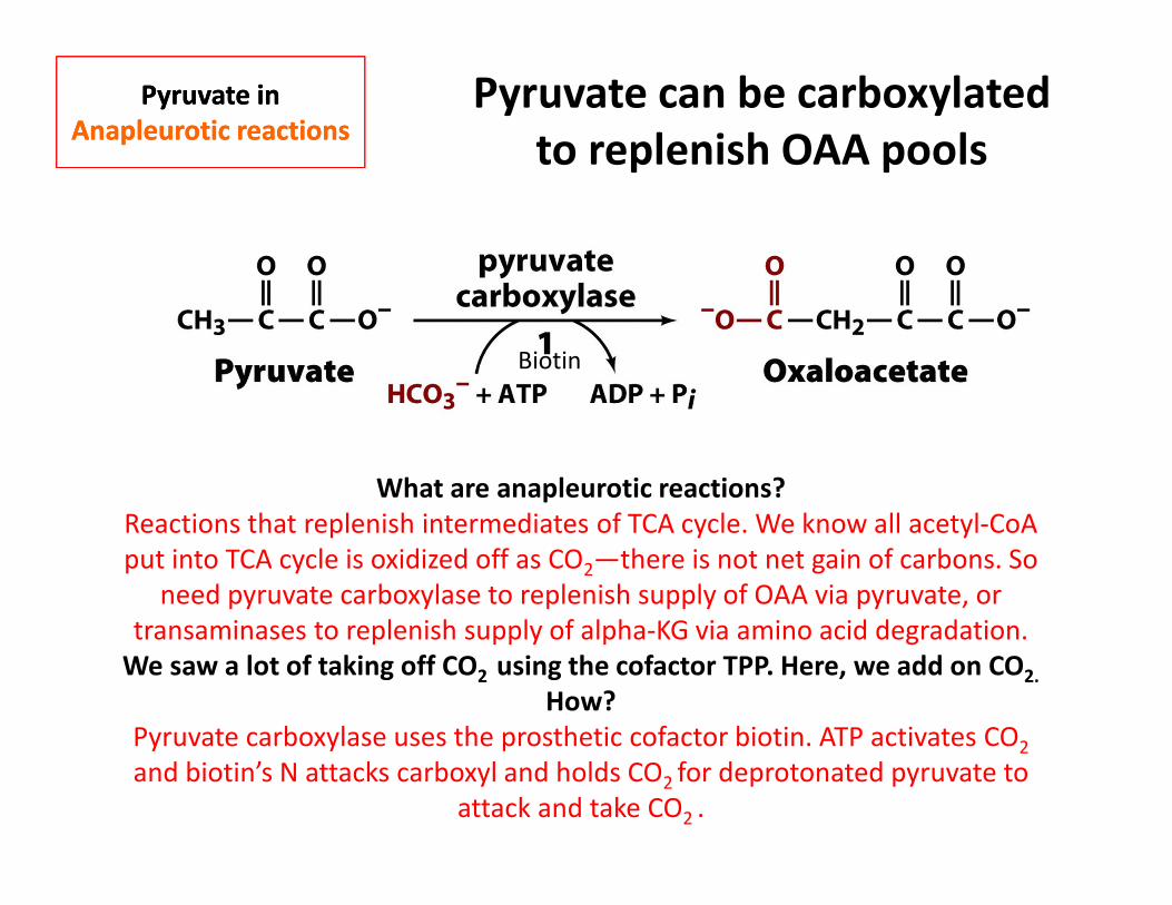
Pyruvate in
Anapleurotic reactions
Pyruvate in
Anapleurotic reactionsPyruvate can be carboxylated
to replenish OAA pools
What are anapleurotic reactions?
Reactions that replenish intermediates of TCA cycle. We know all acetyl-CoA
put into TCA cycle is oxidized off as CO2—there is not net gain of carbons. So
need pyruvate carboxylase to replenish supply of OAA via pyruvate, or
transaminases to replenish supply of alpha-KG via amino acid degradation.
We saw a lot of taking off CO2 using the cofactor TPP. Here, we add on CO2.
How?
Pyruvate carboxylase uses the prosthetic cofactor biotin. ATP activates CO2
and biotin’s N attacks carboxyl and holds CO2 for deprotonated pyruvate to
attack and take CO2 .
Biotin

Pyruvate in
Gluconeogenesis
Pyruvate in
GluconeogenesisPyruvate can start the
gluconeogenesis to make glucose
Why are we carboxylating Pyr and then
decarboxylating OAA?The end goal is to make PEP, a high-energy compound. We
carboxylate to prime Pyr for PEP carboxylkinase (PEPCK) and
also to make more OAA (see anapleurotic rxn). The
decarboxylation catalyzed by PEP carboxylkinase does release
energy, but we still need two energy currencies to make PEP
(1 ATP, 1 GTP)—these first two reactions are that expensive!
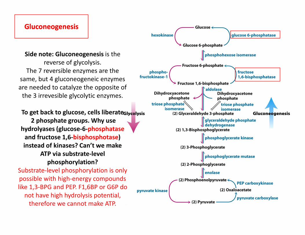
GluconeogenesisGluconeogenesis
Side note: Gluconeogenesis is the
reverse of glycolysis.
The 7 reversible enzymes are the
same, but 4 gluconeogeneic enzymes
are needed to catalyze the opposite of
the 3 irrevesible glycolytic enzymes.
To get back to glucose, cells liberate
2 phosphate groups. Why use
hydrolyases (glucose-6-phosphatase
and fructose 1,6-bisphosphotase)
instead of kinases? Can’t we make
ATP via substrate-level
phosphorylation?
Substrate-level phosphorylation is only
possible with high-energy compounds
like 1,3-BPG and PEP. F1,6BP or G6P do
not have high hydrolysis potential,
therefore we cannot make ATP.
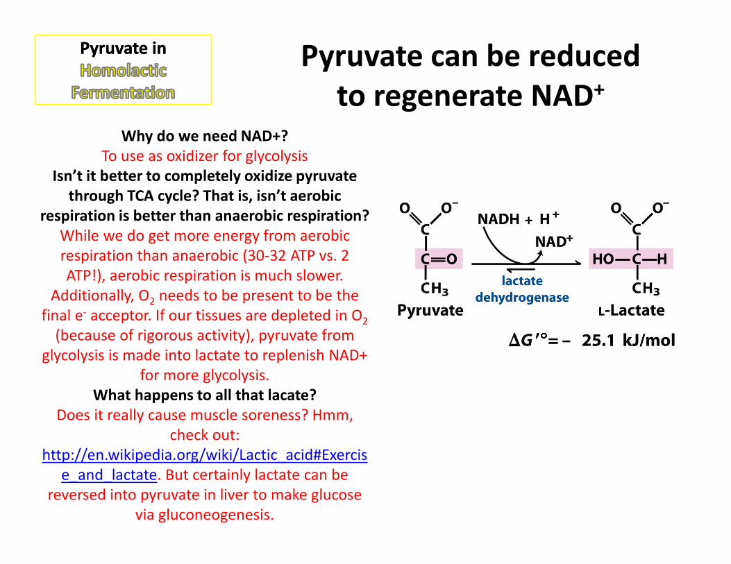
Pyruvate in Pyruvate in Pyruvate can be reduced
to regenerate NAD+
Why do we need NAD+?
To use as oxidizer for glycolysis
Isn’t it better to completely oxidize pyruvate
through TCA cycle? That is, isn’t aerobic
respiration is better than anaerobic respiration?
While we do get more energy from aerobic
respiration than anaerobic (30-32 ATP vs. 2
ATP!), aerobic respiration is much slower.
Additionally, O2 needs to be present to be the
final e- acceptor. If our tissues are depleted in O2
(because of rigorous activity), pyruvate from
glycolysis is made into lactate to replenish NAD+
for more glycolysis.
What happens to all that lacate?
Does it really cause muscle soreness? Hmm,
check out:
http://en.wikipedia.org/wiki/Lactic_acid#Exercis
e_and_lactate. But certainly lactate can be
reversed into pyruvate in liver to make glucose
via gluconeogenesis.
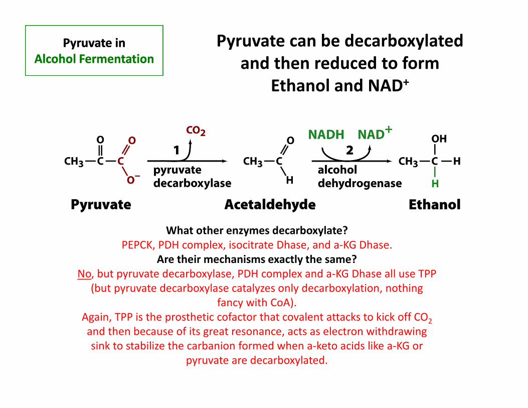
Pyruvate in
Alcohol Fermentation
Pyruvate in
Alcohol Fermentation
Pyruvate can be decarboxylated
and then reduced to form
Ethanol and NAD+
What other enzymes decarboxylate?
PEPCK, PDH complex, isocitrate Dhase, and a-KG Dhase.
Are their mechanisms exactly the same?
No, but pyruvate decarboxylase, PDH complex and a-KG Dhase all use TPP
(but pyruvate decarboxylase catalyzes only decarboxylation, nothing
fancy with CoA).
Again, TPP is the prosthetic cofactor that covalent attacks to kick off CO2
and then because of its great resonance, acts as electron withdrawing
sink to stabilize the carbanion formed when a-keto acids like a-KG or
pyruvate are decarboxylated.
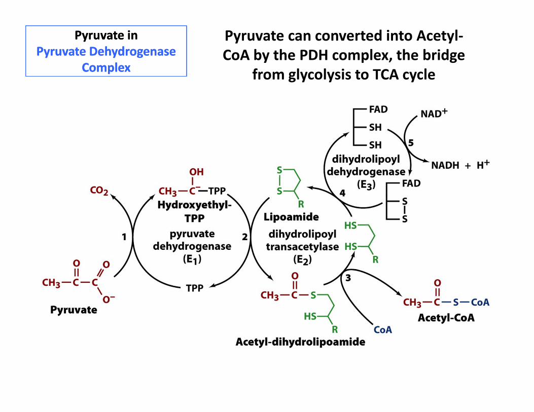
Pyruvate in
Pyruvate Dehydrogenase
Complex
Pyruvate in
Pyruvate Dehydrogenase
Complex
Pyruvate can converted into Acetyl-
CoA by the PDH complex, the bridge
from glycolysis to TCA cycle
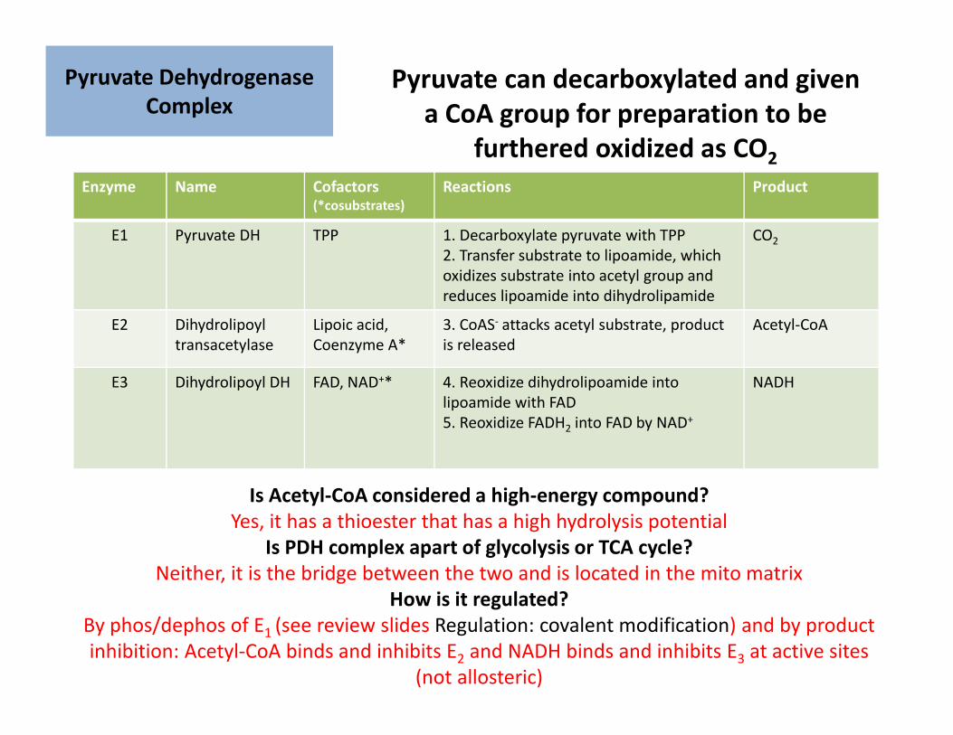
Enzyme Name Cofactors(*cosubstrates)
Reactions Product
E1 Pyruvate DH TPP 1. Decarboxylate pyruvate with TPP
2. Transfer substrate to lipoamide, which
oxidizes substrate into acetyl group and
reduces lipoamide into dihydrolipamide
CO2
E2 Dihydrolipoyl
transacetylase
Lipoic acid,
Coenzyme A*
3. CoAS- attacks acetyl substrate, product
is released
Acetyl-CoA
E3 Dihydrolipoyl DH FAD, NAD+* 4. Reoxidize dihydrolipoamide into
lipoamide with FAD
5. Reoxidize FADH2 into FAD by NAD+
NADH
Pyruvate Dehydrogenase
Complex
Pyruvate Dehydrogenase
Complex
Is Acetyl-CoA considered a high-energy compound?
Yes, it has a thioester that has a high hydrolysis potential
Is PDH complex apart of glycolysis or TCA cycle?
Neither, it is the bridge between the two and is located in the mito matrix
How is it regulated?
By phos/dephos of E1 (see review slides Regulation: covalent modification) and by product
inhibition: Acetyl-CoA binds and inhibits E2 and NADH binds and inhibits E3 at active sites
(not allosteric)
Pyruvate can decarboxylated and given
a CoA group for preparation to be
furthered oxidized as CO2
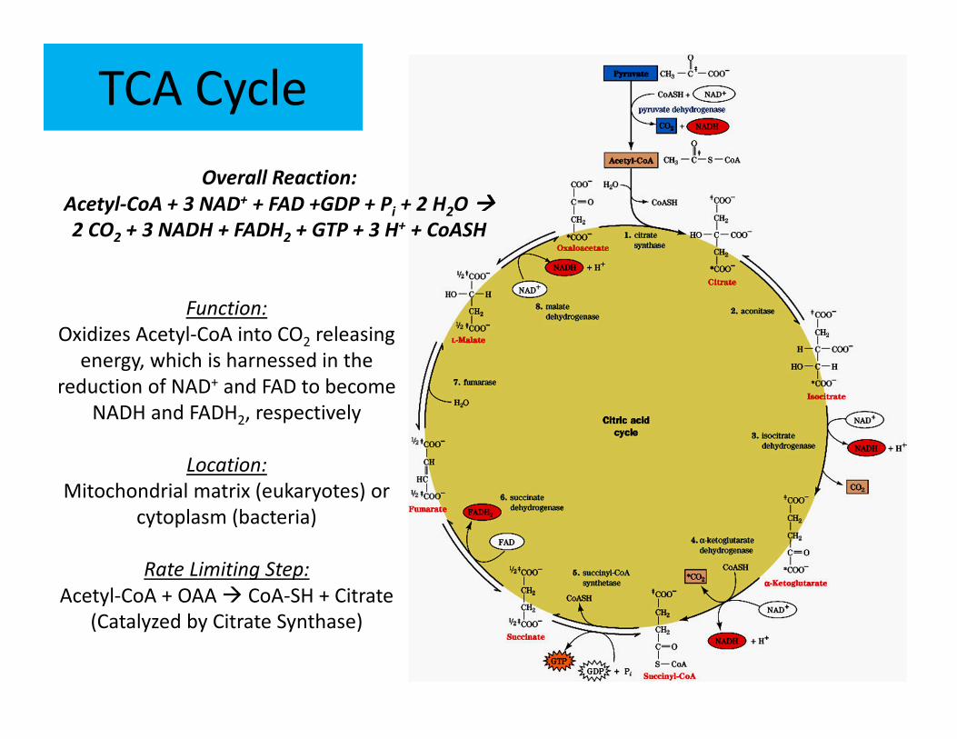
TCA Cycle
Overall Reaction:
Acetyl-CoA + 3 NAD+ + FAD +GDP + Pi + 2 H2O ����
2 CO2 + 3 NADH + FADH2 + GTP + 3 H+ + CoASH
Function:
Oxidizes Acetyl-CoA into CO2 releasing
energy, which is harnessed in the
reduction of NAD+ and FAD to become
NADH and FADH2, respectively
Location:
Mitochondrial matrix (eukaryotes) or
cytoplasm (bacteria)
Rate Limiting Step:
Acetyl-CoA + OAA � CoA-SH + Citrate
(Catalyzed by Citrate Synthase)
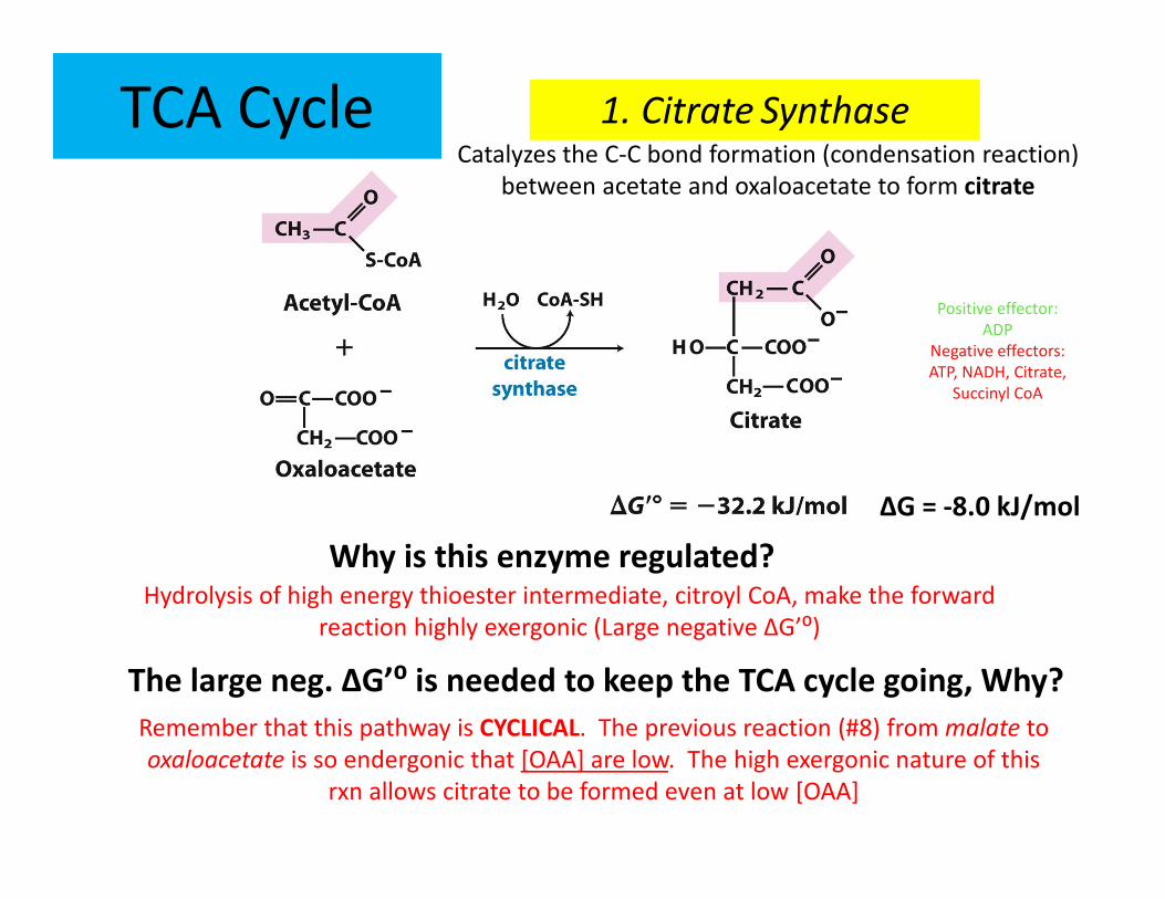
TCA Cycle 1. Citrate SynthaseCatalyzes the C-C bond formation (condensation reaction)
between acetate and oxaloacetate to form citrate
Why is this enzyme regulated?
The large neg. ΔG’⁰ is needed to keep the TCA cycle going, Why?
Remember that this pathway is CYCLICAL. The previous reaction (#8) from malate to
oxaloacetate is so endergonic that [OAA] are low. The high exergonic nature of this
rxn allows citrate to be formed even at low [OAA]
ΔG = -8.0 kJ/mol
Hydrolysis of high energy thioester intermediate, citroyl CoA, make the forward
reaction highly exergonic (Large negative ΔG’⁰)
Positive effector:
ADP
Negative effectors:
ATP, NADH, Citrate,
Succinyl CoA
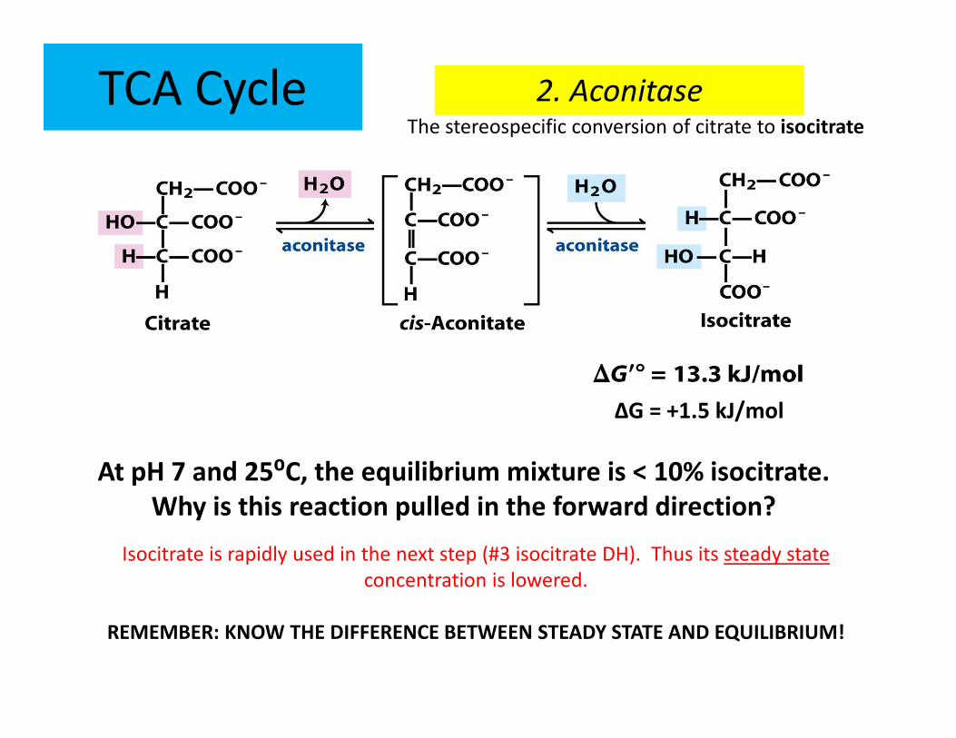
TCA Cycle
At pH 7 and 25⁰C, the equilibrium mixture is < 10% isocitrate.
Why is this reaction pulled in the forward direction?
2. AconitaseThe stereospecific conversion of citrate to isocitrate
ΔG = +1.5 kJ/mol
Isocitrate is rapidly used in the next step (#3 isocitrate DH). Thus its steady state
concentration is lowered.
REMEMBER: KNOW THE DIFFERENCE BETWEEN STEADY STATE AND EQUILIBRIUM!
ΔG'°= -21 kJ/mol
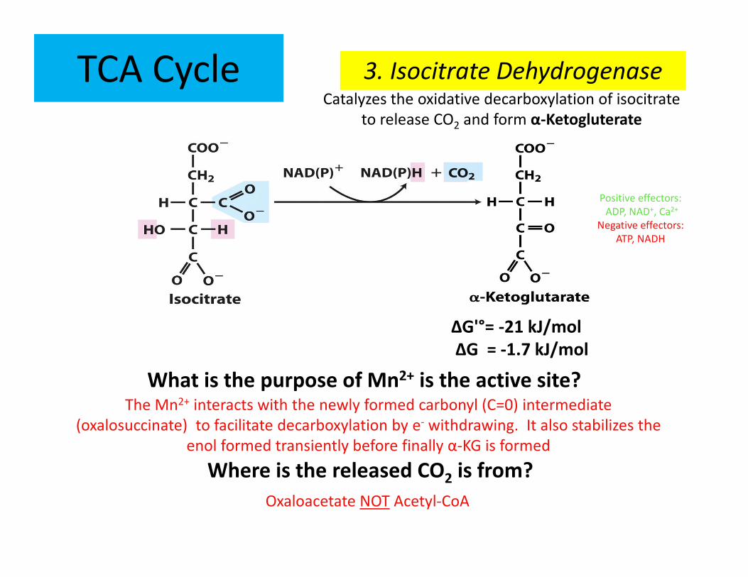
TCA Cycle
What is the purpose of Mn2+ is the active site?
3. Isocitrate DehydrogenaseCatalyzes the oxidative decarboxylation of isocitrate
to release CO2 and form α-Ketogluterate
ΔG = -1.7 kJ/mol
Positive effectors:
ADP, NAD+, Ca2+
Negative effectors:
ATP, NADH
The Mn2+ interacts with the newly formed carbonyl (C=0) intermediate
(oxalosuccinate) to facilitate decarboxylation by e- withdrawing. It also stabilizes the
enol formed transiently before finally α-KG is formed
Where is the released CO2 is from?
Oxaloacetate NOT Acetyl-CoA
Positive effector:
Ca2+
Negative effectors:
ATP, NADH, Succinyl-CoA
ΔG = -8.0 kJ/mol
TCA Cycle
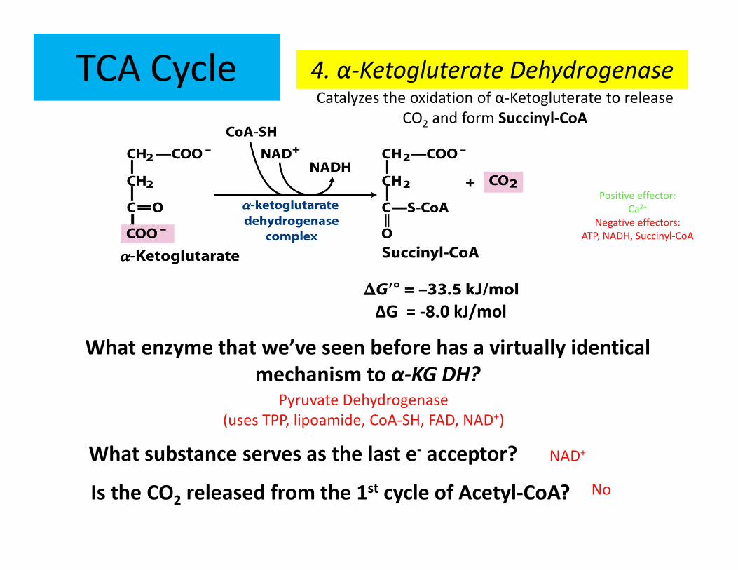
What enzyme that we’ve seen before has a virtually identical
mechanism to α-KG DH?
4. α-Ketogluterate DehydrogenaseCatalyzes the oxidation of α-Ketogluterate to release
CO2 and form Succinyl-CoA
Pyruvate Dehydrogenase
(uses TPP, lipoamide, CoA-SH, FAD, NAD+)
What substance serves as the last e- acceptor? NAD+
Is the CO2 released from the 1st cycle of Acetyl-CoA? No
TCA Cycle
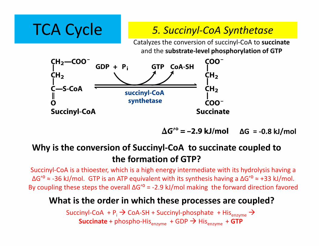
Why is the conversion of Succinyl-CoA to succinate coupled to
the formation of GTP?
5. Succinyl-CoA SynthetaseCatalyzes the conversion of succinyl-CoA to succinate
and the substrate-level phosphorylation of GTP
Succinyl-CoA is a thioester, which is a high energy intermediate with its hydrolysis having a
ΔG’⁰ ≈ -36 kJ/mol. GTP is an ATP equivalent with its synthesis having a ΔG’⁰ ≈ +33 kJ/mol.
By coupling these steps the overall ΔG’⁰ = -2.9 kJ/mol making the forward direction favored
What is the order in which these processes are coupled?
ΔG = -0.8 kJ/mol
Succinyl-CoA + Pi � CoA-SH + Succinyl-phosphate + Hisenzyme �
Succinate + phospho-Hisenzyme + GDP � Hisenzyme + GTP
(tightly bound in enzyme)
TCA Cycle
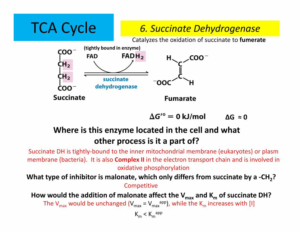
Where is this enzyme located in the cell and what
other process is it a part of?
6. Succinate DehydrogenaseCatalyzes the oxidation of succinate to fumerate
Succinate DH is tightly-bound to the inner mitochondrial membrane (eukaryotes) or plasm
membrane (bacteria). It is also Complex II in the electron transport chain and is involved in
oxidative phosphorylation
What type of inhibitor is malonate, which only differs from succinate by a -CH2?
ΔG ≈ 0
Competitive
How would the addition of malonate affect the Vmax and Km of succinate DH? The Vmax would be unchanged (Vmax = Vmax
app), while the Km increases with [I]
Km < Kmapp
TCA Cycle
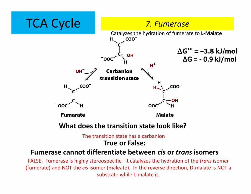
What does the transition state look like?
7. FumeraseCatalyzes the hydration of fumerate to L-Malate
True or False:
Fumerase cannot differentiate between cis or trans isomers
ΔG = - 0.9 kJ/mol
FALSE. Fumerase is highly stereospecific. It catalyzes the hydration of the trans isomer
(fumerate) and NOT the cis isomer (maleate). In the reverse direction, D-malate is NOT a
substrate while L-malate is.
+ H2O
- H2O
The transition state has a carbanion
TCA Cycle
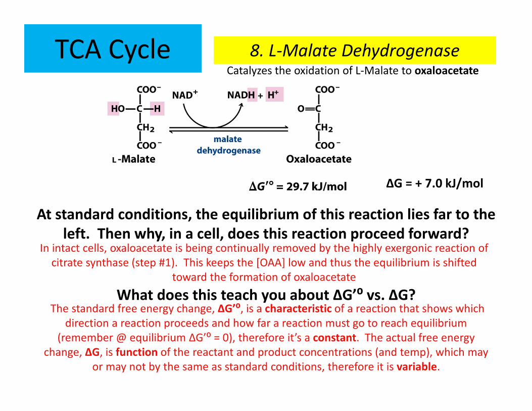
At standard conditions, the equilibrium of this reaction lies far to the
left. Then why, in a cell, does this reaction proceed forward?
8. L-Malate DehydrogenaseCatalyzes the oxidation of L-Malate to oxaloacetate
ΔG = + 7.0 kJ/mol
In intact cells, oxaloacetate is being continually removed by the highly exergonic reaction of
citrate synthase (step #1). This keeps the [OAA] low and thus the equilibrium is shifted
toward the formation of oxaloacetate
What does this teach you about ΔG’⁰ vs. ΔG?The standard free energy change, ΔG’⁰, is a characteristic of a reaction that shows which
direction a reaction proceeds and how far a reaction must go to reach equilibrium
(remember @ equilibrium ΔG’⁰ = 0), therefore it’s a constant. The actual free energy
change, ΔG, is function of the reactant and product concentrations (and temp), which may
or may not by the same as standard conditions, therefore it is variable.
TCA Cycle
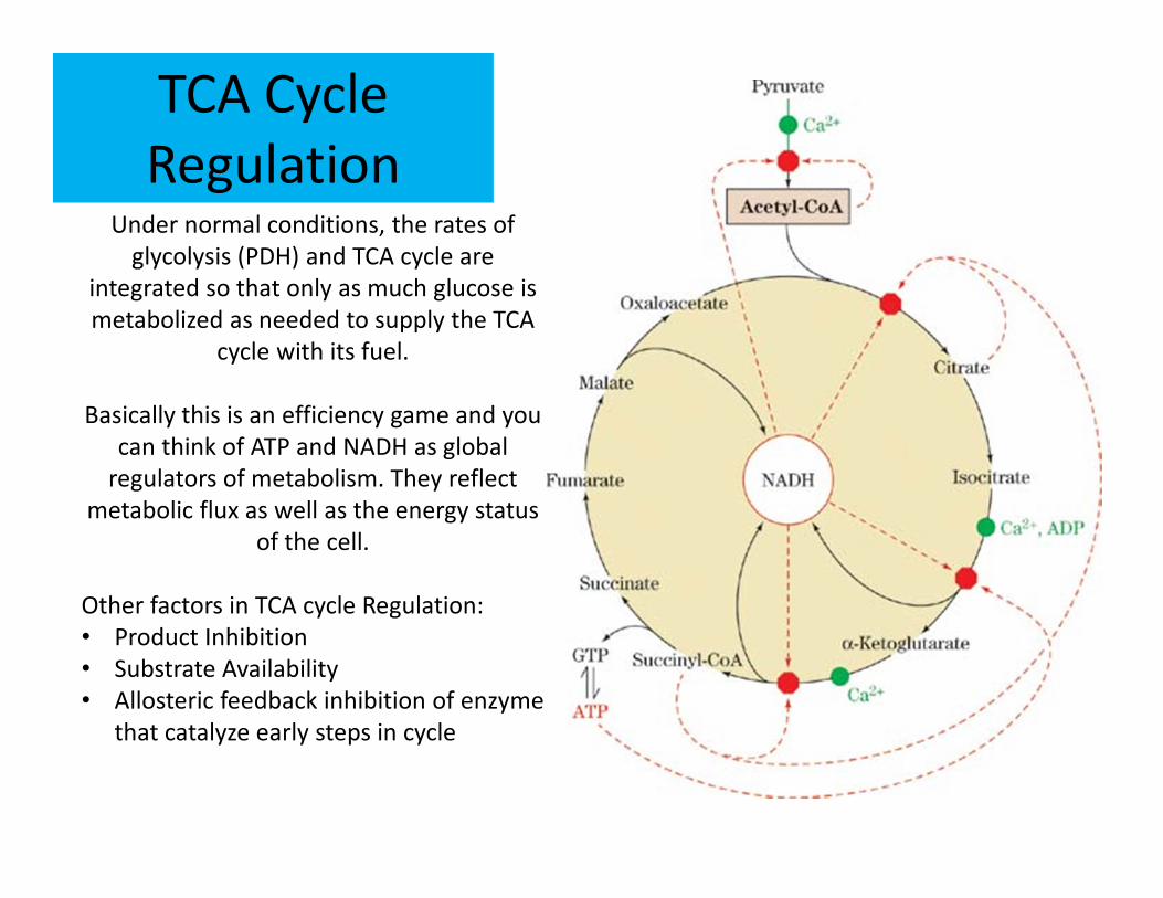
RegulationUnder normal conditions, the rates of
glycolysis (PDH) and TCA cycle are
integrated so that only as much glucose is
metabolized as needed to supply the TCA
cycle with its fuel.
Basically this is an efficiency game and you
can think of ATP and NADH as global
regulators of metabolism. They reflect
metabolic flux as well as the energy status
of the cell.
Other factors in TCA cycle Regulation:
• Product Inhibition
• Substrate Availability
• Allosteric feedback inhibition of enzyme
that catalyze early steps in cycle
TCA Cycle
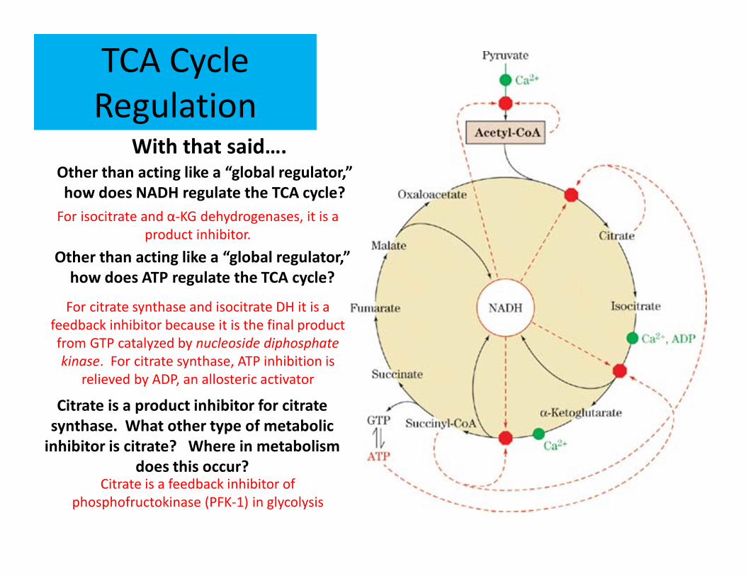
RegulationWith that said….
Other than acting like a “global regulator,”
how does NADH regulate the TCA cycle?
For isocitrate and α-KG dehydrogenases, it is a
product inhibitor.
Other than acting like a “global regulator,”
how does ATP regulate the TCA cycle?
For citrate synthase and isocitrate DH it is a
feedback inhibitor because it is the final product
from GTP catalyzed by nucleoside diphosphate
kinase. For citrate synthase, ATP inhibition is
relieved by ADP, an allosteric activator
Citrate is a product inhibitor for citrate
synthase. What other type of metabolic
inhibitor is citrate? Where in metabolism
does this occur?Citrate is a feedback inhibitor of
phosphofructokinase (PFK-1) in glycolysis
RECAP
So far glycolysis and the TCA cycle have achieved…
• Carbons of glucose has been completely oxidized to CO2
• Substrate-level phosphorylation has conserved some of the energy released from oxidation
• However, most of the energy is conserved (temporarily) in the reducing power of NADH and FADH2
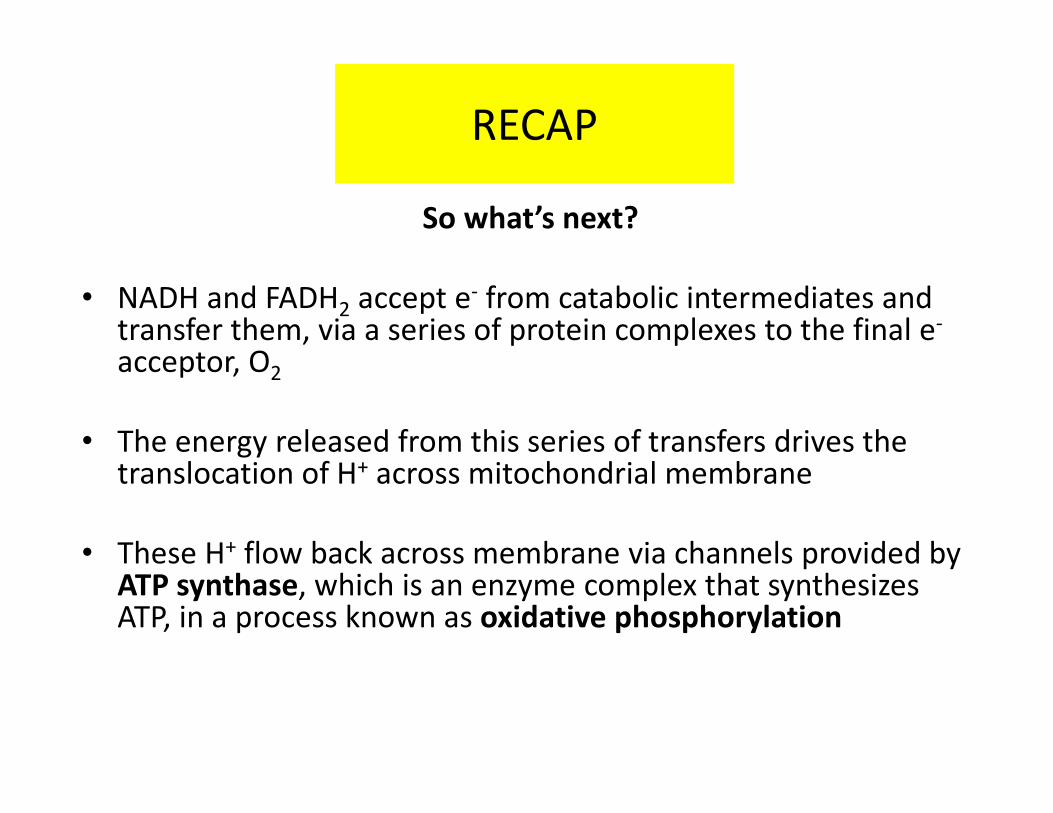
RECAP
So what’s next?
• NADH and FADH2 accept e- from catabolic intermediates and transfer them, via a series of protein complexes to the final e-
acceptor, O2
• The energy released from this series of transfers drives the translocation of H+ across mitochondrial membrane
• These H+ flow back across membrane via channels provided by ATP synthase, which is an enzyme complex that synthesizes ATP, in a process known as oxidative phosphorylation
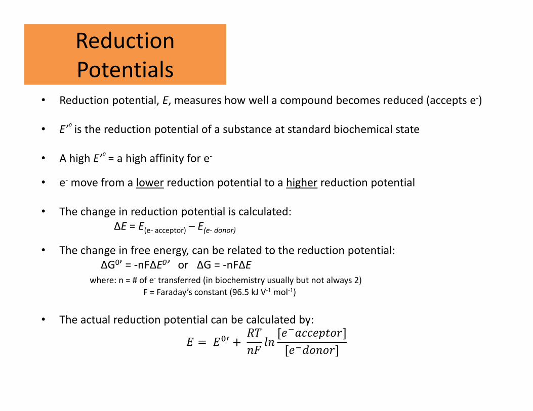
Reduction
Potentials• Reduction potential, E, measures how well a compound becomes reduced (accepts e-)
• E’⁰ is the reduction potential of a substance at standard biochemical state
• A high E’⁰ = a high affinity for e-
• e- move from a lower reduction potential to a higher reduction potential
• The change in reduction potential is calculated:
ΔE = E(e- acceptor) – E(e- donor)
• The change in free energy, can be related to the reduction potential:
ΔG0’ = -nFΔE0’ or ΔG = -nFΔE
where: n = # of e- transferred (in biochemistry usually but not always 2)
F = Faraday’s constant (96.5 kJ V-1 mol-1)
• The actual reduction potential can be calculated by:
� = ��� +��
�[ ���� ����]
[ �����]
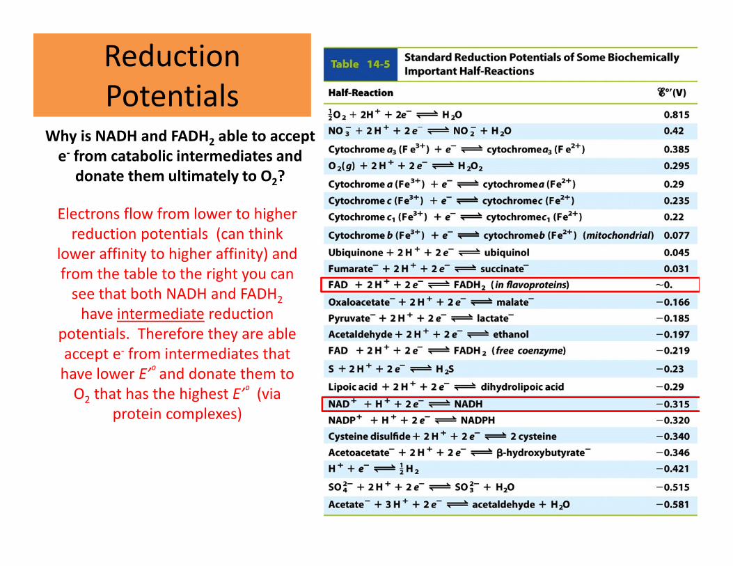
Reduction
PotentialsWhy is NADH and FADH2 able to accept
e- from catabolic intermediates and
donate them ultimately to O2?
Electrons flow from lower to higher
reduction potentials (can think
lower affinity to higher affinity) and
from the table to the right you can
see that both NADH and FADH2
have intermediate reduction
potentials. Therefore they are able
accept e- from intermediates that
have lower E’⁰ and donate them to
O2 that has the highest E’⁰ (via
protein complexes)
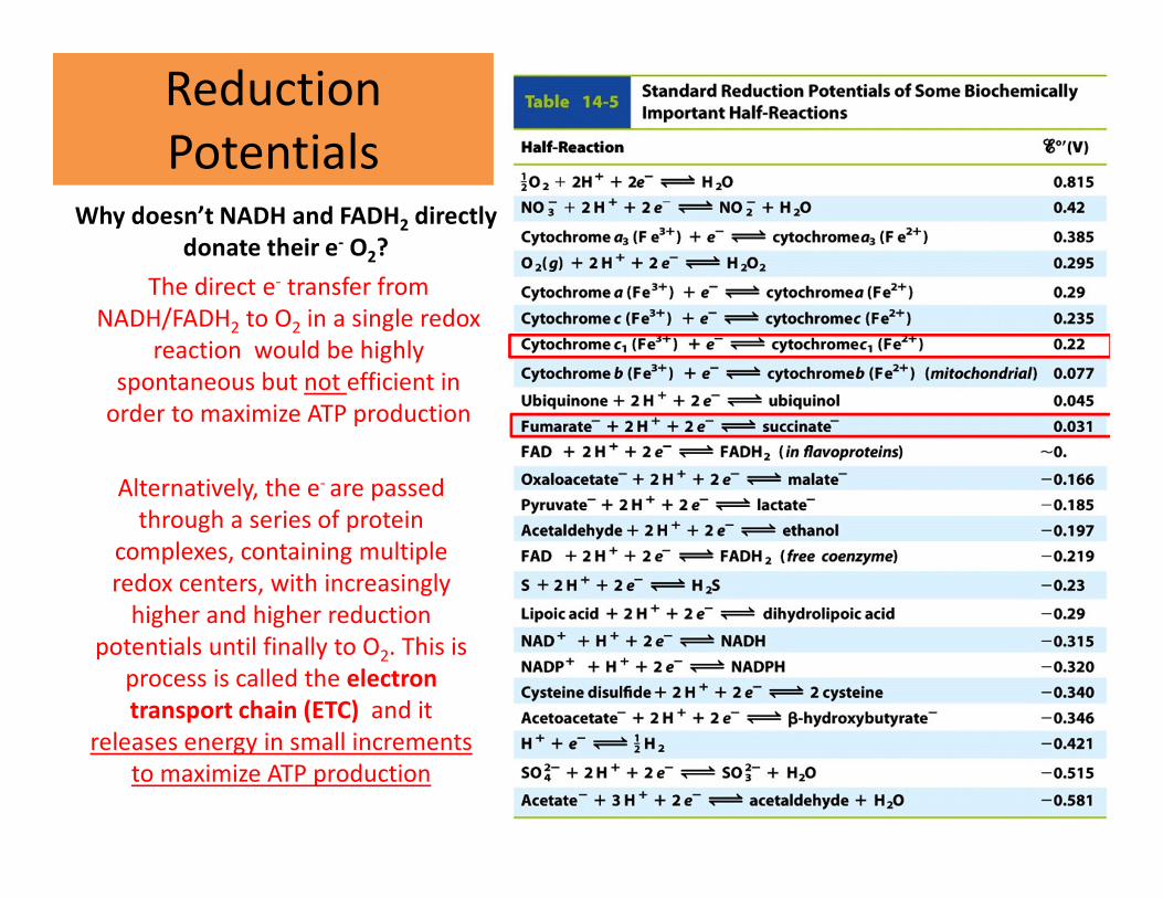
Reduction
PotentialsWhy doesn’t NADH and FADH2 directly
donate their e- O2?
Alternatively, the e- are passed
through a series of protein
complexes, containing multiple
redox centers, with increasingly
higher and higher reduction
potentials until finally to O2. This is
process is called the electron
transport chain (ETC) and it
releases energy in small increments
to maximize ATP production
The direct e- transfer from
NADH/FADH2 to O2 in a single redox
reaction would be highly
spontaneous but not efficient in
order to maximize ATP production
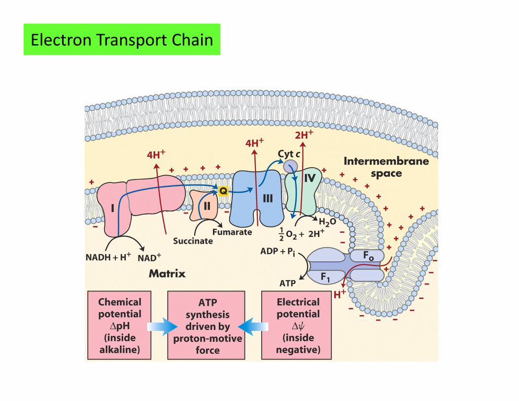
Electron Transport Chain
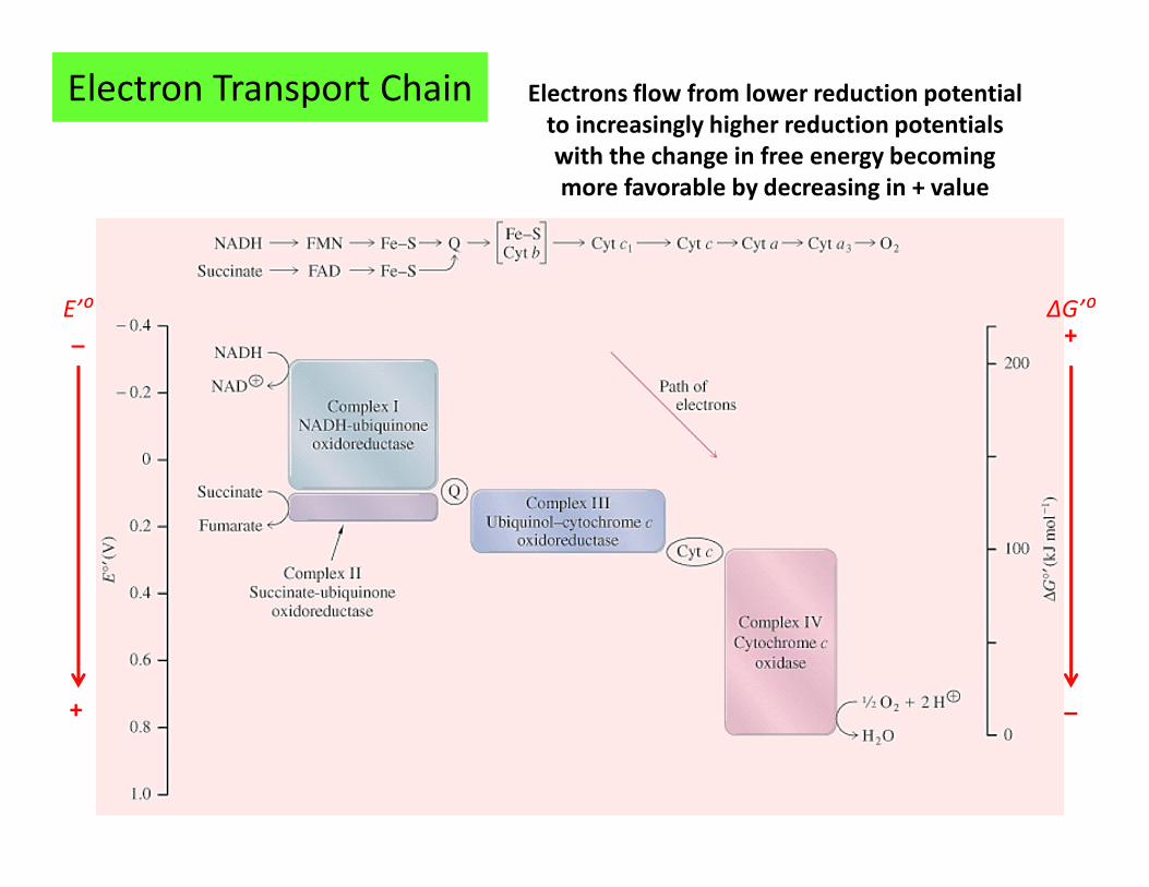
Electrons flow from lower reduction potential
to increasingly higher reduction potentials
with the change in free energy becoming
more favorable by decreasing in + value
Electron Transport Chain
_
+
E’⁰
_
+ΔG’⁰
Electron Transport Chain
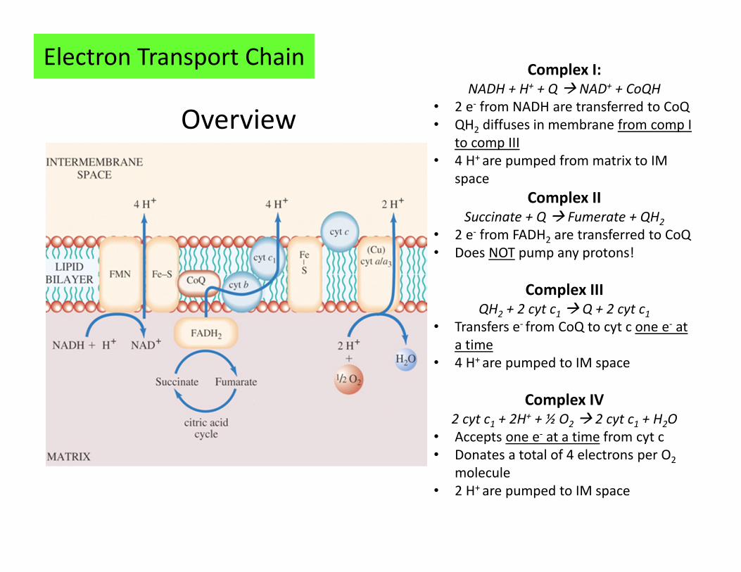
Overview
Complex I: NADH + H+ + Q � NAD+ + CoQH
• 2 e- from NADH are transferred to CoQ
• QH2 diffuses in membrane from comp I
to comp III
• 4 H+ are pumped from matrix to IM
space
Complex IISuccinate + Q � Fumerate + QH2
• 2 e- from FADH2 are transferred to CoQ
• Does NOT pump any protons!
Complex IIIQH2 + 2 cyt c1 � Q + 2 cyt c1
• Transfers e- from CoQ to cyt c one e- at
a time
• 4 H+ are pumped to IM space
Complex IV2 cyt c1 + 2H+ + ½ O2 � 2 cyt c1 + H2O
• Accepts one e- at a time from cyt c
• Donates a total of 4 electrons per O2
molecule
• 2 H+ are pumped to IM space
Electron Transport Chain
Overview
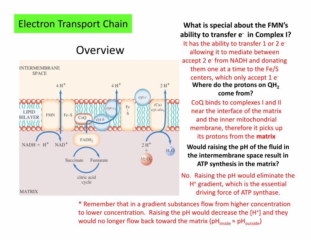
What is special about the FMN’s
ability to transfer e- in Complex I?It has the ability to transfer 1 or 2 e-
allowing it to mediate between
accept 2 e- from NADH and donating
them one at a time to the Fe/S
centers, which only accept 1 e-
Where do the protons on QH2
come from?
CoQ binds to complexes I and II
near the interface of the matrix
and the inner mitochondrial
membrane, therefore it picks up
its protons from the matrix
Would raising the pH of the fluid in
the intermembrane space result in
ATP synthesis in the matrix?
No. Raising the pH would eliminate the
H+ gradient, which is the essential
driving force of ATP synthase.
* Remember that in a gradient substances flow from higher concentration
to lower concentration. Raising the pH would decrease the [H+] and they
would no longer flow back toward the matrix (pHinside ≈ pHoutside)
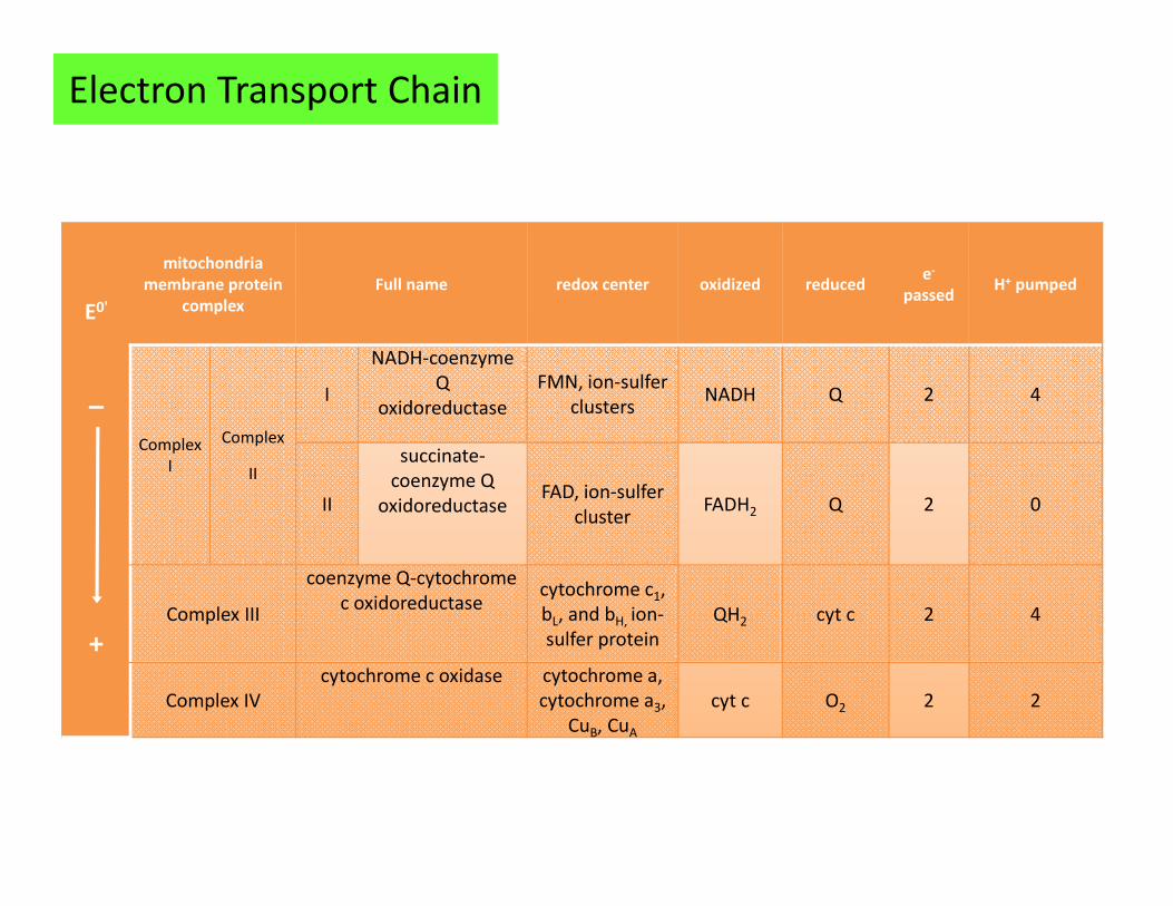
E0'
_
+
mitochondria
membrane protein
complex
Full name redox center oxidized reducede-
passedH+ pumped
Complex
I
Complex
II
I
NADH-coenzyme
Q
oxidoreductase
FMN, ion-sulfer
clustersNADH Q 2 4
II
succinate-
coenzyme Q
oxidoreductaseFAD, ion-sulfer
clusterFADH2 Q 2 0
Complex III
coenzyme Q-cytochrome
c oxidoreductasecytochrome c1,
bL, and bH, ion-
sulfer protein
QH2 cyt c 2 4
Complex IV
cytochrome c oxidase cytochrome a,
cytochrome a3,
CuB, CuA
cyt c O2 2 2
Electron Transport Chain
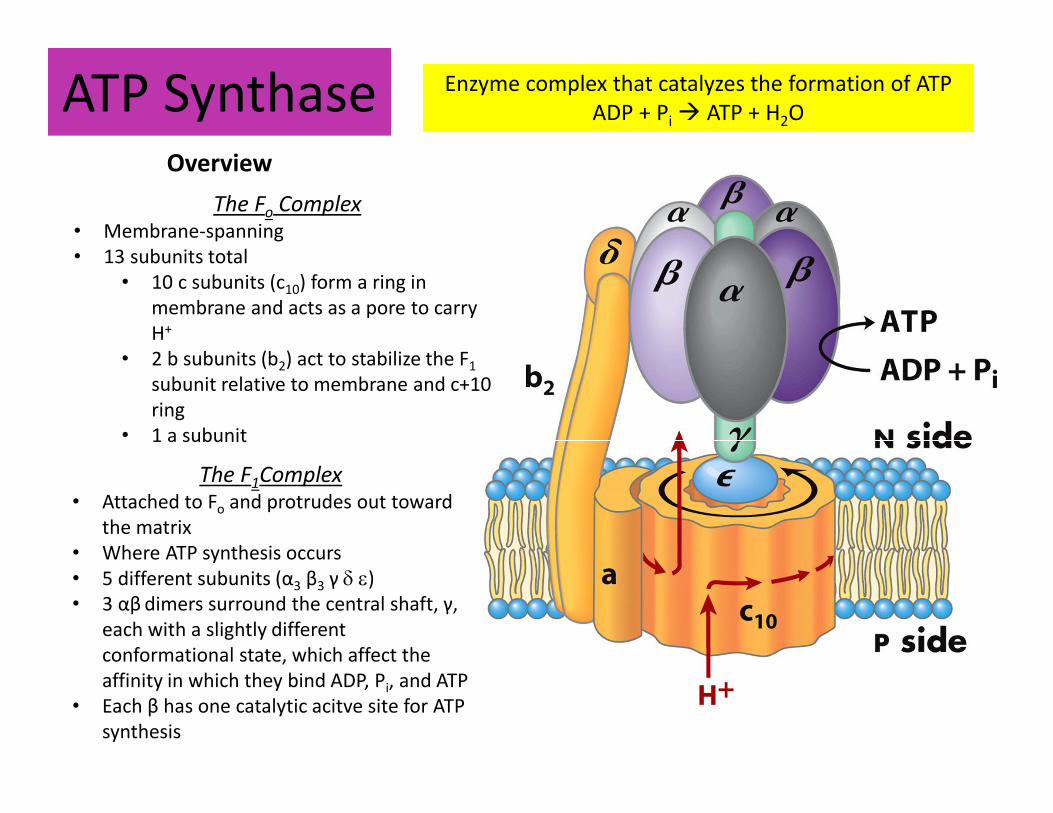
ATP Synthase Enzyme complex that catalyzes the formation of ATP
ADP + Pi � ATP + H2O
The Fo Complex• Membrane-spanning
• 13 subunits total
• 10 c subunits (c10) form a ring in
membrane and acts as a pore to carry
H+
• 2 b subunits (b2) act to stabilize the F1
subunit relative to membrane and c+10
ring
• 1 a subunit
The F1Complex• Attached to Fo and protrudes out toward
the matrix
• Where ATP synthesis occurs
• 5 different subunits (α3 β3 γ δ ε)
• 3 αβ dimers surround the central shaft, γ,
each with a slightly different
conformational state, which affect the
affinity in which they bind ADP, Pi, and ATP
• Each β has one catalytic acitve site for ATP
synthesis
Overview
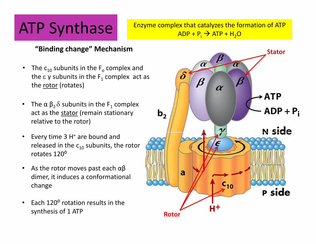
ATP Synthase Enzyme complex that catalyzes the formation of ATP
ADP + Pi � ATP + H2O
“Binding change” Mechanism
• The c10 subunits in the Fo complex and
the ε γ subunits in the F1 complex act as
the rotor (rotates)
Rotor
• The α β2 δ subunits in the F1 complex
act as the stator (remain stationary
relative to the rotor)
Stator
• Every time 3 H+ are bound and
released in the c10 subunits, the rotor
rotates 120⁰
• As the rotor moves past each αβ
dimer, it induces a conformational
change
• Each 120⁰ rotation results in the
synthesis of 1 ATP
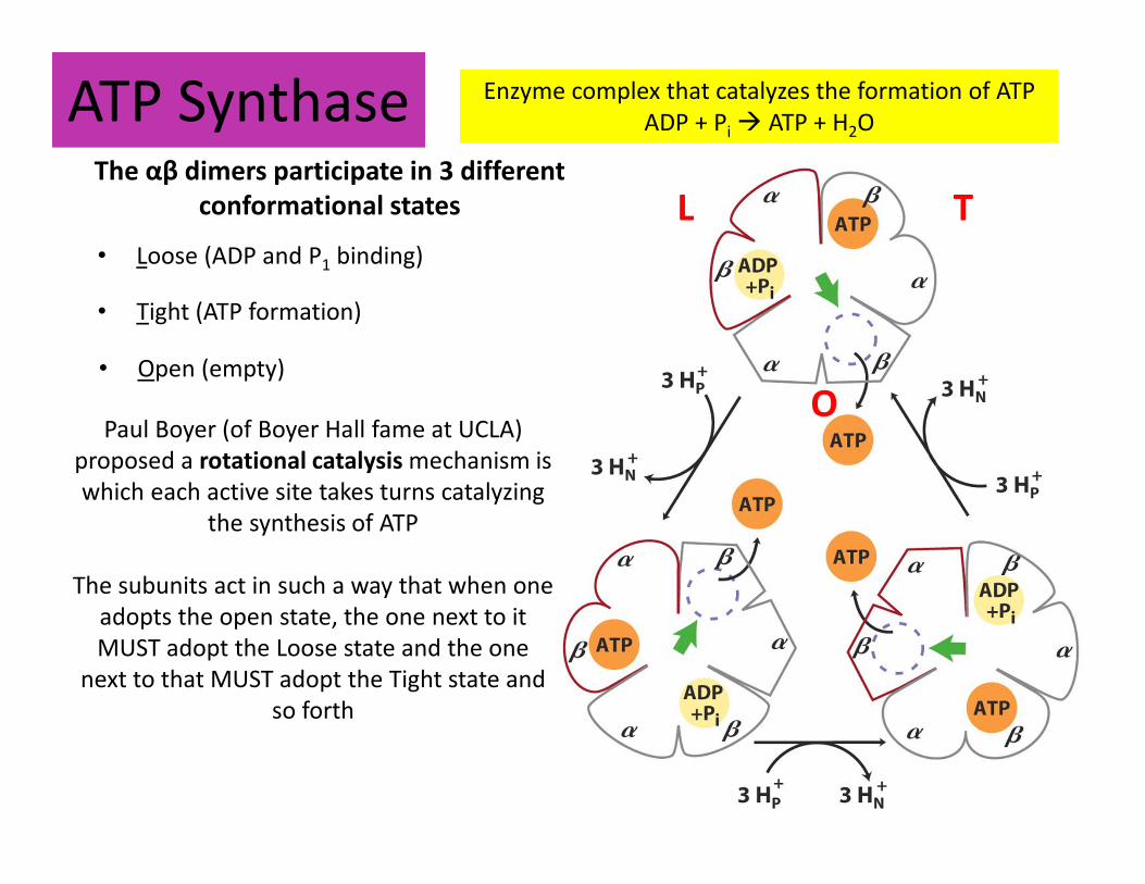
ATP Synthase Enzyme complex that catalyzes the formation of ATP
ADP + Pi � ATP + H2O
The αβ dimers participate in 3 different
conformational states
• Loose (ADP and P1 binding)
L T
O
• Tight (ATP formation)
• Open (empty)
Paul Boyer (of Boyer Hall fame at UCLA)
proposed a rotational catalysis mechanism is
which each active site takes turns catalyzing
the synthesis of ATP
The subunits act in such a way that when one
adopts the open state, the one next to it
MUST adopt the Loose state and the one
next to that MUST adopt the Tight state and
so forth
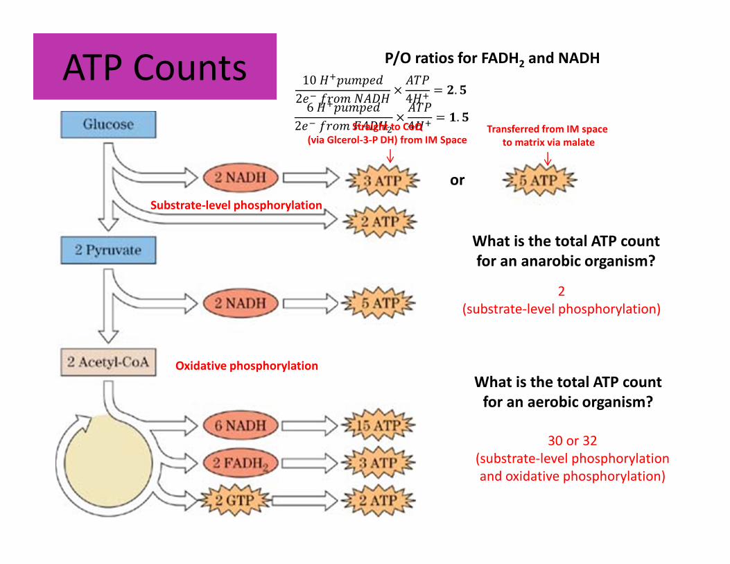
ATP Counts
Substrate-level phosphorylation
or
6������ �
2 �������� !��"
4��= $. &
10������ �
2 �����)���!��"
4��= *. &
P/O ratios for FADH2 and NADH
Straight to CoQ
(via Glcerol-3-P DH) from IM SpaceTransferred from IM space
to matrix via malate
Oxidative phosphorylation
What is the total ATP count
for an anarobic organism?
What is the total ATP count
for an aerobic organism?
2
(substrate-level phosphorylation)
30 or 32
(substrate-level phosphorylation
and oxidative phosphorylation)
ATP Synthase Animation
http://www.dnatube.com/video/104/ATP-synthase-structure-and-mechanism
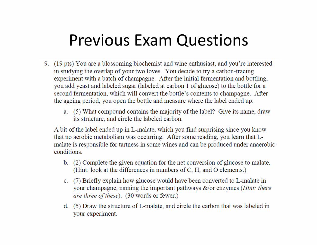
Previous Exam Questions
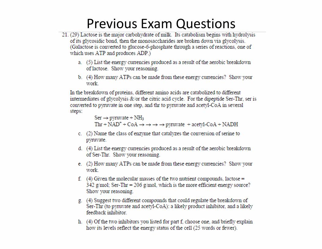
Previous Exam Questions
Great luck studying!
Thanks for a great quarter, biochemists!
P.S. Yes, yes you can ☺