15 - napa valley college pages - napa valley college … 105... · 2015-06-08 · 15-1 the...
TRANSCRIPT
Copyright © 2010 Pearson Education, Inc.
C h a p t e r
15
The Respiratory System
PowerPoint® Lecture Slides
prepared by Jason LaPres
Lone Star College - North Harris
Copyright © 2010 Pearson Education, Inc.
Copyright © 2010 Pearson Education, Inc.
Introduction to the Respiratory System
• The Respiratory System
– Cells produce energy:
• For maintenance, growth, defense, and division
• Through mechanisms that use oxygen and
produce carbon dioxide
Copyright © 2010 Pearson Education, Inc.
Introduction to the Respiratory System
• Oxygen
– Is obtained from the air by diffusion across
delicate exchange surfaces of the lungs
– Is carried to cells by the cardiovascular
system, which also returns carbon dioxide to
the lungs

Copyright © 2010 Pearson Education, Inc.
15-1 The respiratory system,
composed of conducting and
respiratory portions, has
several basic functions
Copyright © 2010 Pearson Education, Inc.
Functions of the Respiratory System
• Provides extensive gas exchange surface area
between air and circulating blood
• Moves air to and from exchange surfaces of
lungs
• Protects respiratory surfaces from outside
environment
• Produces sounds
• Participates in olfactory sense
Copyright © 2010 Pearson Education, Inc.
Components of the Respiratory System
• The Respiratory Tract
– Consists of a conducting portion
• From nasal cavity to terminal bronchioles
– Consists of a respiratory portion
• The respiratory bronchioles and alveoli
The Respiratory Tract
Copyright © 2010 Pearson Education, Inc.
Components of the Respiratory System
• Alveoli
– Are air-filled pockets within the lungs:
• Where all gas exchange takes place
Copyright © 2010 Pearson Education, Inc.
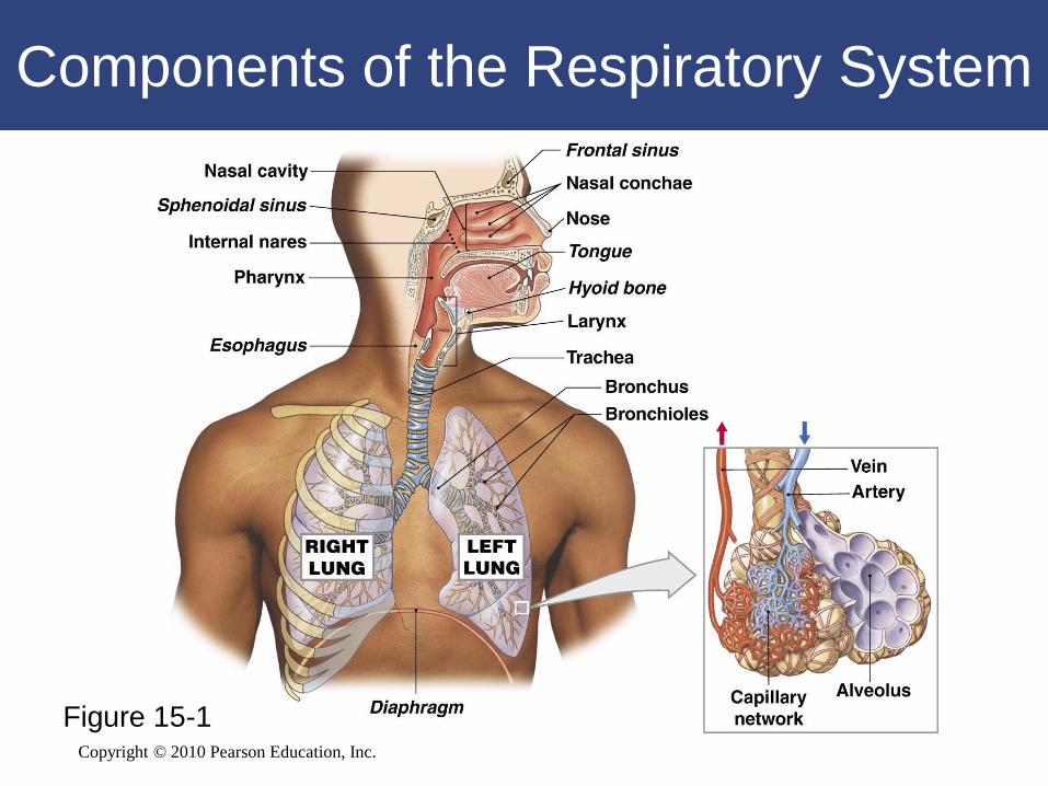
Components of the Respiratory System
Figure 15-1
Copyright © 2010 Pearson Education, Inc.
Components of the Respiratory System
• Structure of Respiratory Epithelium
– Changes along respiratory tract
Copyright © 2010 Pearson Education, Inc.
Components of the Respiratory System
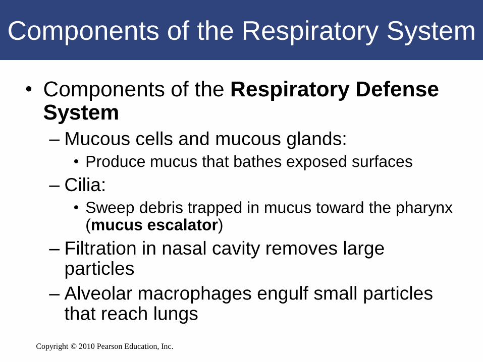
• Components of the Respiratory Defense System
– Mucous cells and mucous glands:• Produce mucus that bathes exposed surfaces
– Cilia:• Sweep debris trapped in mucus toward the pharynx
(mucus escalator)
– Filtration in nasal cavity removes large particles
– Alveolar macrophages engulf small particles that reach lungs
Copyright © 2010 Pearson Education, Inc.
Components of the Respiratory System
• Alveolar Epithelium
– Is a very delicate, simple squamous
epithelium
– Contains scattered and specialized cells
– Lines exchange surfaces of alveoli
Copyright © 2010 Pearson Education, Inc.
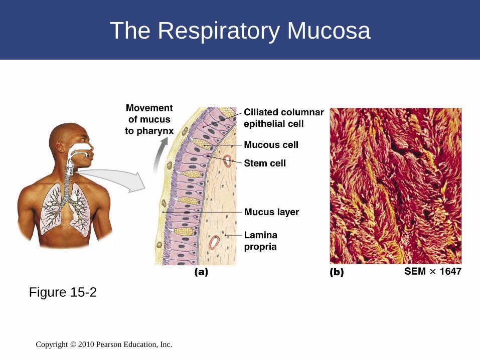
The Respiratory Mucosa
Figure 15-2
Copyright © 2010 Pearson Education, Inc.
15-2 The nose, pharynx,
larynx, trachea, bronchi, and
larger bronchioles conduct air
into the lungs
Copyright © 2010 Pearson Education, Inc.
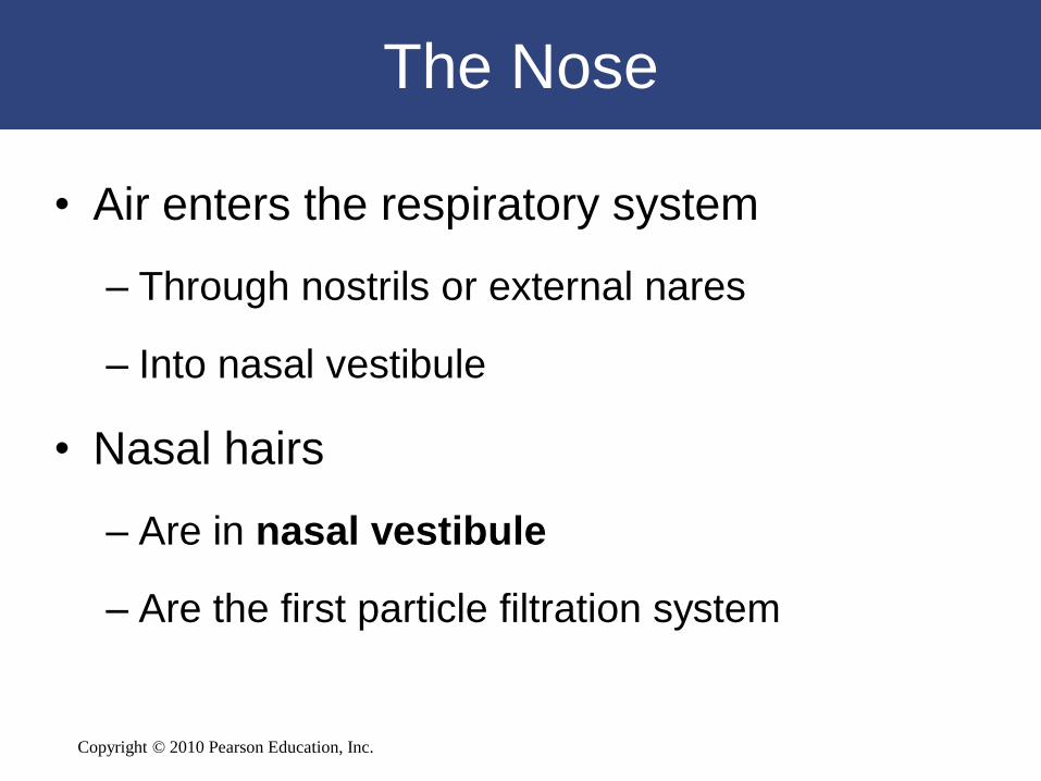
The Nose
• Air enters the respiratory system
– Through nostrils or external nares
– Into nasal vestibule
• Nasal hairs
– Are in nasal vestibule
– Are the first particle filtration system
Copyright © 2010 Pearson Education, Inc.
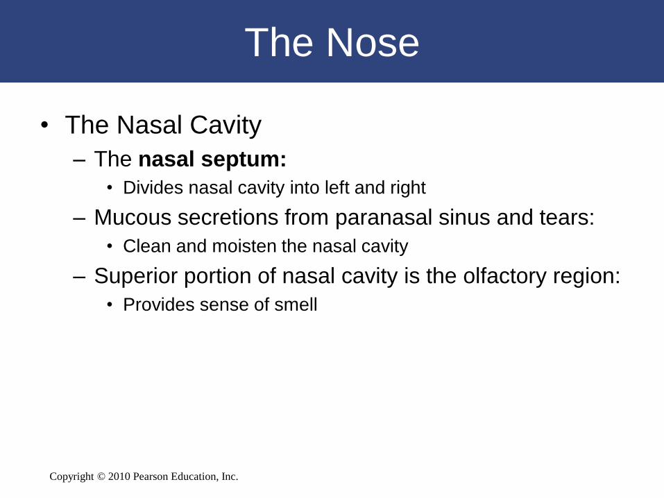
The Nose
• The Nasal Cavity
– The nasal septum:
• Divides nasal cavity into left and right
– Mucous secretions from paranasal sinus and tears:
• Clean and moisten the nasal cavity
– Superior portion of nasal cavity is the olfactory region:
• Provides sense of smell
Copyright © 2010 Pearson Education, Inc.
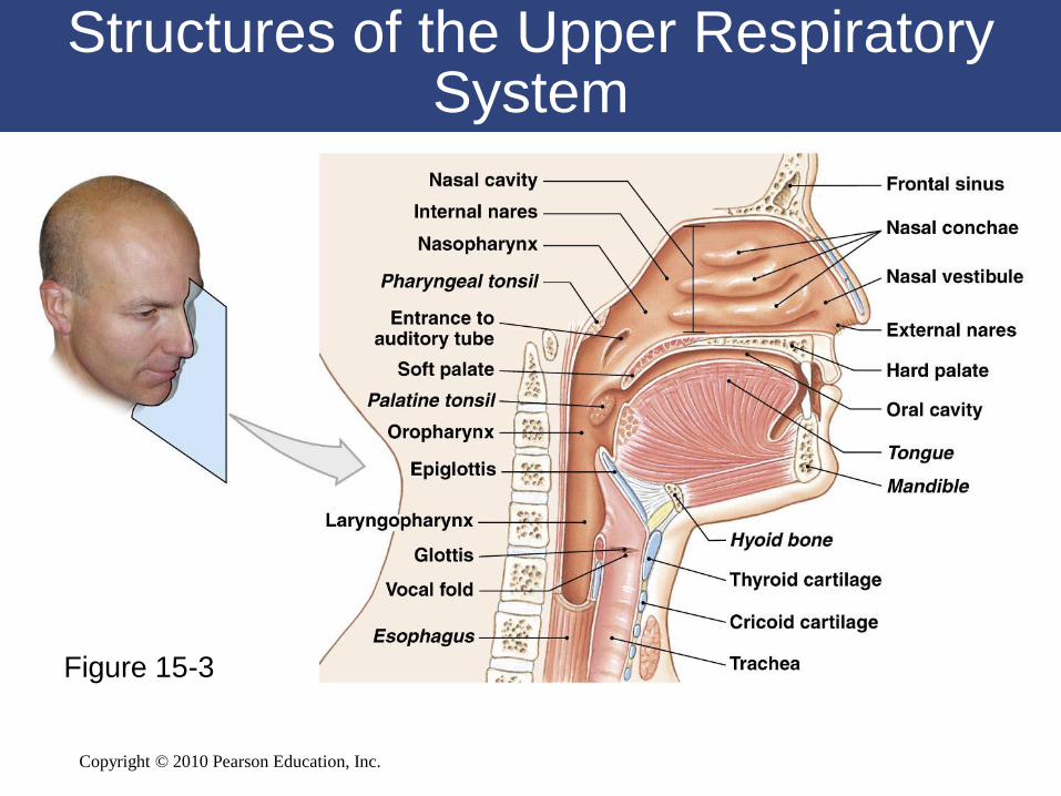
Structures of the Upper Respiratory System
Figure 15-3
Copyright © 2010 Pearson Education, Inc.
The Pharynx
• A chamber shared by digestive and
respiratory systems
• Extends from internal nares to entrances
to larynx and esophagus
• Divided into the nasopharynx, the
oropharynx, and the laryngopharynx
Copyright © 2010 Pearson Education, Inc.
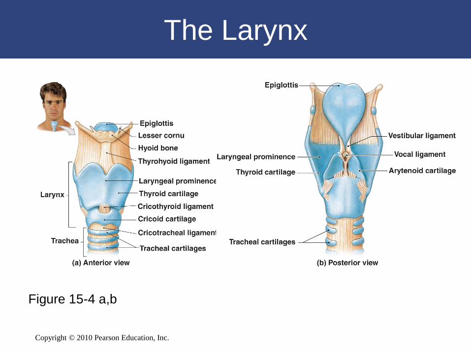
The Larynx
• Cartilages of the Larynx
– Three large, unpaired cartilages form the
larynx:
• Thyroid cartilage
• Cricoid cartilage
• Epiglottis
Copyright © 2010 Pearson Education, Inc.
The Larynx
• The Thyroid Cartilage
– Also called the Adam’s apple
– Is hyaline cartilage
– Forms anterior and lateral walls of larynx
– Ligaments attach to hyoid bone, epiglottis,
and laryngeal cartilages
Copyright © 2010 Pearson Education, Inc.
The Larynx
• The Cricoid Cartilage
– Is hyaline cartilage
– Forms posterior portion of larynx
– Ligaments attach to first tracheal cartilage
– Articulates with arytenoid cartilages
Copyright © 2010 Pearson Education, Inc.
The Larynx
• The Epiglottis
– Composed of elastic cartilage
– Ligaments attach to thyroid cartilage and
hyoid bone
Copyright © 2010 Pearson Education, Inc.
The Larynx
Figure 15-4 a,b
Copyright © 2010 Pearson Education, Inc.
The Larynx
• Sound is varied by
– Tension on vocal folds:
- Vocal folds involved with sound are known as vocal cords
– Voluntary muscles (position arytenoid cartilage relative
to thyroid cartilage)
• Speech is produced by
– Phonation:
• Sound production at the larynx
– Articulation:
• Modification of sound by other structures
Copyright © 2010 Pearson Education, Inc.
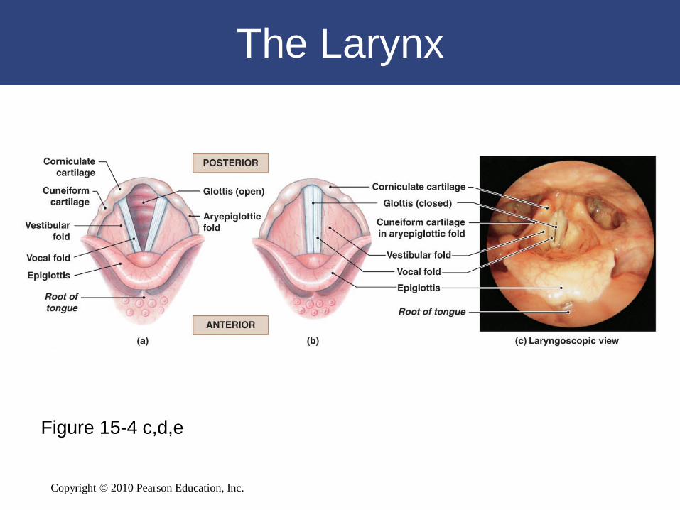
The Larynx
Figure 15-4 c,d,e
Copyright © 2010 Pearson Education, Inc.
The Trachea
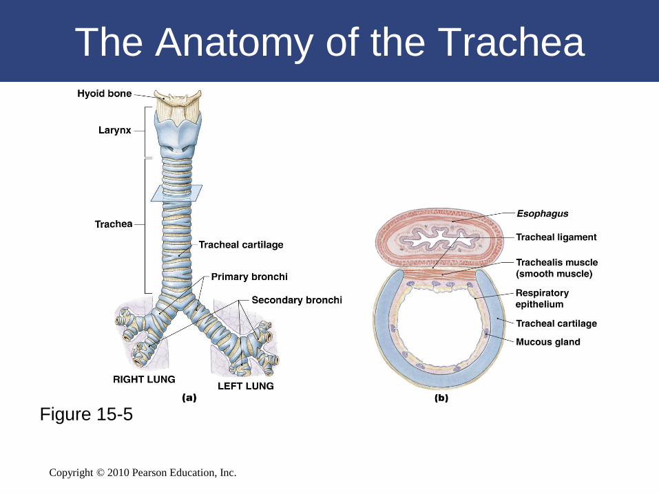
• The Trachea
– Also called the windpipe
– Extends from the cricoid cartilage into mediastinum:
• Where it branches into right and left pulmonary bronchi
• The Submucosa
– Beneath mucosa of trachea
– Contains mucous glands
Copyright © 2010 Pearson Education, Inc.
The Trachea
• The Tracheal Cartilages
– 15–20 tracheal cartilages:
• Strengthen and protect airway
• Discontinuous where trachea contacts esophagus
– Ends of each tracheal cartilage are connected
by:
• An elastic ligament and trachealis muscle
Copyright © 2010 Pearson Education, Inc.
The Anatomy of the Trachea
Figure 15-5
Copyright © 2010 Pearson Education, Inc.
The Bronchi
• The Primary Bronchi
– Right and left primary bronchi:
• Separated by an internal ridge (the carina)
• The Right Primary Bronchus
– Is larger in diameter than the left
– Descends at a steeper angle
Copyright © 2010 Pearson Education, Inc.
The Bronchi
• Structure of Primary Bronchi
– Each primary bronchus:
• Travels to a groove (hilum) along the medial
surface of the lung
Copyright © 2010 Pearson Education, Inc.
The Bronchi
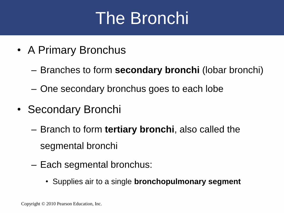
• A Primary Bronchus
– Branches to form secondary bronchi (lobar bronchi)
– One secondary bronchus goes to each lobe
• Secondary Bronchi
– Branch to form tertiary bronchi, also called the
segmental bronchi
– Each segmental bronchus:
• Supplies air to a single bronchopulmonary segment
Copyright © 2010 Pearson Education, Inc.
15-3 The smallest bronchioles
and the alveoli within the
lungs make up the respiratory
portion of the respiratory tract
Copyright © 2010 Pearson Education, Inc.
The Bronchioles
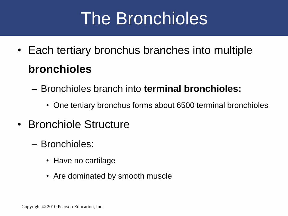
• Each tertiary bronchus branches into multiple
bronchioles
– Bronchioles branch into terminal bronchioles:
• One tertiary bronchus forms about 6500 terminal bronchioles
• Bronchiole Structure
– Bronchioles:
• Have no cartilage
• Are dominated by smooth muscle
Copyright © 2010 Pearson Education, Inc.
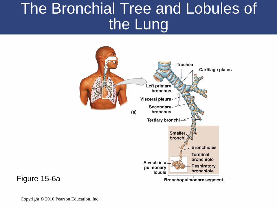
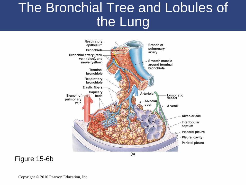
The Bronchial Tree and Lobules of the Lung
Figure 15-6a
Copyright © 2010 Pearson Education, Inc.
Figure 15-6b
The Bronchial Tree and Lobules of the Lung
Copyright © 2010 Pearson Education, Inc.
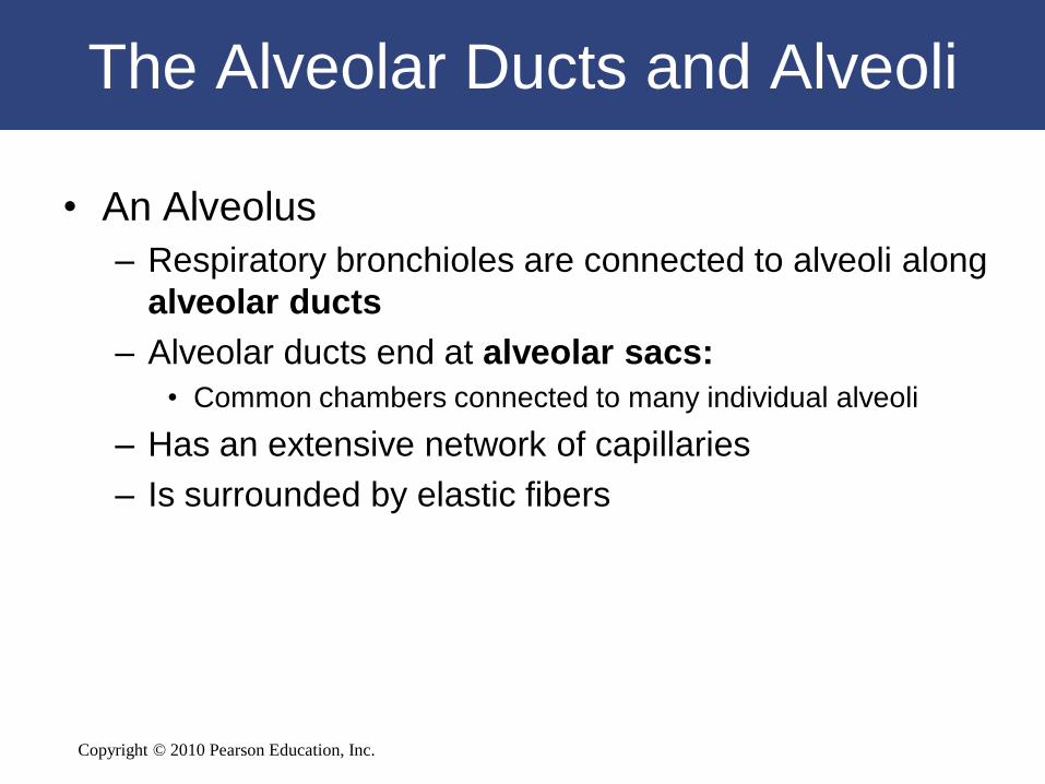
The Alveolar Ducts and Alveoli
• An Alveolus
– Respiratory bronchioles are connected to alveoli along
alveolar ducts
– Alveolar ducts end at alveolar sacs:
• Common chambers connected to many individual alveoli
– Has an extensive network of capillaries
– Is surrounded by elastic fibers

Copyright © 2010 Pearson Education, Inc.
Alveolar Organization
Figure 15-7a
Copyright © 2010 Pearson Education, Inc.
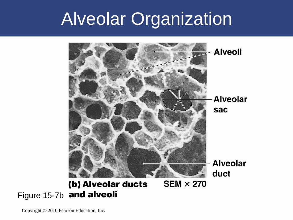
Alveolar Organization
Figure 15-7b
Copyright © 2010 Pearson Education, Inc.
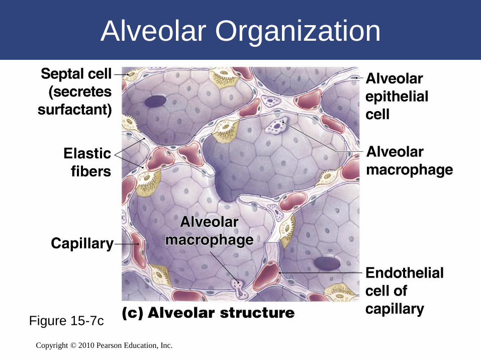
Alveolar Organization
Figure 15-7c
Copyright © 2010 Pearson Education, Inc.
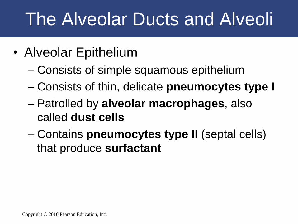
The Alveolar Ducts and Alveoli
• Alveolar Epithelium
– Consists of simple squamous epithelium
– Consists of thin, delicate pneumocytes type I
– Patrolled by alveolar macrophages, also
called dust cells
– Contains pneumocytes type II (septal cells)
that produce surfactant
Copyright © 2010 Pearson Education, Inc.
The Alveolar Ducts and Alveoli
• Surfactant
– Is an oily secretion
– Contains phospholipids and proteins
– Coats alveolar surfaces and reduces surface
tension
Copyright © 2010 Pearson Education, Inc.
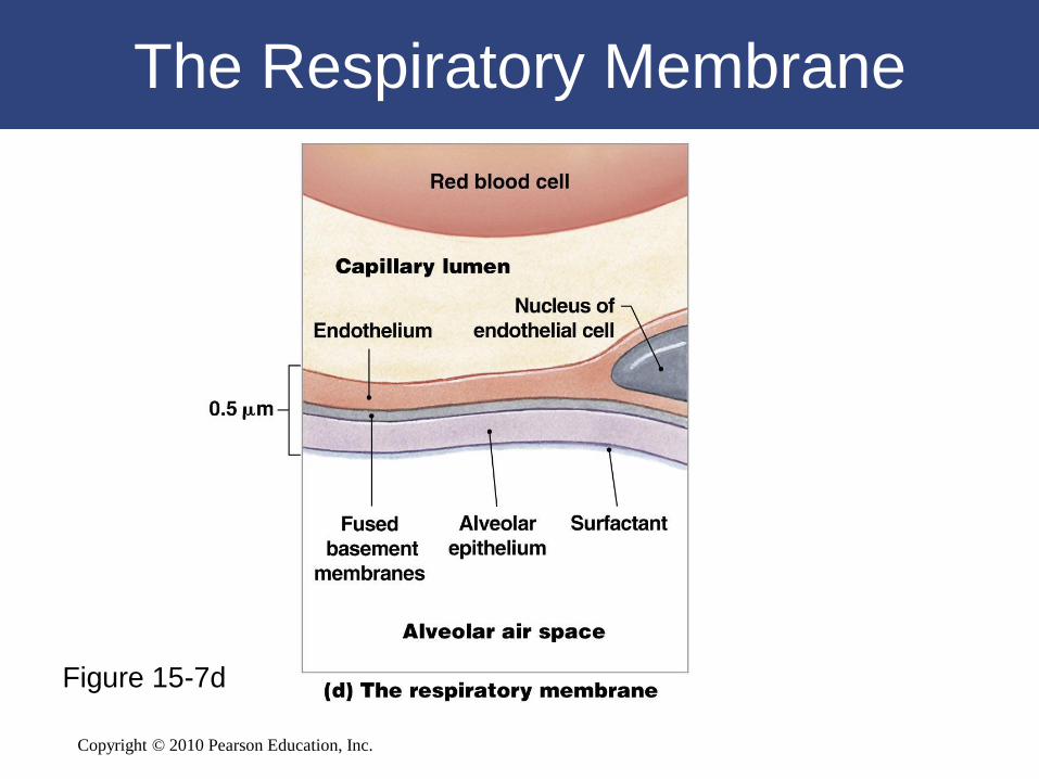
The Respiratory Membrane
• Three Layers of the Respiratory
Membrane
– Squamous epithelial lining of alveolus
– Endothelial cells lining an adjacent capillary
– Fused basal laminae between alveolar and
endothelial cells
Copyright © 2010 Pearson Education, Inc.
The Respiratory Membrane
Figure 15-7d
Copyright © 2010 Pearson Education, Inc.
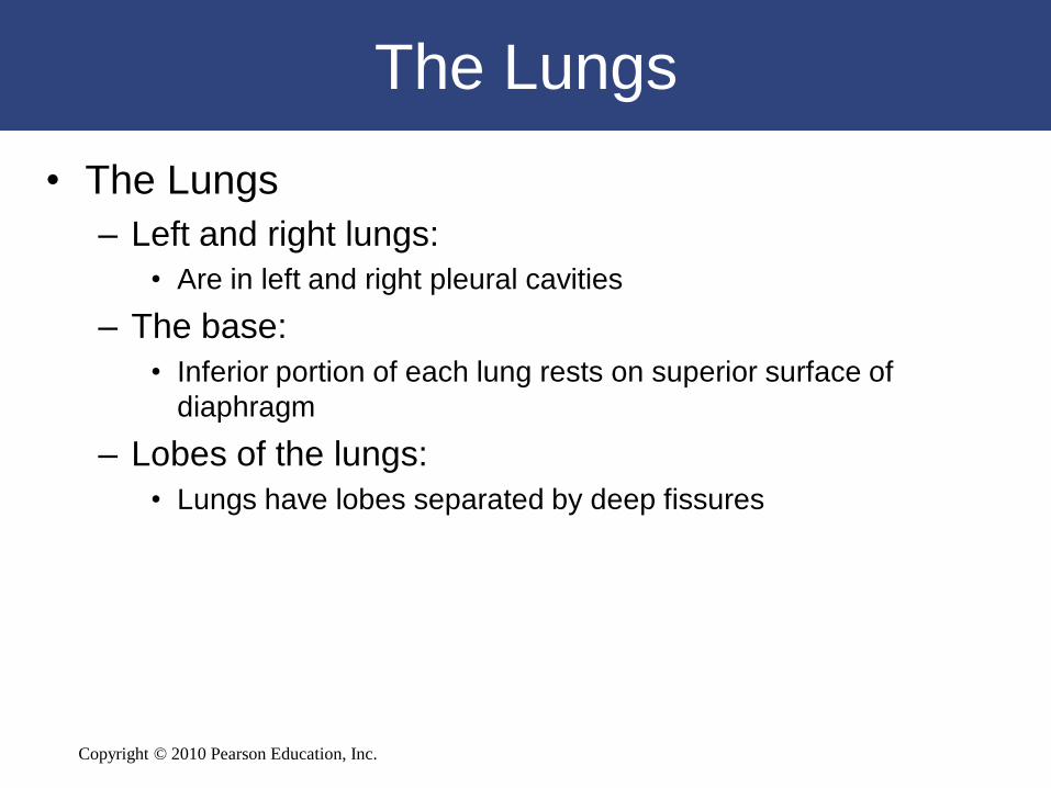
The Lungs
• The Lungs
– Left and right lungs:
• Are in left and right pleural cavities
– The base:
• Inferior portion of each lung rests on superior surface of
diaphragm
– Lobes of the lungs:
• Lungs have lobes separated by deep fissures
Copyright © 2010 Pearson Education, Inc.
The Lungs
• The right lung has three lobes
– Superior, middle, and inferior
– Separated by horizontal and oblique fissures
• The left lung has two lobes
– Superior and inferior
– Separated by an oblique fissure
Copyright © 2010 Pearson Education, Inc.
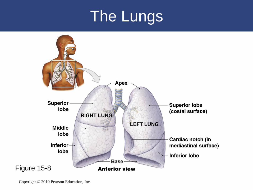
The Lungs
Figure 15-8
Copyright © 2010 Pearson Education, Inc.
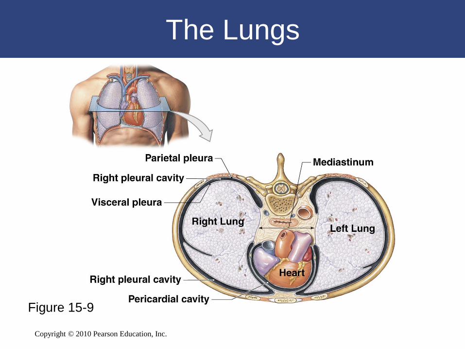
The Lungs
Figure 15-9

Copyright © 2010 Pearson Education, Inc.
The Pleural Cavities
• Two pleural cavities
– Are separated by the mediastinum
• Each pleural cavity
– Holds a lung
– Is lined with a serous membrane (the pleura)
Copyright © 2010 Pearson Education, Inc.
The Pleural Cavities
• The Pleura
– Consists of two layers:
• Parietal pleura
• Visceral pleura
– Pleural fluid:
• Lubricates space between two layers
Copyright © 2010 Pearson Education, Inc.
15-4 External respiration and
internal respiration allow gas
exchange within the body
Copyright © 2010 Pearson Education, Inc.
Introduction to Gas Exchange
• Respiration refers to two integrated processes
– External respiration:
• Includes all processes involved in exchanging O2
and CO2 with the environment
– Internal respiration:
• Also called cellular respiration
• Involves the uptake of O2 and production of CO2
within individual cells

Copyright © 2010 Pearson Education, Inc.
Introduction to Gas Exchange
• Three Processes of External Respiration
1. Pulmonary ventilation (breathing)
2. Gas diffusion:
• Across membranes and capillaries
3. Transport of O2 and CO2:
• Between alveolar capillaries
• Between capillary beds in other tissues
Copyright © 2010 Pearson Education, Inc.
15-5 Pulmonary ventilation —
the exchange of air between
the atmosphere and the lungs
— involves pressure changes
and muscle movement
Copyright © 2010 Pearson Education, Inc.
Pulmonary Ventilation
• Pulmonary Ventilation
– Is the physical movement of air in and out of
the respiratory tract
– Provides alveolar ventilation
• Atmospheric Pressure
– The weight of air:
• Has several important physiological effects
Copyright © 2010 Pearson Education, Inc.
Pressure and Airflow to the Lungs
• Air flows from area of higher pressure to area of
lower pressure
• A Respiratory Cycle
– Consists of:
• An inspiration (inhalation)
• An expiration (exhalation)

Copyright © 2010 Pearson Education, Inc.
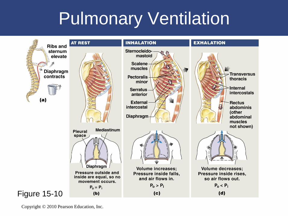
Pulmonary Ventilation
• Pulmonary Ventilation
– Causes volume changes that create changes
in pressure
– Volume of thoracic cavity changes:
• With expansion or contraction of diaphragm or rib
cage
Copyright © 2010 Pearson Education, Inc.
Pulmonary Ventilation
Figure 15-10
Copyright © 2010 Pearson Education, Inc.
Compliance
• An indicator of expandability
• Low compliance requires greater force
• High compliance requires less force
• Factors That Affect Compliance
– Connective tissue structure of the lungs
– Level of surfactant production
– Mobility of the thoracic cage
Copyright © 2010 Pearson Education, Inc.
Modes of Breathing
• Respiratory movements are classified
– By pattern of muscle activity
– Into quiet breathing and forced breathing
Copyright © 2010 Pearson Education, Inc.
Modes of Breathing
• Quiet Breathing (Eupnea)
– Involves active inhalation and passive
exhalation
– Diaphragmatic breathing or deep breathing:
• Is dominated by diaphragm
– Costal breathing or shallow breathing:
• Is dominated by rib cage movements
Copyright © 2010 Pearson Education, Inc.
Lung Volumes and Capacities
• Respiratory Rates and Volumes
– Respiratory system adapts to changing
oxygen demands by varying:
• The number of breaths per minute (respiratory
rate)
• The volume of air moved per breath (tidal volume)

Copyright © 2010 Pearson Education, Inc.
Lung Volumes and Capacities
Figure 15-11

Copyright © 2010 Pearson Education, Inc.
Lung Volumes and Capacities
Figure 15-11
Copyright © 2010 Pearson Education, Inc.
Lung Volumes and Capacities
• Pulmonary Function Tests
– Measure rates and volumes of air movements
Copyright © 2010 Pearson Education, Inc.
15-6 Gas exchange depends
on the partial pressures of
gases and the diffusion
of molecules
Copyright © 2010 Pearson Education, Inc.
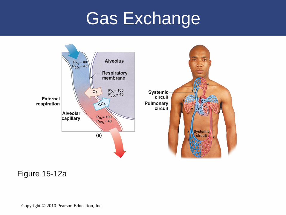
Gas Exchange
• Gas Exchange
– Occurs between blood and alveolar air
– Across the respiratory membrane
• Depends on
– Partial pressures of the gases
– Diffusion of molecules between gas and liquid
Gas Exchange
Copyright © 2010 Pearson Education, Inc.
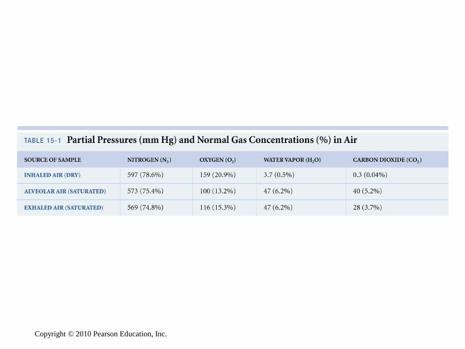
Mixed Gases and Partial Pressures
• Composition of Air
– Nitrogen (N2) is about 78.6%
– Oxygen (O2) is about 20.9%
– Water vapor (H2O) is about 0.5%
– Carbon dioxide (CO2) is about 0.04%
Copyright © 2010 Pearson Education, Inc.
Mixed Gases and Partial Pressures
• Partial Pressures
– Atmospheric pressure (760 mm Hg):
• Produced by air molecules bumping into each
other
– Each gas contributes to the total pressure:
• In proportion to its number of molecules (Dalton’s
law)
Copyright © 2010 Pearson Education, Inc.
Copyright © 2010 Pearson Education, Inc.
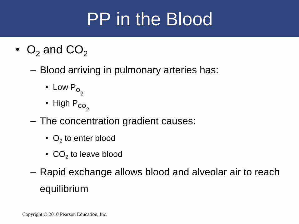
PP in the Blood
• O2 and CO2
– Blood arriving in pulmonary arteries has:
• Low PO2
• High PCO2
– The concentration gradient causes:
• O2 to enter blood
• CO2 to leave blood
– Rapid exchange allows blood and alveolar air to reach
equilibrium
Copyright © 2010 Pearson Education, Inc.
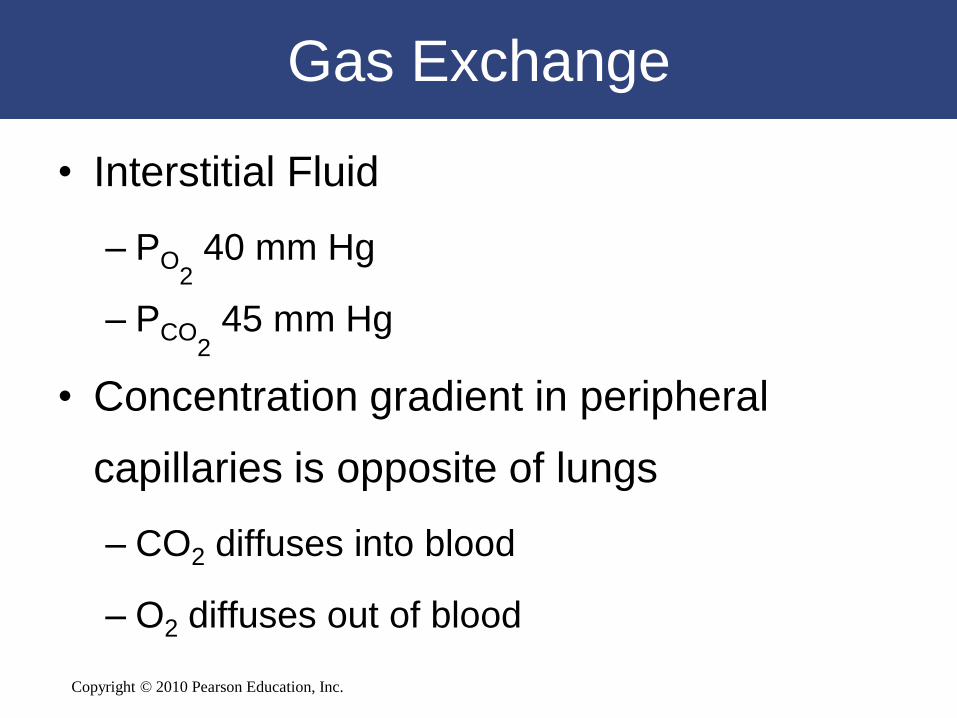
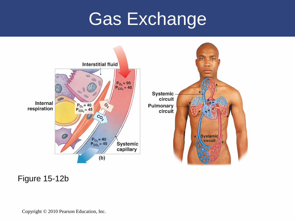
Gas Exchange
• Interstitial Fluid
– PO2
40 mm Hg
– PCO2
45 mm Hg
• Concentration gradient in peripheral
capillaries is opposite of lungs
– CO2 diffuses into blood
– O2 diffuses out of blood
Copyright © 2010 Pearson Education, Inc.
Gas Exchange
Figure 15-12a
Copyright © 2010 Pearson Education, Inc.
Gas Exchange
Figure 15-12b
Copyright © 2010 Pearson Education, Inc.
15-7 Most O2 is transported
bound to hemoglobin (Hb),
whereas CO2 is transported
as carbonic acid, bound to Hb,
or dissolved in plasma
Copyright © 2010 Pearson Education, Inc.
Gas Transport
• Red Blood Cells (RBCs)
– Transport O2 to, and CO2 from, peripheral
tissues
– Remove O2 and CO2 from plasma, allowing
gases to diffuse into blood
Copyright © 2010 Pearson Education, Inc.
Oxygen Transport
• O2 binds to iron ions in hemoglobin (Hb)
molecules
– In a reversible reaction
• Each RBC has about 280 million Hb
molecules
– Each binds four oxygen molecules
Copyright © 2010 Pearson Education, Inc.
Oxygen Transport
• Environmental Factors Affecting Hemoglobin
– PO2
of blood
– Blood pH
– Temperature
Copyright © 2010 Pearson Education, Inc.
Carbon Dioxide Transport
• (CO2)
– Is generated as a by-product of aerobic metabolism
(cellular respiration)
– CO2 in the bloodstream:
• May be:
– converted to carbonic acid
– bound to protein portion of hemoglobin
– dissolved in plasma
Copyright © 2010 Pearson Education, Inc.
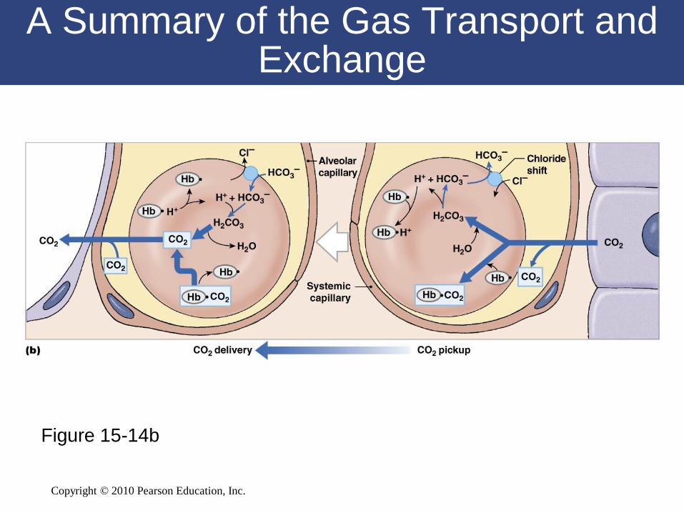
Carbon Dioxide Transport
• Bicarbonate Ions
– Move into plasma by an exchange
mechanism (the chloride shift) that takes in
Cl– ions without using ATP

Copyright © 2010 Pearson Education, Inc.
Carbon Dioxide Transport
Figure 15-13
Copyright © 2010 Pearson Education, Inc.
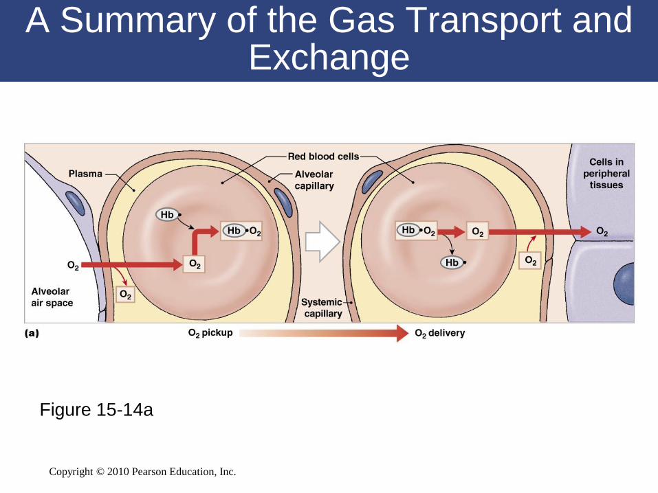
Gas Transport
• CO2 in the Bloodstream
– 70% is transported as carbonic acid (H2CO3):
• Which dissociates into H+ and bicarbonate (HCO3–)
– 23% is bound to amino groups of globular proteins
in Hb molecule
• Forming carbaminohemoglobin
– 7% is transported as CO2 dissolved in plasma
Copyright © 2010 Pearson Education, Inc.
A Summary of the Gas Transport and Exchange
Figure 15-14a
Copyright © 2010 Pearson Education, Inc.
A Summary of the Gas Transport and Exchange
Figure 15-14b

Copyright © 2010 Pearson Education, Inc.
15-8 Neurons in the medulla
and pons, along with
respiratory reflexes, control
respiration
Copyright © 2010 Pearson Education, Inc.
Control of Respiration
• Peripheral and alveolar capillaries
maintain balance during gas diffusion by
– Changes in blood flow and oxygen delivery
– Changes in depth and rate of respiration
Copyright © 2010 Pearson Education, Inc.
Control by the Respiratory Centers of
the Brain
• When oxygen demand rises
– Cardiac output and respiratory rates increase
under neural control:
• Have both voluntary and involuntary components
Copyright © 2010 Pearson Education, Inc.
Control by the Respiratory Centers of
the Brain
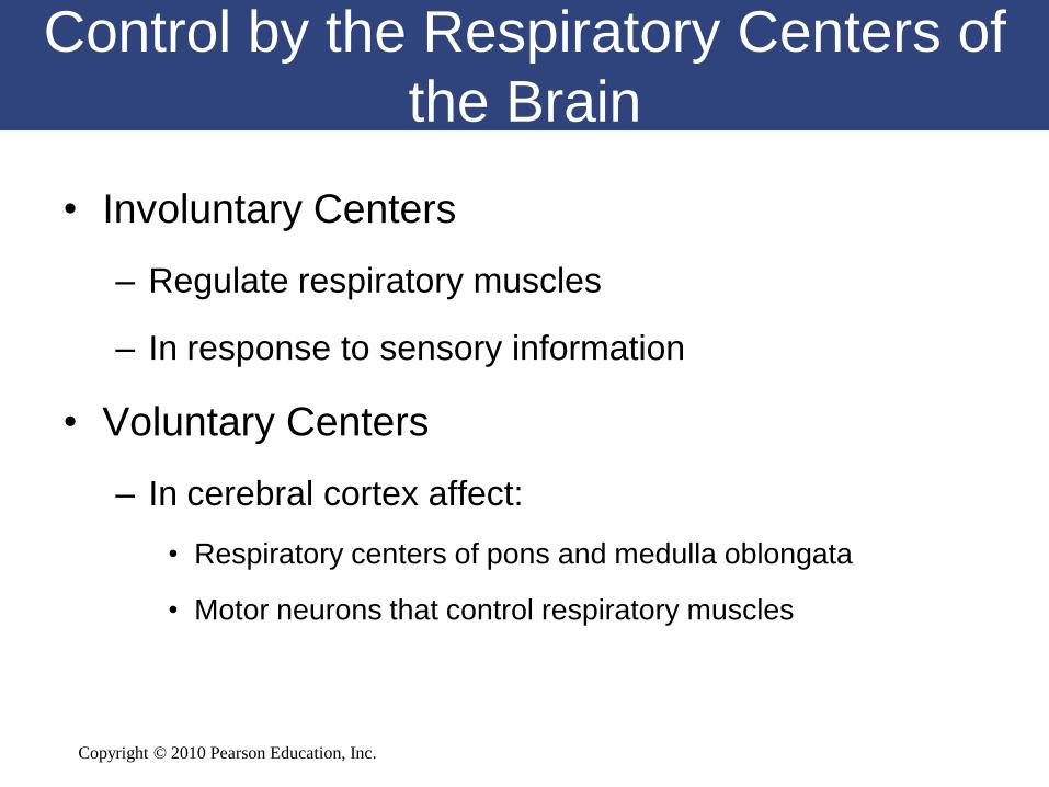
• Involuntary Centers
– Regulate respiratory muscles
– In response to sensory information
• Voluntary Centers
– In cerebral cortex affect:
• Respiratory centers of pons and medulla oblongata
• Motor neurons that control respiratory muscles
Copyright © 2010 Pearson Education, Inc.
Control by the Respiratory Centers of
the Brain
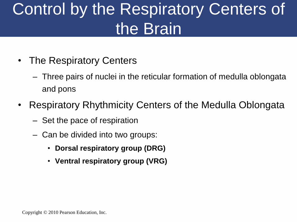
• The Respiratory Centers
– Three pairs of nuclei in the reticular formation of medulla oblongata
and pons
• Respiratory Rhythmicity Centers of the Medulla Oblongata
– Set the pace of respiration
– Can be divided into two groups:
• Dorsal respiratory group (DRG)
• Ventral respiratory group (VRG)
Copyright © 2010 Pearson Education, Inc.
Control by the Respiratory Centers of
the Brain
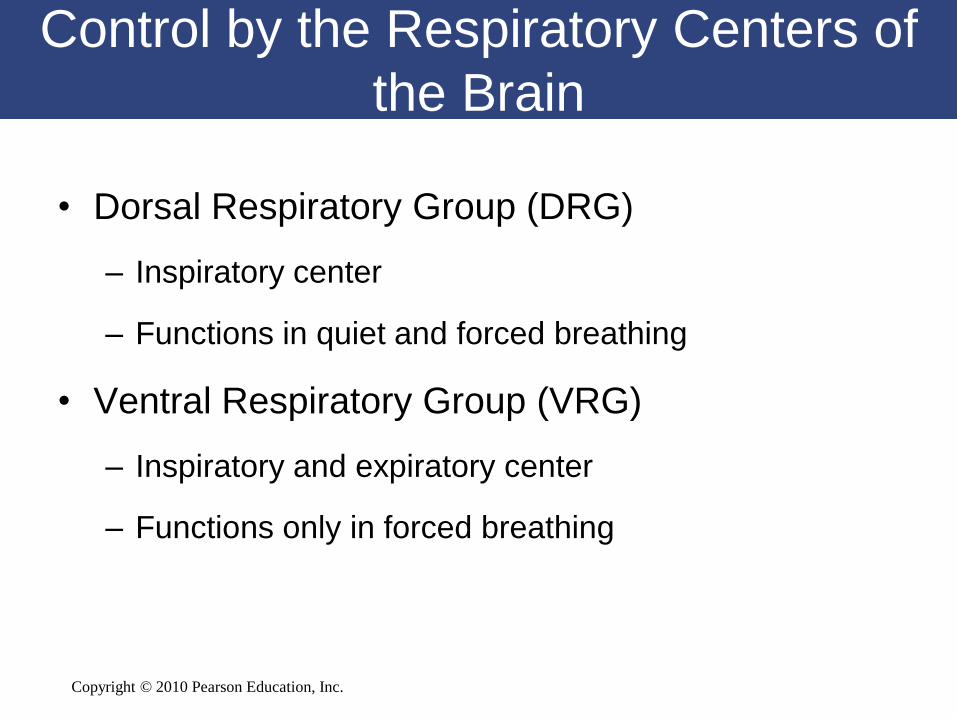
• Dorsal Respiratory Group (DRG)
– Inspiratory center
– Functions in quiet and forced breathing
• Ventral Respiratory Group (VRG)
– Inspiratory and expiratory center
– Functions only in forced breathing
Copyright © 2010 Pearson Education, Inc.
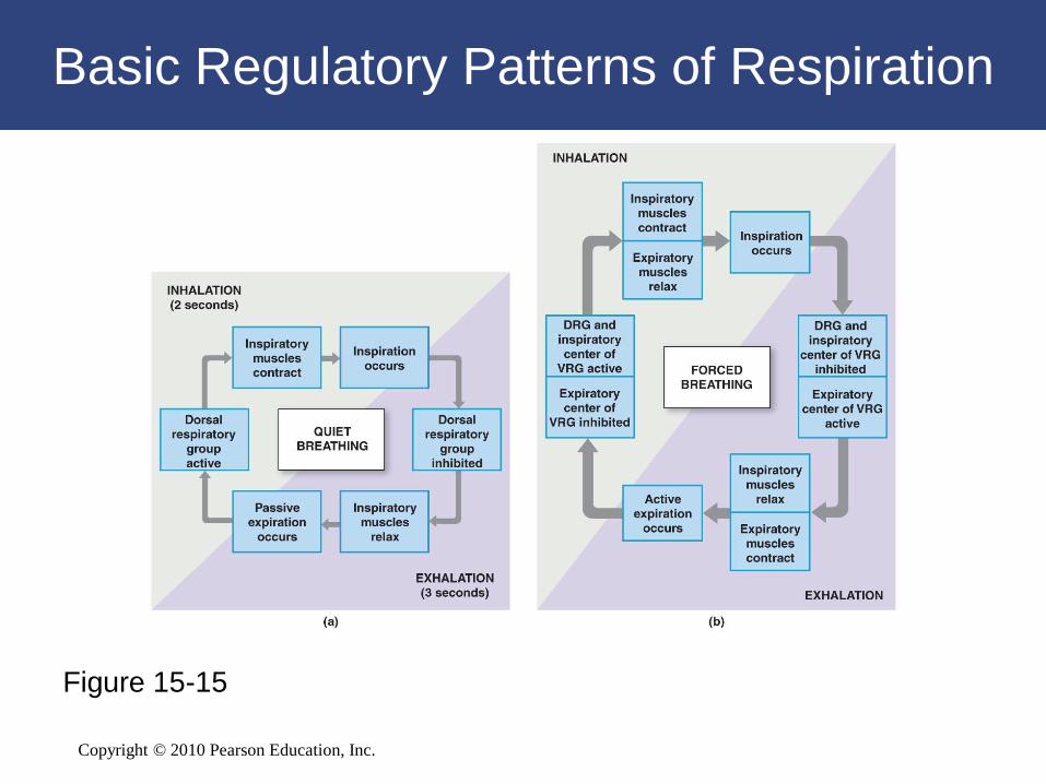
Basic Regulatory Patterns of Respiration
Figure 15-15
Copyright © 2010 Pearson Education, Inc.
Control of Respiration
• Respiratory Reflexes
– Changes in patterns of respiration induced by
sensory input
Copyright © 2010 Pearson Education, Inc.
Control of Respiration
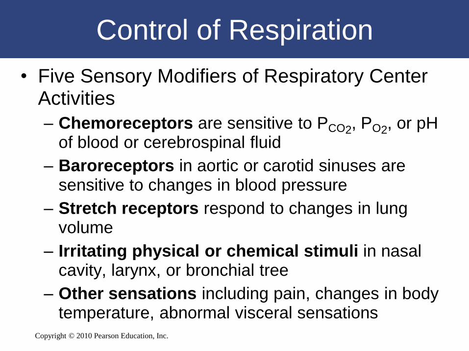
• Five Sensory Modifiers of Respiratory Center Activities
– Chemoreceptors are sensitive to PCO2, PO2, or pH of blood or cerebrospinal fluid
– Baroreceptors in aortic or carotid sinuses are sensitive to changes in blood pressure
– Stretch receptors respond to changes in lung volume
– Irritating physical or chemical stimuli in nasal cavity, larynx, or bronchial tree
– Other sensations including pain, changes in body temperature, abnormal visceral sensations
Copyright © 2010 Pearson Education, Inc.
Control of Respiration
• Baroreceptor Reflexes
– Carotid and aortic baroreceptor stimulation:
• Affects blood pressure and respiratory centers
– When blood pressure falls:
• Respiration increases
– When blood pressure increases:
• Respiration decreases
Copyright © 2010 Pearson Education, Inc.
Control of Respiration
• The Hering–Breuer Reflexes
– Two baroreceptor reflexes involved in forced
breathing
• Inflation reflex:
– prevents overexpansion of lungs
• Deflation reflex:
– inhibits expiratory centers
– stimulates inspiratory centers during lung deflation
Copyright © 2010 Pearson Education, Inc.
Control of Respiration
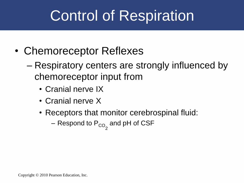
• Chemoreceptor Reflexes
– Respiratory centers are strongly influenced by
chemoreceptor input from
• Cranial nerve IX
• Cranial nerve X
• Receptors that monitor cerebrospinal fluid:
– Respond to PCO2
and pH of CSF
Copyright © 2010 Pearson Education, Inc.
Control of Respiration
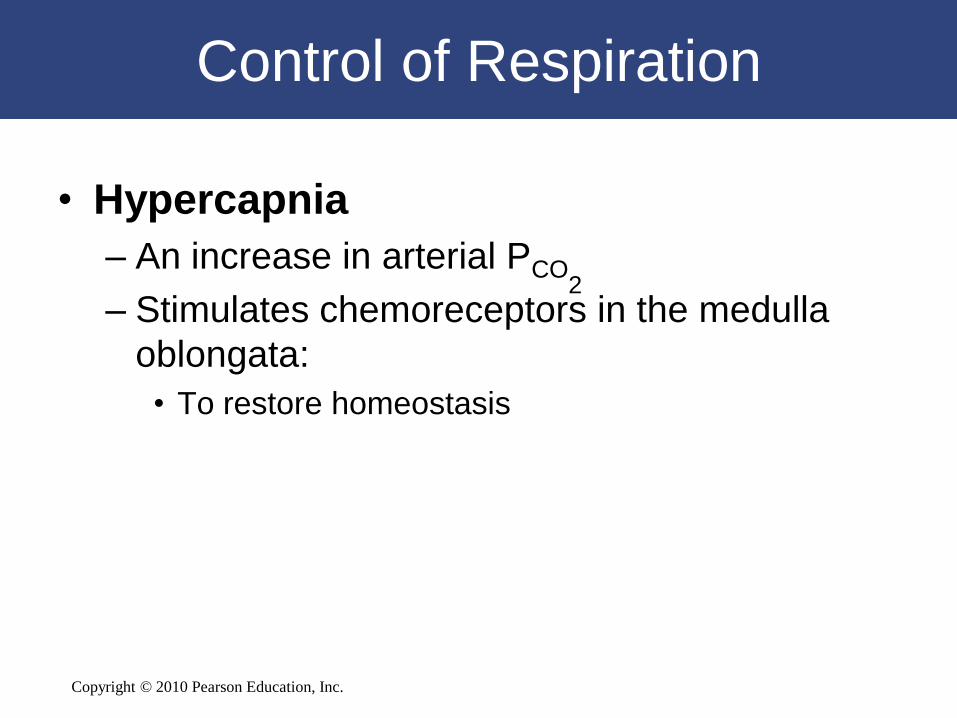
• Hypercapnia
– An increase in arterial PCO2
– Stimulates chemoreceptors in the medulla
oblongata:
• To restore homeostasis
Copyright © 2010 Pearson Education, Inc.
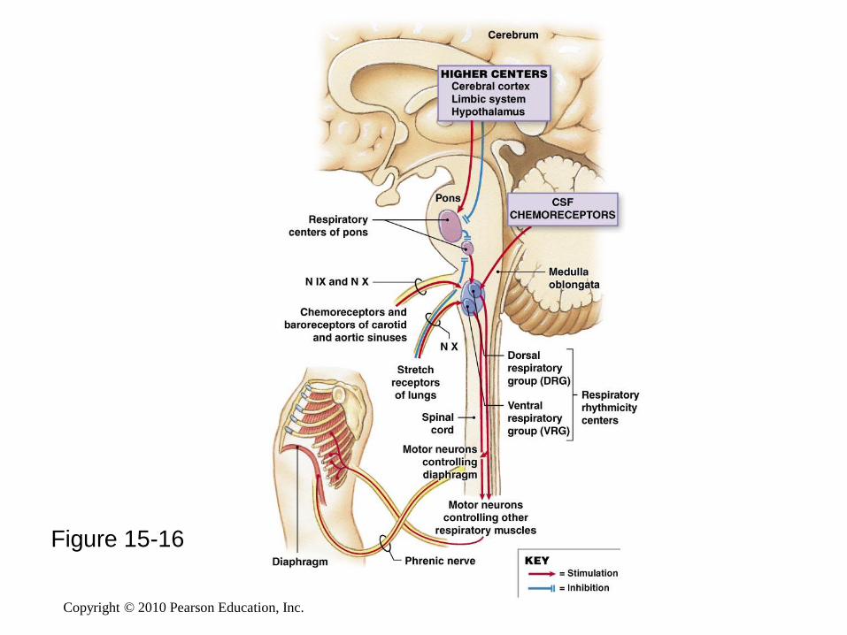
Figure 15-16
Copyright © 2010 Pearson Education, Inc.
Control by Higher Centers
• Voluntary Control of Respiration
1. Strong emotions:
• Can stimulate respiratory centers in hypothalamus
2. Emotional stress:
• Can activate sympathetic or parasympathetic division of ANS
• Causing bronchodilation or bronchoconstriction
3. Anticipation of strenuous exercise:
• Can increase respiratory rate and cardiac output
• By sympathetic stimulation
Copyright © 2010 Pearson Education, Inc.
Respiratory Changes at Birth
1. Before birth
– Pulmonary vessels are collapsed
– Lungs contain no air
2. During delivery
– Placental connection is lost
– Blood PO2 falls
– PCO2 rises
Copyright © 2010 Pearson Education, Inc.
Respiratory Changes at Birth
3. At birth
– Newborn overcomes force of surface tension to inflate
bronchial tree and alveoli and take first breath
4. Large drop in pressure at first breath
– Pulls blood into pulmonary circulation
– Closing foramen ovale and ductus arteriosus
– Redirecting fetal blood circulation patterns
5. Subsequent breaths
– Fully inflate alveoli
Copyright © 2010 Pearson Education, Inc.
15-9 Respiratory performance
declines with age
Copyright © 2010 Pearson Education, Inc.
Respiratory System and Aging
• Elastic tissues deteriorate and arthritis sets in
– Altering lung compliance
– Lowering vital capacity
– Restricting chest movements
– Limiting respiratory minute volume
• Emphysema
– Affects individuals over age 50
– Depending on exposure to respiratory irritants (e.g.,
cigarette smoke)
Copyright © 2010 Pearson Education, Inc.
15-10 The respiratory system
provides oxygen to, and
removes carbon dioxide from,
other organ systems
Copyright © 2010 Pearson Education, Inc.
Integration with Other Systems
• Maintaining homeostatic O2 and CO2
levels in peripheral tissues requires
coordination between several systems
– Particularly the respiratory and
cardiovascular systems
Copyright © 2010 Pearson Education, Inc.
Integration with Other Systems
• Coordination of Respiratory and Cardiovascular
Systems
– Improves efficiency of gas exchange by controlling
lung perfusion
– Increases respiratory drive through chemoreceptor
stimulation
– Raises cardiac output and blood flow through
baroreceptor stimulation
Copyright © 2010 Pearson Education, Inc.
The Respiratory System
in Perspective
Functional Relationships Between
the Respiratory System and Other Systems
Copyright © 2010 Pearson Education, Inc.

Copyright © 2010 Pearson Education, Inc.
• The Integumentary System
protects portions of upper
respiratory tract; hairs guard
entry to external nares
The Integumentary System
Copyright © 2010 Pearson Education, Inc.
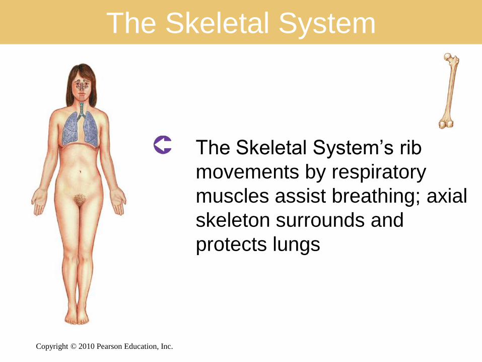
The Skeletal System
• The Skeletal System’s rib
movements by respiratory
muscles assist breathing; axial
skeleton surrounds and
protects lungs
Copyright © 2010 Pearson Education, Inc.
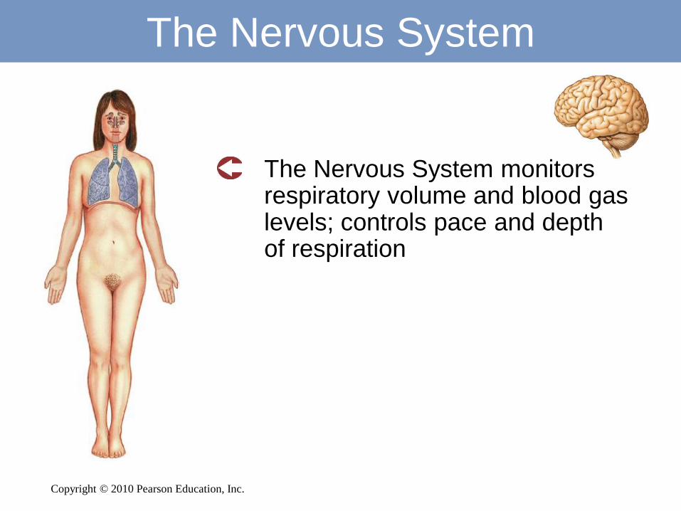
The Nervous System
• The Nervous System monitors respiratory volume and blood gas levels; controls pace and depth of respiration
Copyright © 2010 Pearson Education, Inc.
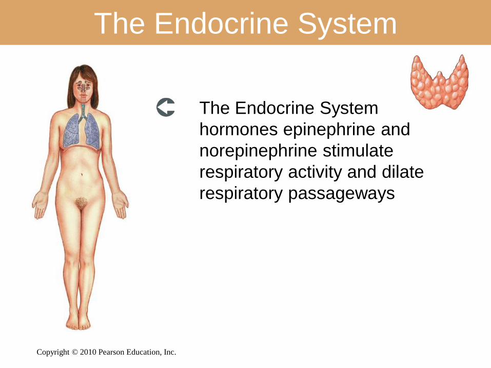
The Endocrine System
• The Endocrine System
hormones epinephrine and
norepinephrine stimulate
respiratory activity and dilate
respiratory passageways
Copyright © 2010 Pearson Education, Inc.
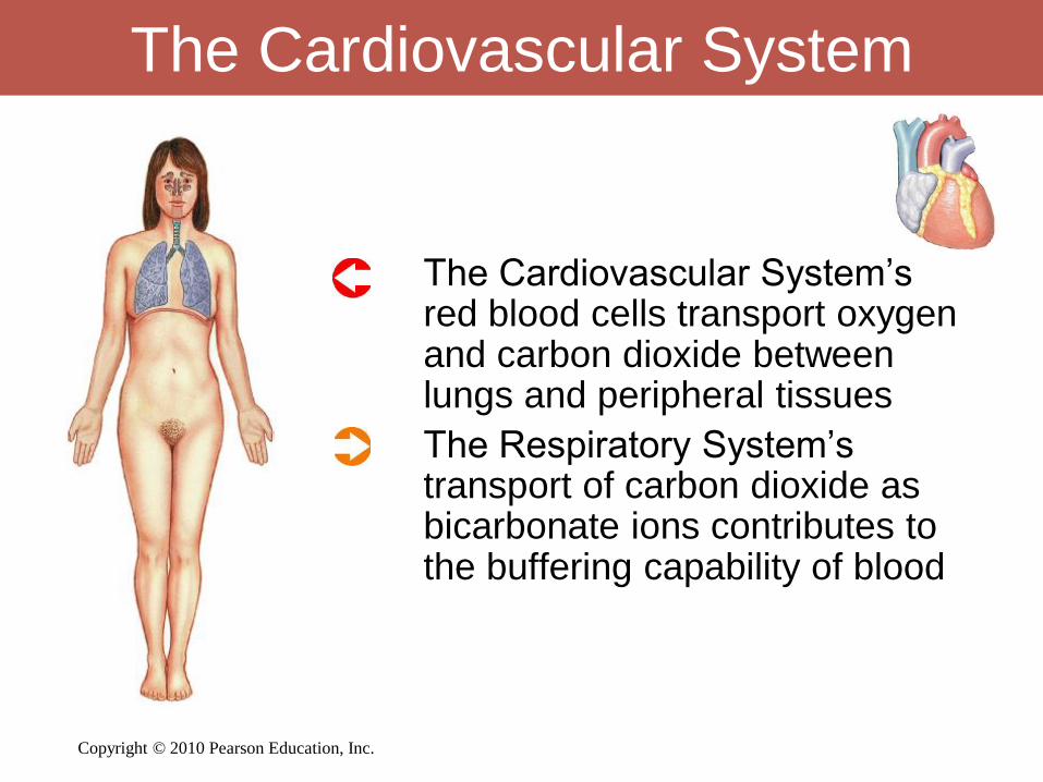
The Cardiovascular System
• The Cardiovascular System’s red blood cells transport oxygen and carbon dioxide between lungs and peripheral tissues
• The Respiratory System’s transport of carbon dioxide as bicarbonate ions contributes to the buffering capability of blood
Copyright © 2010 Pearson Education, Inc.
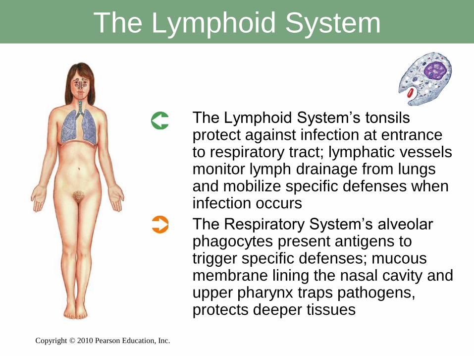
The Lymphoid System
• The Lymphoid System’s tonsils protect against infection at entrance to respiratory tract; lymphatic vessels monitor lymph drainage from lungs and mobilize specific defenses when infection occurs
• The Respiratory System’s alveolar phagocytes present antigens to trigger specific defenses; mucous membrane lining the nasal cavity and upper pharynx traps pathogens, protects deeper tissues
Copyright © 2010 Pearson Education, Inc.
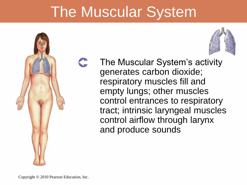
The Muscular System
• The Muscular System’s activity generates carbon dioxide; respiratory muscles fill and empty lungs; other muscles control entrances to respiratory tract; intrinsic laryngeal muscles control airflow through larynx and produce sounds
Copyright © 2010 Pearson Education, Inc.
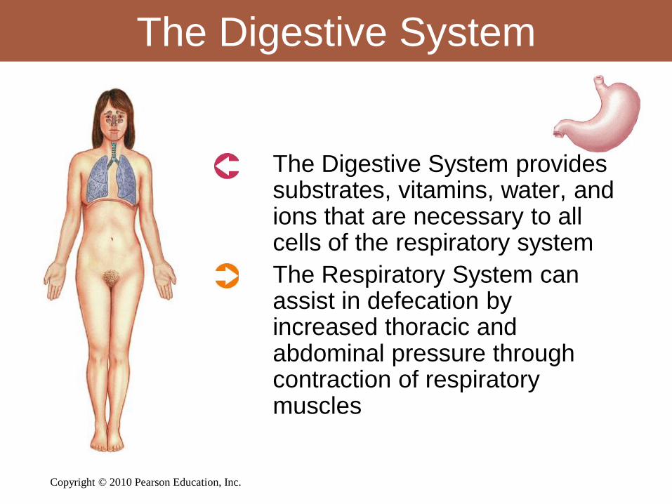
The Digestive System
• The Digestive System provides substrates, vitamins, water, and ions that are necessary to all cells of the respiratory system
• The Respiratory System can assist in defecation by increased thoracic and abdominal pressure through contraction of respiratory muscles
Copyright © 2010 Pearson Education, Inc.
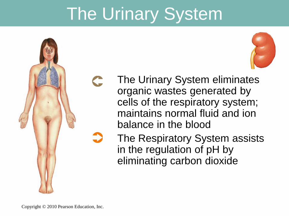
The Urinary System
• The Urinary System eliminates organic wastes generated by cells of the respiratory system; maintains normal fluid and ion balance in the blood
• The Respiratory System assists in the regulation of pH by eliminating carbon dioxide
Copyright © 2010 Pearson Education, Inc.
The Reproductive System
• The Respiratory System
changes respiratory rate and
volume during sexual arousal