11.lumbosacral radicular pain · 11. lumbosacral radicular pain ... terized by a radiating pain in...
TRANSCRIPT

papr_370 339..358
EVIDENCE-BASED MEDICINEEvidence-Based Interventional Pain Medicine
according to Clinical Diagnoses
11. Lumbosacral Radicular Pain
Koen Van Boxem, MD, FIPP*†; Jianguo Cheng, MD, PhD‡;Jacob Patijn, MD, PhD*; Maarten van Kleef, MD, PhD, FIPP*;
Arno Lataster, MSc§; Nagy Mekhail, MD, PhD, FIPP‡;Jan Van Zundert, MD, PhD, FIPP*¶
*Department of Anesthesiology and Pain Management, Maastricht University MedicalCentre, Maastricht, The Netherlands; †Department of Anesthesiology and Pain Management,
Sint-Jozefkliniek, Bornem and Willebroek, Bornem, Belgium; ‡Department of PainManagement, Cleveland Clinic, Cleveland, Ohio, U.S.A.; §Department of Anatomy and
Embryology, Maastricht University, Maastricht, the Netherlands; ¶Department ofAnesthesiology and Multidisciplinary Pain Centre, Ziekenhuis Oost-Limburg, Genk, Belgium
� Abstract: Lumbosacral radicular pain is characterized bya radiating pain in one or more lumbar or sacral der-matomes; it may or may not be accompanied by other radicu-lar irritation symptoms and/or symptoms of decreasedfunction. The annual prevalence in the general population,described as low back pain with leg pain traveling below theknee, varied from 9.9% to 25%, which means that it is pre-sumably the most commonly occurring form of neuropathicpain.
The patient’s history may give a suggestion of lumbosac-ral radicular pain. The best known clinical investigation is thestraight-leg raising test. Final diagnosis is made based on acombination of clinical examination and potentially addi-tional tests. Medical imaging studies are indicated to excludepossible serious pathologies and to confirm the affected levelin patients suffering lumbosacral radicular pain for longerthan 3 months. Magnetic resonance imaging is preferred.Selective diagnostic blocks help confirming the affectedlevel.
There is controversy concerning the effectiveness of con-servative management (physical therapy, exercise) and phar-macological treatment.
When conservative treatment fails, in subacute lumbosac-ral radicular pain under the level L3 as the result of acontained herniation, transforaminal corticosteroid adminis-tration is recommended (2 B+). In chronic lumbosacral radicu-lar pain, (pulsed) radiofrequency treatment adjacent to thespinal ganglion (DRG) can be considered (2 C+). For refractorylumbosacral radicular pain, adhesiolysis and epiduroscopycan be considered (2 B1), preferentially study-related.
In patients with a therapy-resistant radicular pain in thecontext of a Failed Back Surgery Syndrome, spinal cord stimu-lation is recommended (2 A+). This treatment should be per-formed in specialized centers. �
Key Words: lumbosacral radicular pain, epiduralcorticosteroids, pulsed radiofrequency treatment,evidence-based medicine
INTRODUCTIONThis review on lumbosacral radicular pain is part of theseries “Evidence-based Interventional Pain MedicineAccording to Clinical Diagnoses.” Recommendationsformulated in this article are based on “Gradingstrength of recommendations and quality of evidence in
Address correspondence and reprint requests to: Koen Van Boxem,MD, FIPP, Department of Anesthesiology and Pain Therapy, St.-Jozefkliniek, Bornem and Willebroek, Kasteelstraat 23, 2880 Bornem,Belgium. E-mail: [email protected].
DOI. 10.1111/j.1533-2500.2010.00370.x
© 2010 World Institute of Pain, 1530-7085/10/$15.00Pain Practice, Volume 10, Issue 4, 2010 339–358

clinical guidelines” described by Guyatt et al.,1 andadapted by van Kleef et al.2 in the editorial accompany-ing the first article of this series (Table 1).
The latest literature update was performed in Decem-ber 2009.
A lumbosacral radicular syndrome (LSR) is charac-terized by a radiating pain in one or more lumbar orsacral dermatomes; it may or may not be accompaniedby other radicular irritation symptoms and/or symp-toms of decreased function. In the literature, this disor-der can also be referred to as sciatica, ischias, or nerveroot pain. A consensus approach toward standardiza-tion of back pain definitions clearly highlights hugedifferences in the description of low back pain, whichmakes comparison of epidemiological data extremelydifficult.3 The terms radicular pain and radiculopathyare also sometimes used interchangeably, although theycertainly are not synonyms. In the case of radicular pain,only radiating pain is present, while in the case ofradiculopathy, sensory and/or motor loss that can beobjectified can be observed. Both syndromes frequentlyoccur together and radiculopathy can be a continuumof radicular pain. In this review, lumbosacral radicu-lar pain is considered as pain radiating into one ormore dermatomes caused by nerve root irritation/inflammation and/or compression.
The annual prevalence in the general population,described as low back pain with leg pain traveling belowthe knee, varied from 9.9% to 25%. Also the point
prevalence (4.6% to 13.4%) and lifetime prevalence(1.2% to 43%) are very high,4 which means that lum-bosacral radicular pain is presumably the most com-monly occurring form of neuropathic pain.5,6 The mostimportant risk factors are: being male, obesity, smoking,history of lumbalgia, anxiety and depression, workwhich requires lengthy periods of standing and bendingforward, heavy manual labor, lifting heavy objects, andbeing exposed to vibration.7
Pain completely or partially resolves in 60% of thepatients within 12 weeks of onset.8 However, about30% of the patients still have pain after 3 months to 1year. Apparently, the female population with LRS has aconsiderably worse outcome compared with the malepopulation. The estimated unadjusted odd for a long-term poor outcome was 3.3 times higher for femalepatients than for males.9
In patients under 50 years of age, a herniated disc isthe most frequent cause of an LSR. After the age of 50,radicular pain is often caused by degenerative changes inthe spine (eg, stenosis of the foramen intervertebrale).10
I. DIAGNOSIS
I.A HISTORY
The patient may experience the radiating pain as sharp,dull, piercing, throbbing, or burning. Pain caused by aherniated disc classically increases by bending forward,sitting, coughing, or (excessive) stress on the lumbar
Table 1. Summary of Evidence Scores and Implications for Recommendation
Score Description Implication
1 A+ Effectiveness demonstrated in various RCTs of good quality. The benefits clearly outweigh risk and burdens
Positive recommendation1 B+ One RCT or more RCTs with methodologic weaknesses, demonstrate effectiveness. The benefits clearly
outweigh risk and burdens2 B+ One or more RCTs with methodologic weaknesses, demonstrate effectiveness. Benefits closely balanced
with risk and burdens
2 B� Multiple RCTs, with methodologic weaknesses, yield contradictory results better or worse than the controltreatment. Benefits closely balanced with risk and burdens, or uncertainty in the estimates of benefits,risk and burdens.
Considered, preferablystudy-related
2 C+ Effectiveness only demonstrated in observational studies. Given that there is no conclusive evidence of theeffect, benefits closely balanced with risk and burdens
0 There is no literature or there are case reports available, but these are insufficient to suggest effectivenessand/or safety. These treatments should only be applied in relation to studies.
Only study-related
2 C- Observational studies indicate no or too short-lived effectiveness. Given that there is no positive clinicaleffect, risk and burdens outweigh the benefit
Negative recommendation2 B- One or more RCTs with methodologic weaknesses, or large observational studies that do not indicate any
superiority to the control treatment. Given that there is no positive clinical effect, risk and burdensoutweigh the benefit
2 A- RCT of a good quality which does not exhibit any clinical effect. Given that there is no positive clinicaleffect, risk and burdens outweigh the benefit
RCT, randomized controlled trial.
340 • van boxem et al.

discs and can be avoided by lying down or sometimesby walking.7 Inversely, pain from a lumbar spinalcanal stenosis can typically increase when walking andimprove immediately upon bending forward.10 Inaddition to the pain, the patients also often report par-esthesia in the affected dermatome. The distribution ofpain along a dermatome can be indicative in the deter-mination of the level involved; however, there is a largevariation in radiation pattern. The S1 dermatome seemsthe most reliable.11 If present, the dermatomal distribu-tion of paresthesia is more specific.10
I.B PHYSICAL EXAMINATION
The diagnostic value of anamnesis and physical exami-nation has as yet been insufficiently studied. Only paindistribution is considered to be a meaningful parameterfrom anamnesis.12 The clinical test described most oftenfor the LSR is the Lasègue test. If radicular pain can beelicited under 60°, there is a large chance that a lumbarherniated disc is present. However, the sensitivity of thistest for the detection of LSR due to a herniated discvaries sharply: the global sensitivity is 0.91 with a speci-ficity of 0.26.9,13 This specificity drops even more whenthe test is positive above 60°. The crossed Lasègue test isthe only examination with good specificity (0.88), butthis comes at the expense of the sensitivity (0.29).13 Bothtests are described in Table 2.
There is no consensus about the specificity of theother neurological signs (paresis, sensory loss, or loss of
reflexes).12 In practice, the presence of signs that areindicative of an L4 involvement (lessened patellar reflex,foot inversion) or an L5-S1 hernia (Achilles tendonreflex) are checked in a neurological examination. AnL5 motor paresis will probably be characterized clini-cally by the “stomping foot,” decreased ankle dorsiflex-ion and/or extension of the toes and an S1 paresis due toa decrease in plantar flexion, among other things10
(Table 3).In summary, a diagnosis of LSR appears to be justi-
fied if the patient reports radicular pain in one leg,combined with one or more positive neurological signsthat indicate a nerve root irritation or neurological lossof function.14
I.C ADDITIONAL TESTS
Imaging Studies
Given that the natural course of lumbosacral radicularpain is favorable in 60% to 80% of patients and that thepain improves spontaneously or even disappears com-pletely after 6 to 12 weeks, additional examination haslittle value in the acute phase.8,15 Medical imaging, pri-marily magnetic resonance imaging (MRI), can confirmthe presence of a herniated disc; this technique is pre-ferred because of the better visualization of soft tissuesand the lower radiation dose.14 The specificity of MRIand computer tomography (CT) is very low given thata herniated disc was identified by CT or MRI in 20% to
Table 2. Lasègue and Crossed Lasègue Test
The Lasègue test is performed by placing the patient in a supine position and having the patient lift up the affected leg (with a straight knee). Thetest is positive if this maneuver reproduces the symptoms. Rotation, abduction and adduction in the hip should be avoided, since these movementscan have an effect on the result
The crossed Lasègue test is performed by a patient in the supine position lifting up the contralateral leg. The test is positive if lifting is accompaniedby a pain reaction in the affected leg which follows the same pattern that appeared in the regular Lasègue test.
Table 3. Neurological Examination of the Lumbosacral Radicular Syndrome
Level Pain Sensory Loss Paresthesia Motor Disturbances or Weakness Disturbances in Reflexes
L3 Front of the thigh to the knee medial portion thigh and knee quadriceps femoris, iliopsoas, hipadductors
patellar reflex, adductor reflex
L4 Medial portion leg medial portion leg anterior tibialis, quadriceps, hipadductors
patellar reflex
L5 Lateral portion thigh and leg,dorsum of the foot
lateral portion leg, dorsum of foot,first toe
toe extensors and flexors, ankledorsiflexors, eversion andinversion of the ankle, hipabductors
S1 Posterior portion thigh, calf andheel
sole of the foot, lateral portionfoot and ankle, two most lateraltoes
gastrocnemius, biceps femoris,gluteus maximus, toe flexors
Achilles reflexes
Adapted from: Tarulli AW, Raynor EM: Lumbosacral radiculopathy. Neurol Clin. 2007; 25 (2): 387–405.
Lumbosacral Radicular Pain • 341

36% of the asymptomatic population,16 and thereis little correlation between the severity of a possibleradiculopathy and the magnitude of the spinal discherniation. Incidentally, the symptoms can disappearafter a conservative therapy without a correspondingdecrease in the volume of the herniated disc.17–19
In addition to this, a hernia could not be demon-strated on the scans of some patients with clinicalsymptoms of a radicular syndrome.20,21 In the eventof an unclear clinical picture or in the absence ofradiological arguments for radicular complaints, elec-tromyography (EMG)/nerve conduction studies (NCS)can be performed to differentiate lumbar radicular syn-drome from peripheral neuropathy (sensitivity 0.45 to0.65).22 Other common causes of lumbar radicularpain, such as stenosis of the foramen intervertebrale,may be revealed by MRI or CT. Entrapment ofthe sciatic nerve such as piriformis syndrome is notincluded in this chapter.
Selective Segmental Nerve Blocks
Although the diagnostic nerve root block is a commonlyused technique for determining the level of the radicularpain, there is uncertainty concerning its sensitivity andspecificity. In a LSR without clear signs of a focal neu-rological deficit, there appears to have been a variablehypoesthesia already present in the majority of thepatients before the execution of a diagnostic nerve rootblock.23 These changes in sensory function can also varyin time and location.
With an intraforaminal block, there is also a realchance of a simultaneous block of the nervus sinuverte-bralis. This nerve is responsible for the afferent input ofthe nearby disci intervertebrales (superficial annulusfibrosus), ligamentum longitudinale posterius, and theventral dura mater and nerve root sleeve. In addition,the sensory fibers of the ramus dorsalis of the segmentalnerve pass through the ganglion spinale (dorsal rootganglion, DRG) which is also blocked. This nerve inner-vates local back muscles and nearby facet joints. Fur-thermore, it is known that if the etiology of the pain islocated proximally to a nerve block, this pain can bereduced by a peripheral nerve block. As a result, painthat originates from proximal spinal nerve root irrita-tion with corresponding pain in the leg and back can infact be influenced by a more peripheral block.24 Thiswas confirmed in a study by North25 in which patientswith radicular pain as their chief complaint had, in arandomized sequence, 4 different blocks with local
anesthetic. Paraspinal lumbosacral root blocks andmedial branch posterior primary ramus blocks (at thesame level or proximally) as well as nervus ischiadicus(sciatic nerve) blocks (collaterally or distal to the pathol-ogy) with 3 mL bupivacaine 0.5% provide a temporarygreater pain reduction in the majority of cases, in com-parison with a lumbar subcutaneous administration ofthe same product in an identical volume. The specificityof a single-level diagnostic block is further influenced bythe injected volume, as 0.5 mL of contrast alreadyreaches the adjacent level in 30% of cases, and 1.0 mLeven in 67% of cases.26 As a result, it appears that thespecificity of diagnostic nerve root blocks is limited: anegative block has a specific predictive value, but iso-lated positive blocks are nonspecific.27
An example of the variability of the effect ofnerve root blocks in patients with LSR withoutneurological deficit is the incidence, location,and extent of the dermatomal areas witha hypoesthesia. Namely, the total area in whichhypoesthesia can be found is very extensive, yet it isexceptional that in some patients, absolutely no hypo-esthesia develops even though the technique performedis identical.27 This pattern of hypoesthesia and radicu-lar pain usually surpasses the boundaries of standarddermatomal charts, but is better understood if anoverlap with the adjacent dermatomes is taken intoaccount. The resulting adapted dermatomes are twiceas large as those in standard dermatomal charts, but asa result, the sensory effects of diagnostic nerve rootblocks lie more within the limits of the (adapted) der-matomal charts.27
Conversely, the variability of paresthesia as a resultof electro-stimulation appears to be much smaller; itis usually registered in the central sections of the stan-dard dermatomes. The reproducibility of paresthesiaby electro-stimulation also appears to be high: 80% ofthe paresthesia can be traced to within the borders of thestandard dermatomal charts, and 98% to within theborders of the adapted dermatomal charts. In spite ofthis, the relationship to pain remains unclear. When painis reported in an “adapted” dermatome, in only 1/3 ofcases can a corresponding reduction in pain, paresthe-sia, and hypoesthesia be induced by electro-stimulationand nerve root blocks.
After a nerve root block, the average muscle force isreduced within the corresponding myotome, but themuscle force within the myotome is increased if theblock has reduced the pain.28 A possible explanation forthe increase in muscle force in patients with a chronic
342 • van boxem et al.

lumbar radicular syndrome is the finding that pain hasan inhibiting effect on the muscle force (diffuse noxiousinhibitory control or DNIC).29 After pain reduction, theinhibition lessens which results in a normalization of themuscle force.30
In practice, the most rational method used to confirmthe suspected level of radicular complaints is still theuse of one or more selective diagnostic blocks. Theseselective infiltrations must occur with a limited amountof local anesthetic (max. 1 mL) per level and in separatesessions.
I.D DIFFERENTIAL DIAGNOSIS
In cases of acute low back pain, physical abnormalities,which can account for the complaints, are ruled out firston the basis of the so-called “red flags”; yet in cases ofchronic low back pain, we recommend also checkingwhether there are signs which could indicate underly-ing pathology such as tumors and infections, amongothers (Table 4). When making a differential diagnosis,inflammatory/metabolic causes (diabetes, ankylosingspondylitis, Paget’s disease, arachnoiditis, sarcoidosis)must also be taken into account; these must be ruled outfirst.10
The acute cauda equina syndrome is usually theresult of a large, central disc herniation with compres-sion of the low lumbar and sacral nerve roots, usually atthe L4-L5 level. As a result of the sacral polyradiculopa-thy, a significant bowel and micturition dysfunction canarise with a characteristic saddle anesthesia. If thelumbar nerve roots are also involved, this leads to weak-ness in the legs that can possibly lead to paraplegia.Rapid recognition of these symptoms and referral foremergency surgery is recommended.10
II. TREATMENT OPTIONS
II.A CONSERVATIVE MANAGEMENT
(Sub)Acute Radicular Complaints
Controversy exists concerning the conservativeapproach to LSR since there is no strong evidence of theeffectiveness of most treatments.31 Providing adequateinformation to the patient about the causes and prog-nosis of LSR can be a logical step in the management ofthis problem, but this has not yet been studied in ran-domized, controlled studies.14
There is no difference between the advice for bed restwhen compared with the advice to remain active.32
The use of Non-Steroidal Anti-Inflammatory Drugsor Cox-2 inhibitors can have a significant effect on acuteradicular pain compared with placebo.33,34 There arehowever no long-term results on the evolution of LRS.
Exercise therapy can possibly have a beneficial effect.For this reason, it is often considered a first-line treat-ment. However, until now, evidential value for this islacking.12,31 A randomized study was able to demon-strate a better outcome after 52 weeks in patients whoreceived physiotherapy in the form of exercise therapycombined with a conservative therapy from the generalpractitioner in comparison with patients who receivedonly the conservative therapy (79% versus 56% GlobalPerceived Effect, respectively). However, this does notappear to be cost-effective.35 For a selected population,a surgical intervention results in a more rapid lesseningof the acute radicular complaints in comparison with aconservative approach, but the outcomes after 1 to 2years are equivalent.36–38 Furthermore, the effect ofsurgery on the natural course of the herniated discdisease is unclear and there are no proven arguments foran optimal time period for surgery.39
For patients with a neurological loss of function dueto a herniated disc, immediate surgical treatment isusually recommended. From the available studies, itappears that this loss of function remains steady ini-tially, but after surgery it can still regress (up to 50% ofthe patients).40,41 It can therefore be stated that theoutcome in cases of herniated disc with regard to neu-rological loss of function is determined by the severity ofthe lesion at the outset and not by whether an interven-tion occurs sooner or later.42
In patients with a spinal canal stenosis with second-ary neurological loss of function on which surgery hasbeen performed, reflex disturbances and sensory andmotor deficits will be permanent or will only very slowly
Table 4. Red Flags
First appearance of back complaints before 20th or after the 55th yearTraumaConstant progressive back painMalignant disorder in the medical historyLong-term use of corticosteroidsDrug use, immunosupression, HIV(Frequent) general malaiseUnexplained weight lossStructural deformities of the spinal columnInfectious disorders (eg, herpes zoster, epidural abscess, HIV, Lyme
disease)Neurological loss of function (motor weakness, sensory disturbances,
and/or micturition disturbances)
Lumbosacral Radicular Pain • 343

be partially restored. Up to 70% of the patients willcontinue to have residual neurological abnormalitiesafter decompression43 and the risk of permanent neur-opathy is larger in central spinal canal stenosis in com-parison with lateral spinal canal stenosis.44
Chronic Radicular Complaints
The place of physiotherapy in these cases is also unclear,since there are no randomized studies available.45 Forchronic LSR, a trial period with medication is indicated.Classically, neuropathic pain is treated by prescribingtricyclic antidepressants (TCAs) such as amitriptyline.46
Although a medicinal treatment policy is still in theforeground, in practice, this is not always evident. Thus,for these neurogenic conditions, less than 1/3 of thepatients will experience a reduction in pain that is betterthan “moderate”.46 Furthermore, various reviews wereperformed concerning the place of the TCAs47 and anti-convulsants6,46 in the treatment of neuropathic pain. It isstriking that the included studies were mostly performedin patients with diabetic neuropathy and postherpeticneuralgia. The extension of these results to patients withLRS, with a physiopathology based more on compres-sion and inflammation of the nerve root and the ganglionspinale (DRG) has not yet been scientifically proven.5
Anticonvulsants are a possible alternative for thetreatment of neuropathic pain if tricyclic antidepres-sants cannot be tolerated or are contraindicated. Gaba-pentin has been studied most often in this indication andis supported by a randomized controlled trial (RCT).48
The results are variable and optimization of the dosageis frequently hindered by side effects. The role of opioidsin the treatment of neuropathic pain has long beenconsidered controversial. Recent guidelines concerningthe treatment of neuropathic pain mention tramadoland oxycodone as possible therapeutic options.6 In anopen-label trial using transdermal fentanyl in 18patients with radicular pain, an average pain reductionof 32% was achieved.49
II.B INTERVENTIONAL MANAGEMENT
Anesthesiological treatment techniques are indicated forpatients with radicular pain. Epidural administration ofcorticosteroids is generally indicated in cases of sub-acute radicular pain. In patients with chronic radicularcomplaints, corticosteroids will not provide anyimprovement in the outcome in comparison with localanesthetics alone. This indicates that epidural corticos-teroids are more effective for (sub)acute radicular pain
where a significant inflammatory pain component ispresent.50 (Pulsed) radiofrequency (PRF) treatment is atreatment option for chronic radicular pain.
Epidural Corticosteroid Administration
The logic of epidural corticosteroid administration restson the anti-inflammatory effect of the corticosteroids,which are administered directly onto the inflamed nerveroot. There are three approaches: interlaminar, transfo-raminal, and caudal.
Interlaminar Corticosteroids. The available evidenceconcerning interlaminar corticosteroid administrationhas been studied in systematic reviews. The conclusionsof these reviews are divergent depending on the chosenevaluation parameters. McQuay and Moore calculatedthe Number Needed to Treat (NNT). To achieve 50%pain reduction in the short term (1 day to 3 months), anNNT of 3 is obtained and an NNT of 13 for long-termpain relief (3 months to 1 year).51 A systematic review ofRCTs concluded that there is insufficient proof of theefficacy of this technique. If there are benefits, then theyare of short duration.52 A recent systematic review ofRCTs showed that among the 11 RCTs of interlaminarsteroid injection for radiculopathy, four trials are ratedhigh quality.53 Three of the four trials used ligamantuminterspinale (interspinous ligament) saline injection ascontrol intervention. All three trials showed positiveresults for short-term benefits (2 1 months).54–56 Theother trial used epidural saline injection as control anddid not show any benefit.57
Transforaminal Corticosteroids. The variable resultsof corticosteroids administered interlaminarly areascribed to the fact that there is no certainty that theneedle reaches the epidural space and even if it did,there is no certainty that the medication reaches theventral section of the epidural space.58 Transforaminaladministration allows a more precise application of thecorticosteroids at the level of the inflamed nerve root.Three high quality, placebo controlled trials evaluatingtransforaminal approach reported mixed results.53 Oneshowed long-term benefits in one year,59 one showedmixed short-term benefits,60 and one showed nobenefit.50
In a double-blind, randomized study, patients whowere scheduled for surgical intervention received an epi-dural injection with local anesthetic only or local anes-thetic with corticosteroid at random. By the follow-up(13 to 28 months), 20/28 patients in the local anesthetic
344 • van boxem et al.

with corticosteroid group had decided not to undergosurgery, while in the local anesthetic only group, 9/27decided to forego a surgical intervention.59 The majority(81%) of the patients who had not yet had surgery 1year after infiltration were able to avoid the operationafter 5 years.61 There was no statistical differencebetween the treatment groups.
A prospective controlled study of transforaminal epi-dural corticosteroids showed superiority of this proce-dure over trigger-point injection in patients with discherniation.62 Karpinnen’s group60 carried out a random-ized, controlled study in patients with radicular painand disc herniation documented by MRI, in which thetransforaminal administration of local anesthetic withcorticosteroid was compared with transforaminal injec-tions of normal saline solution. Two weeks after thetreatment, the clinical result in the corticosteroid groupwas better than that of the group treated with normalsaline solution. After 3 to 6 months, on the other hand,patients in the group with normal saline were in bettercondition owing to a rebound effect that was noted inthe corticosteroid group. A subanalysis in which theresults of patients with a “contained” herniation werecompared with those of patients with an “extruded”herniation showed that in the first group, corticosteroidinjections were superior to placebo while in the groupwith “extruded” herniation, the opposite was found.63
In this study, “contained herniation” was definedas a herniation with a broad base, which is still con-tained within the ligamentum longitudinale posterius.“Extruded herniation” is a herniation that breaksthrough the ligamentum longitudinale posterius.
In a comparative study, the effectiveness of caudal,interlaminar, and transforaminal corticosteroid admin-istration in the epidural space was compared in patientswith radicular pain as a result of disc herniation. Thetransforaminal approach gave the best clinical results.64
A double-blind, randomized study compared the effi-cacy of interlaminar and transforaminal corticosteroidadministration in patients with lumbar radicular pain asa result of CT- or MRI-confirmed herniated disc thatlasted less than 30 days. Six months after the treatment,the results in the transforaminal-treatment group wassignificantly better than that of the group that wastreated interlaminarly in the areas of pain reduction,daily activity, free-time and work activities, and anxietyand depression.65
Caudal Corticosteroids. Four placebo-controlledtrials were conducted, but none were rated high quality.53
The results are mixed and no definitive conclusions canbe drawn from these studies.
In summary, one can state that the transforaminalepidural corticosteroid administration is preferable. Inpractice, due to the not-yet-completely elucidated, rareneurological complications associated with the transfo-raminal administration route, the interlaminar andcaudal approaches can also still be considered.
PRF
The application of conventional RF treatment (at 67°C)adjacent to the lumbar ganglion spinale (DRG) has lostinterest because no extra value could be shown in com-parison with a sham procedure in a randomized,double-blind, sham-controlled study.66
PRF treatment adjacent to the lumbar ganglionspinale (DRG) was studied in a retrospective study. In agroup of 13 patients for which a surgical interventionwas planned, the PRF treatment adjacent to the gan-glion spinale (DRG) of the nerve involved precluded theintervention in 11 patients. One patient had a discoperation and 1 underwent a spinal fusion 1 year afterthe treatment without having radicular pain at the timeof the operation.67 In another retrospective study, PRFtreatments were carried out in patients with a radicularsyndrome as a result of disc herniation, spinal canalstenosis, or failed back surgery syndrome (FBSS). Asignificant reduction in pain and in analgesic consump-tion was attained in the patients with a disc herniation(NNT: 1.38) and spinal canal stenosis (NNT: 1.19), butnot in those with FBSS (NNT: 6.5).68 An RCT aimed atidentifying the potential additional effect of a conven-tional RF treatment directly after a PRF treatment adja-cent to the lumbar ganglion spinale (DRG). Thirty-sevenpatients were treated with PRF and 39 patients withPRF and RF. A marked decrease in VAS pain score wasobserved in both groups, but no significant differencebetween groups in pain reduction and duration of actioncould be identified.69
Adhesiolysis and Epiduroscopy
The goal of lysis of epidural adhesions is to removebarriers in the epidural space that may contribute topain generation and prevent delivery of pain relievingdrugs to target sites.
The development of a navigable, radio-opaque, kink-resistant, soft-tipped catheter has allowed placement ator near this target site in most patients. In the literature,adhesiolysis with or without endoscopic control issometimes assessed together. There are 2 RCTs on
Lumbosacral Radicular Pain • 345

fluoroscopic-guided adhesiolysis. Patients included inthe RCTs suffered chronic low back pain and sciaticaand might have undergone previous back surgery, fur-thermore the treatment protocols differed. Heavneret al.70 compared the effect of mechanical adhesiolysiswith (1) a combination of hyaluronidase and hypertonicsaline; (2) hypertonic saline solution; (3) isotonic salinesolution; and (4) hyaluronidase and isotonic saline solu-tion. The treatment consisted of a 3-day procedurewhere the catheter was inserted and the drugs wereinjected on three consecutive days. Manchikanti et al.71
assessed a one-day procedure in 3 patient groups: acontrol group treated with injection of local anestheticcorticosteroid and normal saline without adhesiolysis;the second group consisting of patients undergoingadhesiolysis, with injection of local anesthetic, steroid,and normal saline; and the third group consisting ofpatients undergoing adhesiolysis, with an injection of10% sodium chloride solution, in addition to local anes-thetic and steroid. The third trial compared the effect ofadhesiolysis and injection of corticosteroid and localanesthetic followed, 30 minutes later, by an injection ofhypertonic saline (10%) with conservative treatment.72
These trials and all the observational trials but onefound positive short- and long-term outcome. The trialon the effect of adhesiolysis with hypertonic salinefound only short-term positive outcome.73
Epiduroscopy, which is also called spinal endoscopy,is an alternative way to perform adhesiolysis undervisual control. It couples the possibility of diagnosticand therapeutic interventions in one session. Thistechnique was evaluated in 2 systematic reviews.74,75
A prospective randomized trial showed significantimprovement without adverse effects in 80% of thepatients receiving epiduroscopy at 3 months, 56% at 6months, and 48% at 12 months, compared with 33% ofthe patients in the control group showing improvementat one month and none thereafter.76 In an RCT, 60patients with a 6-to-18-month history of sciaticareceived either targeted epidural local anesthetic andsteroid placement with manipulation of the adhesionsusing a spinal endoscope or caudal epidural local anes-thetic and steroid treatment. No significant differenceswere found between the groups for any of the measuresat any time.77 Observational studies showed good short-and long-term pain relief.78–83
Spinal Cord Stimulation in FBSS
FBSS is a persistent back pain that may or may notinclude pain radiating to the leg after one or more back
operations. Spinal cord stimulation (SCS) consists of thepercutaneous application of electrodes at the level of thespinal cord segment involved. These electrodes are thenconnected to a generator that delivers electrical pulses tostimulate the painful dermatome and to induce alteredpain conductivity, transmissibility, and perception. Asystematic review of the effectiveness of SCS for thetreatment of chronic low back and leg pain in patientswith FBSS included an RCT, a cohort study and 72 casereports. The RCT demonstrated clear advantage of SCSin comparison with repeat surgery. However, the resultsof the case reports are very heterogeneous.84 A random-ized study that compared SCS with conventional treat-ment in FBSS patients showed that fewer patients fromthe SCS group switched over to conventional treatmentthan did patients who initially received a conventionaltreatment and then switched over to SCS. The numberof patients satisfied with the treatment was higher in theSCS group.85
II.C COMPLICATIONS OF INTERVENTIONALMANAGEMENT
Complications and Side Effects ofEpidural Corticosteroids
Interlaminar Epidural Corticosteroids. The most fre-quent side effect is a dural puncture (2.5%) with orwithout a transient headache (2.3%).86 Minor sideeffects, such as transient increase in complaints or theappearance of new neurological symptoms more than24 hours after the infiltration, occur in 4% of thepatients; the median duration of the complaints was 3days (1–20 days).87 In a study examining side effects in4,722 infiltrations with betamethasone dipropionateand betamethasone sodium phosphate, 14 (0.7%)serious side effects were reported (cardiovascular, gas-trointestinal, allergy), 7 of which were attributed to theproduct.88 More serious complications are cases ofaseptic meningitis, arachnoiditis, and conus medullarissyndrome, but these typically occur after multiple acci-dental subarachnoidal injections. Two cases of epiduralabscess, 1 case of bacterial meningitis, and 1 case ofaseptic meningitis were also reported.89
Transforaminal Epidural Corticosteroids. At the timeof preparing this manuscript, 7 publications report 9cases of neurological complications such as paraplegiafollowing lumbar transforaminal epidural corticosteroidadministration.90–96 The probable mechanism is aninjury to an unusually low dominant radiculomedullary
346 • van boxem et al.

artery.90 The largest radicular artery is the arteriaradicularis magna (artery of Adamkiewicz); in 80% ofthe population, this artery is present in the spinal canalbetween T9 and L1. However, in a minority of cases, itcan occur between T7 and L4, which results in thepossibility that the artery is in the vicinity of the endposition of the needle in a transforaminal infiltration.Depot injections can then mimic an embolism; if thisoccurs in a critical artery which supplies the anteriorspinal artery, spinal cord ischemia may result.97 Of thereported cases of neurologic complications, 1 occurredafter Th12-L1, 1 case at L1-L2, 2 cases at L2-L3, 3 casesat L3-L4, 1 after simultaneous L3-L4 and L4-L5 injec-tion, and finally, 1 case after an S1 injection.
A retroperitoneal hematoma was reported in apatient having anticoagulant therapy who received atransforaminal injection.60 Two cases of dural punc-ture,98 one disc entry,99 one case of cauda equina100 andone case of transient blindness attributed to the tempo-rarily intra-epidural pressure increase.101 Infectiouscomplications such as epidural abscess caused by MRSA(1 case),102 discitis (1 case)103 and one case of vertebralosteomyelitis104 are reported.
The recently reported cases of serious complicationwith the transforaminal approach warrant a cautiouspolicy. It is recommended to only perform transforami-nal infiltrations under the L3 level and to always admin-ister the injection fluid during real-time imaging, theadditional use of digital subtraction angiography maybe of value. It is also recommended to first administer atest dosage of local anesthetic before infiltrating thedepot corticosteroid after waiting 1 to 2 minutes toobserve potential neurologic signs.105 Neurological com-plications rarely occur when using the correct techniqueand when sedation is avoided. If a significant increase inpain is reported during the injection of contrast agent,local anesthetic and/or corticosteroids, the proceduremust be immediately stopped in order to ascertain thecause of the pain.
Endocrine Side Effects. Cushing’s syndrome wasreported in the prospective study of the side effects ofepidurally administered betamethasone dipropionateand betamethasone sodium phosphate.88
Side Effects and Complications ofRF Treatments
Conventional RF Treatment. A burning pain wasfound to occur in 60% of RF-treated patients, and ahyposensitivity in the associated dermatome in 35% of
RF-treated patients.106 These side effects disappearedspontaneously after 6 weeks. However, in a later study,there was no difference in side effects and complicationsbetween a classic RF group and a sham group.66
PRF Treatment. In an extensive review of the litera-ture on the use of PRF covering over 1,200 patients noneurological complication was identified.107 Twelvepublications are currently available regarding PRFtreatment adjacent to the ganglion spinale (DRG).Eight of those publications specifically report PRF treat-ment adjacent to the lumbar ganglion spinale(DRG).67–69,108–112 In total information on 295 PRF pro-cedures is listed and no side-effects or complications arementioned.
Side Effects and Complications of EpiduralAdhesiolysis and Epiduroscopy
Four studies look specifically into the complicationsof epidural adhesiolysis.113–116 The most commonlyreported complications of percutaneous adhesiolysisare dural puncture, catheter shearing, and infection.Other potential complications include intravascularinjection, vascular injury, cerebral vascular or pulmo-nary embolus, reaction to the steroids, hypertonicsaline, or hyaluronidase, and administration of highvolumes of fluids potentially, resulting in excessive epi-dural hydrostatic pressures, brain damage, and death.
Talu and Erdine113 reviewed percutaneous adhesioly-sis complications in 250 patients. Three patients (1.2%)developed epidural abscesses, and 1 patient developed asevere headache. Retained sheared adhesiolysis catheterwas described in a patient who underwent percutaneousadhesiolysis to treat persistent back and leg pain after 2previous lumbar surgeries.114
Unintended subarachnoid or subdural puncture withinjection of local anesthetic or hypertonic saline is oneof the major complications of the procedure with cath-eter adhesiolysis.
For epiduroscopy, side effects and complications arecomparable to those of adhesiolysis without endoscopiccontrol. There is however an additional potential ofincreased pressure in the epidural space due to the con-tinuous pressurized liquid injection, necessary to obtaina clear image. Up till now, only one report of visualdisturbances due to increased liquor pressure has beenreported. Careful monitoring of pressure fluctuations iswarranted to reduce the risk of prolonged increasedliquor pressure and the duration of the procedureshould be limited to maximum 60 minutes.
Lumbosacral Radicular Pain • 347

Side Effects and Complications of SCS
In a review of the complications of SCS, 18 studies on112 patients receiving SCS for FBSS were identified.Forty-eight patients (42%) reported a side effect or com-plication. Complications can be subdivided in: techni-cal, biological (postoperative), and others. The majority(> 25%) of the complications are of technical order suchas lead migration, lead breakage, hardware malfunc-tion, battery failure, and loose connection. Postsurgicalcomplications can be infection, cerebrospinal fluidleakage, and hematoma. Undesirable stimulation, painover the implant, skin erosion, and allergy have alsobeen reported.117
II.D EVIDENCE FORINTERVENTIONAL MANAGEMENT
The summary of the evidence for interventional man-agement of lumbosacral radicular pain is given inTable 5.
III. RECOMMENDATIONSBased on the evidence available regarding effects andcomplications, we recommend the following techniquesfor the treatment of LRS:
• Since epidural corticosteroid injections havemainly short-term effects; these techniques arerecommended for patients with subacute radicu-lar pain symptoms.
• In patients with pain at the lumbosacral level(L4, L5, S1) as a result of a “contained hernia-tion,” a transforaminal epidural injection withlocal anesthetic and corticosteroids is recom-mended. A preference seems to exist for transfo-
raminal epidural corticosteroid administrationover caudal and interlaminar corticosteroidsbelow level L3.
• RF treatment adjacent to the ganglion spinale(DRG) is not recommended. A PRF treatmentadjacent to the ganglion spinale (DRG) can beconsidered.
• Spinal cord stimulation is recommended forpatients with a therapy-resistant radicular syn-drome, but only in specialized centers.
• Epiduroscopy and adhesiolysis can be consideredin the context of research and only in specializedcenters.
III.A CLINICAL PRACTICE ALGORITHM
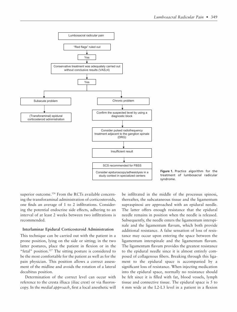
Figure 1 represents the treatment algorithm based onthe available evidence.
III.B TECHNIQUES
Practical Recommendations EpiduralCorticosteroid Administration
There are 7 systematic reviews concerning epidural cor-ticosteroid administration for the treatment of LRS.With regard to short-term effectiveness, 6 of the 7 sys-tematic reviews give a positive assessment and 1 gives anegative assessment (conflicting evidence).86,118–122 Thereare no comparative studies available for the effective-ness and/or complications of the various depot corticos-teroids, which means that a distinction between theseproducts cannot be verified.
It is possible that the particle size of the depot corti-costeroid is related to the reported neurological compli-cations, but the literature concerning this possibility isalso inconclusive.123 Up till now, no reported neurologiccomplications were noted with the nonparticulate cor-ticosteroid dexamethasone. One abstract has prospec-tively compared the transforaminal use of triamcinolonewith dexamethasone in 50 patients.124 A significantgreater reduction in pain was noted after 2 weeks inpatients treated with triamcinolone, so this far evidenceabout its efficacy at the lumbar level is lacking. Cur-rently, there is no evidence that a higher corticosteroiddosage produces a better clinical effect,125 yet the risk ofendocrine side effects is substantially higher. It is for thisreason that the lowest dosage of depot corticosteroid iscurrently recommended.
With regard to the number of infiltrations, there areno comparative studies that have shown that the sys-tematic implementation of 3 infiltrations would result in
Table 5. Summary of the Evidence for InterventionalManagement
Technique Assessment
Interlaminar corticosteroid administration 2 B1Transforaminal corticosteroid administration in “contained
herniation”2 B+
Transforaminal corticosteroid administration in “extrudedherniation”
2 B-
Radiofrequency lesioning at the level of the spinalganglion (DRG)
2 A-
Pulsed radiofrequency treatment at the level of the spinalganglion
2 C+
Spinal cord stimulation (FBSS only) 2 A+Adhesiolysis—epiduroscopy 2 B1
348 • van boxem et al.
superior outcome.126 From the RCTs available concern-ing the transforaminal administration of corticosteroids,one finds an average of 1 to 2 infiltrations. Consider-ing the potential endocrine side effects, adhering to aninterval of at least 2 weeks between two infiltrations isrecommended.
Interlaminar Epidural Corticosteroid Administration
This technique can be carried out with the patient in aprone position, lying on the side or sitting; in the twolatter postures, place the patient in flexion or in the“fetal” position.127 The sitting posture is considered tobe the most comfortable for the patient as well as for thepain physician. This position allows a correct assess-ment of the midline and avoids the rotation of a lateraldecubitus position.
Determination of the correct level can occur withreference to the cresta illiaca (iliac crest) or via fluoros-copy. In the medial approach, first a local anesthetic will
be infiltrated in the middle of the processus spinosi,thereafter, the subcutaneous tissue and the ligamentumsupraspinosi are approached with an epidural needle.The latter offers enough resistance that the epiduralneedle remains in position when the needle is released.Subsequently, the needle enters the ligamentum interspi-nale and the ligamentum flavum, which both provideadditional resistance. A false sensation of loss of resis-tance may occur upon entering the space between theligamentum interspinale and the ligamentum flavum.The ligamentum flavum provides the greatest resistanceto the epidural needle since it is almost entirely com-posed of collagenous fibers. Breaking through this liga-ment to the epidural space is accompanied by asignificant loss of resistance. When injecting medicationinto the epidural space, normally no resistance shouldbe felt since it is filled with fat, blood vessels, lymphtissue and connective tissue. The epidural space is 5 to6 mm wide at the L2-L3 level in a patient in a flexion
Figure 1. Practice algorithm for thetreatment of lumbosacral radicularsyndrome.
Lumbosacral radicular pain
“Red flags” ruled out
Conservative treatment was adequately carried out
without conclusive results (VAS≥4)
Yes
(Transforaminal) epidural corticosteroid administration
Chronic problem
Confirm the suspected level by using a diagnostic block
Consider pulsed radiofrequency treatment adjacent to the ganglion spinale
(DRG)
Insufficient result
SCS recommended for FBSS
Consider epiduroscopy/adhesiolysis in a study context in specialized centers
Yes
Subacute problem
Lumbosacral Radicular Pain • 349

position. In addition, the injection of contrast agent canverify the correct positioning in the epidural space.
In the case of aspiration of blood, the needle must bereoriented; in the case of aspiration of cerebrospinalfluid, the procedure must be repeated at another level. Inthe latter case, an overflow to the cerebrospinal fluid ispossible; therefore, this procedure must be carried outwith caution.
Classically, an infiltration consists of an injection of alocal anesthetic with a corticosteroid. There is a ten-dency to perform this procedure under fluoroscopy, yetthus far, no advantages of fluoroscopic control havebeen demonstrated.128,129
Transforaminal EpiduralCorticosteroid Administration
In a transforaminal approach, the C-arm is adjusted insuch a way that the X-rays run parallel to the coverplates of the relevant level. Thereafter, the C-arm isrotated until the processus spinosus projects over thecontralateral facet column. With the C-arm in this pro-jection, the injection point is found by projecting a metalruler over the medial part of the foramen interverte-brale. If there is a superposition of the processus articu-laris superius (superior articular process) of theunderlying joint, the C-arm must be rotated cranially.
A 10-cm long, 25-G or 22-G needle with connectiontubing that is first flushed with contrast medium isinserted here locally in the direction of the radiationbeam. Thereafter, the direction is corrected such that theneedle is projected as a point on the screen (Figure 2).Then, in a lateral view, the depth of the needle tip ischecked. A classical approach is in the dorsocranialquadrant, care should be taken that no arterial/venousflow is noticed during real time imaging of contrastinjection. We recommend avoiding that the needle elicitsparesthesia in the patient. Paresthesia is consideredunpleasant by the patient and, in addition, segmentalmedullary blood vessels may be hit.91,130 Therefore, the“safe triangle” should be taken into account (Figure 3).This triangle is formed cranially by the underside of theupper pediculus, laterally by a line between the lateraledges of the upper and lower pediculus and medially bythe spinal nerve root (as the tangential base of the tri-angle). This is considered to be a safe zone; if a radiatingpain still occurs during the procedure, the needle mustbe pulled back several millimeters.
The direction of the radiation beam is now modifiedto forward-backward (A-P view); as a result, the pointof the needle should be located between the lateral edge
and the middle of the facet column. After the injectionof a small quantity of contrast agent during real-timeimaging, the course of the ramus anterior (spinal nerve),in the epidural or laterocaudal direction becomesvisible. If this image is not attained due to a position thatis too lateral, the needle must be more deeply insertedtoward the ganglion spinale (DRG). The execution ofthis procedure during real-time imaging allows the dis-tinction to be made between an accidental intrathecal,intra-arterial or intravenous injection.
After a correct visualization of the ramus anterior(spinal nerve), a test is carried out with 1 mL bupiv-acaine 0.5% or xylocaine, 1 to 2 minutes thereafter, thepatient is asked to move the legs to rule out a suddenparesthesia based on medullary ischemia.91,97 The corti-costeroid dosage can then be injected.
S1 Transforaminal Epidural Procedure
The technique used at the S1 level is analogous with thatused for the lumbar levels; however, this time the needleis positioned through the foramen sacrale dorsale of S1on the S1 pedicle. For this, the target lies on the caudaledge of the S1 pediculus on a location homologous tothat in the case of the lumbar transforaminal infiltra-tions. Radiologically, this foramen cannot be thatclearly distinguished, but by reorienting the C-armcephalo-caudally and rotating it ipsilaterally, one can
Figure 2. Lumbar transforaminal epidural injection: injectionpoint (oblique insertion).
350 • van boxem et al.

cause the foramen sacrale ventrale and the foramensacrale dorsale of S1 to overlap. The puncture point ischosen at the level of the lateral edge of the dorsal sacralforamen of S1. In an optimal position, the needle pointis positioned at 5 mm from the floor of the canalissacralis in a lateral view.
PRF Treatment
Diagnostic Block. In a diagnostic block, the C-arm isadjusted in such a way that the X-rays run parallel tothe end plates of the relevant level. Thereafter, theC-arm is rotated until the processus spinosus projectsover the contralateral facet column. With the C-arm inthis projection, the injection point is found by project-ing a metal ruler over the lateral part of the foramenintervertebrale. A 10-cm long, 22-G needle is insertedhere locally in the direction of the rays. Thereafter, thedirection is corrected such that the needle is projectedas a point on the screen (Figure 4). The direction ofthe radiation beam is now modified to a profile(lateral) view, and the needle inserted until the point islocated in the craniodorsal part of the foramen inter-vertebrale (Figure 5).
In an AP view, the course of a small amount ofcontrast agent is followed with “real-time imaging”; itspreads out laterocaudally along the spinal nerve(Figure 6). Finally, a maximum of 1 mL lidocaine 2% orbupivacaine 0.5% is injected.
A prognostic block is considered positive if there is a50% reduction in symptoms 20 to 30 minutes after the
intervention. The level that best satisfies the aforemen-tioned criteria is chosen for PRF treatment.
Lumbar Percutaneous PRF. The insertion point forPRF treatment is determined in the same way as for thediagnostic block; this time, the projection is kept asmedial as possible in order to maximally reach the gan-glion spinale (DRG). The cannula is inserted in the
Figure 4. Lumbar DRG: oblique insertion.
Figure 3. “Safe triangle” for the inser-tion of the needle in transfor-aminal epidural injection (illustration:Rogier Trompert Medical Art. http://www.medical-art.nl).
Lumbosacral Radicular Pain • 351

direction of the radiation beam. While the cannula isstill located in the superficial layers, the direction iscorrected so that the cannula is projected as a point onthe screen. Thereafter, the cannula is carefully inserted
further until the point is located in the middle on theforamen intervertebrale in lateral view.
The stylet is removed and exchanged for the RFprobe. The impedance is checked, and thereafter, stimu-lation at 50 Hz is done. The patient should now feeltingling at a voltage of < 0.5 V.
If these criteria are met, the position of the cannula isrecorded in two directions on a video printer. Thereafter,a pulsed current (routinely 20 ms current and 480 mswithout current) is applied for 120 s with an output of45 V; during this procedure, the temperature at the tipof the electrode may not surpass 42°C. The output mayneed to be reduced.
The target is an impedance of less than 500 W. If it ishigher, fluid injection can reduce this value. There arereports that the injection of a contrast agent can para-doxically increase the impedance. After repositioning,one can search for a lower stimulation threshold foradditional treatment.
Adhesiolysis.131 Under fluoroscopic control the targetlevel is identified. The C-arm is then rotated 15 to 20°oblique to the ipsilateral side of the targeted foramenintervertebrale. Once a “Scotty dog” image is obtained,the fluoroscope is rotated in a caudal-cephalad direc-tion for 15 to 20°. A caudal-cephalad rotation elon-gates the superior articular process (“ear of the Scottydog”). The tip of the ear, or superior articular process,in the “gun barrel” technique is marked on the skin asentry point. An 18-G needle is used to make a puncturewound. Through this wound, a 16-G Epimed R-K epi-dural needle is advanced anteriorly until bone is con-tacted. A lateral fluoroscopic image is obtained beforefurther introduction of the needle. To facilitate passageof the needle past the articular process, the epiduralneedle is turned laterally to slide past the bone and stopjust after a “pop” is felt. The needle tip on a lateralview should be in the posterior aspect of the foramen.An Epimed Tun-L-XL epidural catheter is then insertedthrough the epidural needle. Occasionally, the epiduralneedle must be tilted at the hub laterally to aid entryof the epidural catheter into the anterior epiduralspace. The catheter is advanced medial to the pedicle.After catheter placement is confirmed to be in the ante-rior epidural space under anteroposterior and lateralviews, the stylet is removed from the catheter and aconnector is placed on the proximal end of the epiduralcatheter.
Aspiration should be negative before 3 mL radio-graphic contrast is injected. The contrast injection
Figure 5. Lumbar DRG lateral insertion.
Figure 6. Lumbar DRG: spread of contrast fluid along the seg-mental nerve.
352 • van boxem et al.

should show opening of the entered neuroforamen, withcontrast exiting along the path of the nerve root.
Lysis is commonly performed with hypertonic salinebut remains controversial due to its potential neuro-toxocity should intrathecal spread occur.
After performing the lysis, local anesthetic and corti-costeroid is injected.
When performing adhesiolysis according to the Raczprocedure, the catheter is kept in place and lysis isrepeated on 3 consecutive days.132 Manchikanti on theother hand advocates a one-day procedure.133
Epiduroscopy.79 Epiduroscopy is performed with thepatient in the prone position on a translucent table.Intravenous access, electrocardiographic, blood pres-sure, and oxygen saturation monitoring must be estab-lished. The patient is lightly sedated, making sure thatcommunication is possible throughout the procedure.
The sacral cornua are identified. When this proves tobe difficult, internal rotation of the feet will widen thegluteal cleft, thus facilitating the identification of thesacral hiatus. After anesthesia of the skin and underlyingtissues, an 18-G Tuohy needle is advanced 2 to 3 cminto the sacral canal. Care must be taken not to exceedthe level of S3 to prevent intradural placement of theneedle and subsequent equipment. Through the Tuohyneedle a guide-wire is directed cranially, as close aspossible to the target area. The Manchikanti group rec-ommends not to position the guidewire beyond the S3level. In this case, however, there is an increased risk ofdislocation when placing the introducer and performingdilation. A small incision is made at the introduction siteand after removal of the Tuohy needle, a dilator ispassed over the guide wire followed by the introducersheath. The side arm of the introducer sheath is left opento allow drainage of excess saline. A flexible 0.9 mm(outer diameter) fiberoptic endocscope (magnification¥45) is introduced through one of two main access portsof a disposable 2.2 mm (outer diameter) steering cath-eter. The steering catheter also contains 2 side channelsfor fluid instillation. One side channel of the steeringcatheter is used for the intermittent flush of normalsaline. The other side channel is connected to an auto-matic monitoring system by means of a standard arterialpressure monitoring system, to allow for continuousmonitoring of epidural/saline delivery pressure. Afterdistention of the sacral epidural space with normalsaline, the steering catheter with the fiberoptic endo-scope is slowly advanced to the target area. The epiduralspace is kept distended with normal saline, but the pres-
sure should be limited to minimize the risks of compro-mised perfusion. Total saline volume ranges between 50and 250 mL. When fibrosis or adhesions become visibleduring epiduroscopy, these can be mobilized with the tipof the endoscope. It is recommended to limit the dura-tion of the procedure to maximum 60 minutes
IV. SUMMARYThere is no gold standard for the diagnosis of lumbosac-ral radicular pain.
• History and clinical examination are the cor-nerstones of the diagnostic process.
• In case red flags are present or if an interven-tional treatment is being considered, medicalimaging is recommended with a slight prefer-ence for MRI.
• When conservative treatment fails:• in (sub)acute lumbosacral radicular pain
under the L3 level as a result of a containedherniation, transforaminal corticosteroidadministration is recommended.
• In chronic lumbosacral radicular pain, PRFtreatment at the level of the spinal ganglioncan be considered.
• For refractory lumbosacral radicular pain,adhesiolysis and epiduroscopy can be consid-ered, preferentially study-related.
• In patients with a therapy-resistant radicularpain in the context of an FBSS, spinal cordstimulation is recommended in a studydesign.
ACKNOWLEDGEMENTS
This review was initially based on practice guidelines,written by Dutch and Flemish (Belgian) experts thatare assembled in a handbook for the Dutch-speakingpain physicians. After translation, the manuscriptwas updated and edited in cooperation with U.S./International pain specialists. The authors thank NicoleVan den Hecke for coordination and suggestions regard-ing the manuscript.
CONFLICT OF INTEREST
None of the authors have conflict of interest.
Lumbosacral Radicular Pain • 353

REFERENCES
1. Guyatt G, Gutterman D, Baumann MH, et al.Grading strength of recommendations and quality of evidencein clinical guidelines: report from an american college of chestphysicians task force. Chest. 2006;129:174–181.
2. van Kleef M, Mekhail N, van Zundert J. Evidence-based guidelines for interventional pain medicine according toclinical diagnoses. Pain Pract. 2009;9:247–251.
3. Dionne CE, Dunn KM, Croft PR, et al. A consensusapproach toward the standardization of back pain definitionsfor use in prevalence studies. Spine. 2008;33:95–103.
4. Konstantinou K, Du nn KM. Sciatica: review of epi-demiological studies and prevalence estimates. Spine (Phila Pa1976). 2008;33:2464–2472.
5. Khoromi S, Patsalides A, Parada S, et al. Topiramatein chronic lumbar radicular pain. J Pain. 2005;6:829–836.
6. Dworkin RH, O’Connor AB, Backonja M, et al.Pharmacologic management of neuropathic pain: evidence-based recommendations. Pain. 2007;132:237–251.
7. Younes M, Bejia I, Aguir Z, et al. Prevalence and riskfactors of disk-related sciatica in an urban population inTunisia. Joint Bone Spine. 2006;73:538–542.
8. Weber H. The natural course of disc herniation. ActaOrthop Scand Suppl. 1993;251:19–20.
9. Peul WC, Brand R, Thomeer RT, Koes BW. Influenceof gender and other prognostic factors on outcome of sciatica.Pain. 2008;138:180–191.
10. Tarulli AW, Raynor EM. Lumbosacral radiculopa-thy. Neurol Clin. 2007;25:387–405.
11. Murphy DR, Hurwitz EL, Gerrard JK, Clary R. Painpatterns and descriptions in patients with radicular pain: doesthe pain necessarily follow a specific dermatome? ChiroprOsteopat. 2009;17:9.
12. Vroomen PC, de Krom MC, Knottnerus JA. Diag-nostic value of history and physical examination in patientssuspected of sciatica due to disc herniation: a systematicreview. J Neurol. 1999;246:899–906.
13. Deville WL, van der Windt DA, Dzaferagic A,Bezemer PD, Bouter LM. The test of Lasegue: systematicreview of the accuracy in diagnosing herniated discs. Spine.2000;25:1140–1147.
14. Koes BW, van Tulder MW, Peul WC. Diagnosis andtreatment of sciatica. BMJ. 2007;334:1313–1317.
15. Hofstee DJ, Gijtenbeek JM, Hoogland PH, et al.Westeinde sciatica trial: randomized controlled study of bedrest and physiotherapy for acute sciatica. J Neurosurg.2002;96:45–49.
16. Jensen MC, Brant-Zawadzki MN, Obuchowski N,et al. Magnetic resonance imaging of the lumbar spinein people without back pain. N Engl J Med. 1994;331:69–73.
17. Delauche-Cavallier MC, Budet C, Laredo JD, et al.Lumbar disc herniation. Computed tomography scan changes
after conservative treatment of nerve root compression. Spine.1992;17:927–933.
18. Wiesel SW, Tsourmas N, Feffer HL, Citrin CM, Pat-ronas N. A study of computer-assisted tomography. I. Theincidence of positive CAT scans in an asymptomatic group ofpatients. Spine. 1984;9:549–551.
19. Maigne JY, Rime B, Deligne B. Computed tomo-graphic follow-up study of forty-eight cases of nonoperativelytreated lumbar intervertebral disc herniation. Spine. 1992;17:1071–1074.
20. Modic MT, Obuchowski NA, Ross JS, et al. Acutelow back pain and radiculopathy: MR imaging findings andtheir prognostic role and effect on outcome. Radiology.2005;237:597–604.
21. Modic MT, Ross JS, Obuchowski NA, et al.Contrast-enhanced MR imaging in acute lumbar radiculopa-thy: a pilot study of the natural history. Radiology. 1995;195:429–435.
22. Tullberg T, Svanborg E, Isaccsson J, Grane P. Apreoperative and postoperative study of the accuracy andvalue of electrodiagnosis in patients with lumbosacral discherniation. Spine. 1993;18:837–842.
23. Wolff AP, Groen GJ, Wilder-Smith OH. Influence ofneedle position on lumbar segmental nerve root block selec-tivity. Reg Anesth Pain Med. 2006;31:523–530.
24. Xavier AV, Farrell CE, McDanal J, Kissin I. Doesantidromic activation of nociceptors play a role in sciaticradicular pain? Pain. 1990;40:77–79.
25. North RB, Kidd DH, Zahurak M, Piantadosi S.Specificity of diagnostic nerve blocks: a prospective, random-ized study of sciatica due to lumbosacral spine disease. Pain.1996;65:77–85.
26. Furman MB, Lee TS, Mehta A, Simon JI, Cano WG.Contrast flow selectivity during transforaminal lumbosacralepidural steroid injections. Pain Physician. 2008;11:855–861.
27. Wolff AP, Groen GJ, Crul BJ. Diagnostic lumbosac-ral segmental nerve blocks with local anesthetics: a prospectivedouble-blind study on the variability and interpretation ofsegmental effects. Reg Anesth Pain Med. 2001;26:147–155.
28. Wolff AP, Wilder Smith OH, Crul BJ, van de HeijdenMP, Groen GJ. Lumbar segmental nerve blocks with localanesthetics, pain relief, and motor function: a prospectivedouble-blind study between lidocaine and ropivacaine. AnesthAnalg. 2004;99:496–501, table of contents.
29. Le Bars D. The whole body receptive field of dorsalhorn multireceptive neurones. Brain Res Brain Res Rev. 2002;40:29–44.
30. Wolff A, Wilder-Smith O. Diagnosis in patients withchronic radiating low back pain without overt focal neuro-logical deficits: what is the value of segmental nerve rootblocks? Therapy. 2005;2:577–585.
31. Luijsterburg PA, Lamers LM, Verhagen AP, et al.Cost-effectiveness of physical therapy and general practitionercare for sciatica. Spine. 2007;32:1942–1948.
354 • van boxem et al.

32. Hagen KB, Jamtvedt G, Hilde G, Winnem MF. Theupdated cochrane review of bed rest for low back pain andsciatica. Spine. 2005;30:542–546.
33. Amlie E, Weber H, Holme I. Treatment of acutelow-back pain with piroxicam: results of a double-blindplacebo-controlled trial. Spine (Phila Pa 1976). 1987;12:473–476.
34. Dreiser RL, Le Parc JM, Velicitat P, Lleu PL. Oralmeloxicam is effective in acute sciatica: two randomised,double-blind trials versus placebo or diclofenac. Inflamm Res.2001;50(suppl 1):S17–S23.
35. Luijsterburg PA, Verhagen AP, Ostelo RW, et al.Physical therapy plus general practitioners’ care versus generalpractitioners’ care alone for sciatica: a randomised clinicaltrial with a 12-month follow-up. Eur Spine J. 2008;17:509–517.
36. Atlas SJ, Keller RB, Wu YA, Deyo RA, SingerDE. Long-term outcomes of surgical and nonsurgicalmanagement of lumbar spinal stenosis: 8–10 year resultsfrom the maine lumbar spine study. Spine. 2005;30:936–943.
37. Weinstein JN, Tosteson TD, Lurie JD, et al. Surgicalvs nonoperative treatment for lumbar disk herniation: theSpine Patient Outcomes Research Trial (SPORT): a random-ized trial. JAMA. 2006;296:2441–2450.
38. Peul WC, van Houwelingen HC, van den Hout WB,et al. Surgery versus prolonged conservative treatment for sci-atica. N Engl J Med. 2007;356:2245–2256.
39. Gibson JN, Waddell G. Surgical interventions forlumbar disc prolapse: updated Cochrane Review. Spine.2007;32:1735–1747.
40. Jonsson B, Stromqvist B. Clinical characteristics ofrecurrent sciatica after lumbar discectomy. Spine. 1996;21:500–505.
41. Postacchini F, Giannicola G, Cinotti G. Recovery ofmotor deficits after microdiscectomy for lumbar disc hernia-tion. J Bone Joint Surg Br. 2002;84:1040–1045.
42. CBO. Het Lumbosacrale radiculaire syndroom. In:Toetsing CbvdI, ed. Consensus Richtlijnen. Utrecht: CBO;1996.
43. Guigui P, Cardinne L, Rillardon L, et al. Per- andpostoperative complications of surgical treatment of lumbarspinal stenosis. Prospective study of 306 patients. Rev ChirOrthop Reparatrice Appar Mot. 2002;88:669–677.
44. Jonsson B, Stromqvist B. Motor affliction of the L5nerve root in lumbar nerve root compression syndromes.Spine. 1995;20:2012–2015.
45. Hahne AJ, Ford JJ. Functional restoration for achronic lumbar disk extrusion with associated radiculopathy.Phys Ther. 2006;86:1668–1680.
46. Finnerup NB, Otto M, McQuay HJ, JensenTS, Sindrup SH. Algorithm for neuropathic paintreatment: an evidence based proposal. Pain. 2005;118:289–305.
47. Saarto T, Wiffen PJ. Antidepressants for neu-ropathic pain. Cochrane Database Syst Rev. 2007;4:CD005454.
48. Yildirim K, Kataray S. The effectiveness of gabapen-tin n patients with chronic radiculopathy. Pain Clin. 2003;15:213–218.
49. Dellemijn PL, van Duijn H, Vanneste JA. Prolongedtreatment with transdermal fentanyl in neuropathic pain. JPain Symptom Manage. 1998;16:220–229.
50. Ng L, Chaudhary N, Sell P. The efficacy of corticos-teroids in periradicular infiltration for chronic radicular pain:a randomized, double-blind, controlled trial. Spine. 2005;30:857–862.
51. McQuay HJ, Moore RA. Epidural Corticosteroidsfor Sciatica. Oxford, New York, Tokyo: Oxford UniversityPress; 1998.
52. Koes BW, Scholten RJPM, Mens JMA, Bouter LM.Epidural steroid injections for low back pain and sciatica: anupdated systematic review of randomized clinical trials. PainDigest. 1999;9:241–247.
53. Chou R, Atlas SJ, Stanos SP, Rosenquist RW. Non-surgical interventional therapies for low back pain: a review ofthe evidence for an American Pain Society clinical practiceguideline. Spine (Phila Pa 1976). 2009;34:1078–1093.
54. Arden NK, Price C, Reading I, et al. A multicentrerandomized controlled trial of epidural corticosteroid injec-tions for sciatica: the WEST study. Rheumatology (Oxford).2005;44:1399–1406.
55. Dilke TF, Burry HC, Grahame R. Extradural corti-costeroid injection in management of lumbar nerve root com-pression. Br Med J. 1973;2:635–637.
56. Wilson-MacDonald J, Burt G, Griffin D, Glynn C.Epidural steroid injection for nerve root compression. A ran-domised, controlled trial. J Bone Joint Surg Br. 2005;87:352–355.
57. Carette S, Leclaire R, Marcoux S, et al. Epiduralcorticosteroid injections for sciatica due to herniated nucleuspulposus. N Engl J Med. 1997;336:1634–1640.
58. Bogduk N. Epidural steroids. Spine. 1995;20:845–848.
59. Riew KD, Yin Y, Gilula L, et al. The effect of nerve-root injections on the need for operative treatment of lumbarradicular pain. A prospective, randomized, controlled, double-blind study. J Bone Joint Surg Am. 2000;82-A:1589–1593.
60. Karppinen J, Malmivaara A, Kurunlahti M, et al.Periradicular infiltration for sciatica: a randomized controlledtrial. Spine. 2001;26:1059–1067.
61. Riew KD, Park JB, Cho YS, et al. Nerve root blocksin the treatment of lumbar radicular pain. A minimumfive-year follow-up. J Bone Joint Surg Am. 2006;88:1722–1725.
62. Vad VB, Bhat AL, Lutz GE, Cammisa F. Transfo-raminal epidural steroid injections in lumbosacral radiculopa-thy: a prospective randomized study. Spine. 2002;27:11–16.
Lumbosacral Radicular Pain • 355

63. Karppinen J, Ohinmaa A, Malmivaara A, et al. Costeffectiveness of periradicular infiltration for sciatica: subgroupanalysis of a randomized controlled trial. Spine. 2001;26:2587–2595.
64. Ackerman WE, 3rd, Ahmad M. The efficacy oflumbar epidural steroid injections in patients with lumbar discherniations. Anesth Analg. 2007;104:1217–1222, tables ofcontents.
65. Thomas E, Cyteval C, Abiad L, et al. Efficacy oftransforaminal versus interspinous corticosteroid injectionindiscal radiculalgia—a prospective, randomised, double-blindstudy. Clin Rheumatol. 2003;22:299–304.
66. Geurts JW, van Wijk RM, Wynne HJ, et al. Radiof-requency lesioning of dorsal root ganglia for chroniclumbosacral radicular pain: a randomised, double-blind, con-trolled trial. Lancet. 2003;361:21–26.
67. Teixeira A, Grandinson M, Sluijter M. Pulsed radiof-requency for radicular pain due to a herniated intervertebraldisc—an initial report. Pain Prac. 2005;5:111–115.
68. Abejon D, Garcia-del-Valle S, Fuentes ML, et al.Pulsed radiofrequency in lumbar radicular pain: clinicaleffects in various etiological groups. Pain Pract. 2007;7:21–26.
69. Simopoulos TT, Kraemer J, Nagda JV, Aner M,Bajwa ZH. Response to pulsed and continuous radiofrequencylesioning of the dorsal root ganglion and segmental nerves inpatients with chronic lumbar radicular pain. Pain Physician.2008;11:137–144.
70. Heavner JE, Racz GB, Raj P. Percutaneous epiduralneuroplasty: prospective evaluation of 0.9% NaCl versus 10%NaCl with or without hyaluronidase. Reg Anesth Pain Med.1999;24:202–207.
71. Manchikanti L, Rivera JJ, Pampati V, et al. One daylumbar epidural adhesiolysis and hypertonic saline neurolysisin treatment of chronic low back pain: a randomized, double-blind trial. Pain Physician. 2004;7:177–186.
72. Veihelmann A, Devens C, Trouillier H, et al. Epidu-ral neuroplasty versus physiotherapy to relieve pain in patientswith sciatica: a prospective randomized blinded clinical trial. JOrthop Sci. 2006;11:365–369.
73. Manchikanti L, Pakanati R, Bakhit CE, Pampati V.Role of adhesiolysis and hypertonic saline neurolysis in man-agement of low back pain. Evaluation of modification of Raczprotocol. Pain Digest. 1999;9:91–96.
74. Gillespie G, MacKenzie P. Epiduroscopy—a review.Scott Med J. 2004;49:79–81.
75. Boswell MV, Trescot AM, Datta S, et al. Interven-tional techniques: evidence-based practice guidelines in themanagement of chronic spinal pain. Pain Physician. 2007;10:7–111.
76. Manchikanti L, Boswell MV, Rivera JJ, et al.[ISRCTN 16558617] A randomized, controlled trial of spinalendoscopic adhesiolysis in chronic refractory low back andlower extremity pain. BMC Anesthesiol. 2005;5:10.
77. Dashfield AK, Taylor MB, Cleaver JS, Farrow D.Comparison of caudal steroid epidural with targeted steroidplacement during spinal endoscopy for chronic sciatica: aprospective, randomized, double-blind trial. Br J Anaesth.2005;94:514–519.
78. Manchikanti L, Pampati V, Bakhit CE, PakanatiRR. Non-endoscopic and endoscopic adhesiolysis in post-lumbar laminectomy syndrome: a one-year outcome studyand cost effectiveness analysis. Pain Physician. 1999;2:52–58.
79. Geurts JW, Kallewaard JW, Richardson J, Groen GJ.Targeted methylprednisolone acetate/hyaluronidase/clonidineinjection after diagnostic epiduroscopy for chronic sciatica: aprospective, 1-year follow-up study. Reg Anesth Pain Med.2002;27:343–352.
80. Ruetten S, Meyer O, Godolias G. Endoscopic surgeryof the lumbar epidural space (epiduroscopy): results of thera-peutic intervention in 93 patients. Minim Invasive Neurosurg.2003;46:1–4.
81. Igarashi T, Hirabayashi Y, Seo N, et al. Lysis ofadhesions and epidural injection of steroid/local anaestheticduring epiduroscopy potentially alleviate low back and legpain in elderly patients with lumbar spinal stenosis. Br JAnaesth. 2004;93:181–187.
82. Raffaeli W, Righetti D. Surgical radio-frequencyepiduroscopy technique (R-ResAblator) and FBSS treatment:preliminary evaluations. Acta Neurochir Suppl. 2005;92:121–125.
83. Sakai T, Aoki H, Hojo M, et al. Adhesiolysis andtargeted steroid/local anesthetic injection during epiduros-copy alleviates pain and reduces sensory nerve dysfunctionin patients with chronic sciatica. J Anesth. 2008;22:242–247.
84. Taylor RS, Van Buyten JP, Buchser E. Spinal cordstimulation for chronic back and leg pain and failed backsurgery syndrome: a systematic review and analysis of prog-nostic factors. Spine. 2005;30:152–160.
85. Kumar K, Taylor RS, Jacques L, et al. Spinal cordstimulation versus conventional medical management for neu-ropathic pain: a multicentre randomised controlled trialin patients with failed back surgery syndrome. Pain. 2007;132:179–188.
86. Watts RW, Silagy CA. A Meta-Analgysis on the effi-cacy of epidural corticosteroids in the treatment of sciatica.Anaesth Intensive Care. 1995;23:564–569.
87. Armon C, Argoff CE, Samuels J, Backonja MM.Assessment: use of epidural steroid injections to treat radicularlumbosacral pain: report of the Therapeutics and TechnologyAssessment Subcommittee of the American Academy of Neu-rology. Neurology. 2007;68:723–729.
88. Van Zundert J, le Polain de Waroux B. Safety ofepidural steroids in daily practice: evaluation of more than4,000 administrations. In: Monitor TI, ed. XX Annual ESRAMeeting. Rome: ESRA; 2000:122.
356 • van boxem et al.

89. Abram SE, O’Connor TC. Complications associatedwith epidural steroid injections. Reg Anesth. 1996;21:149–162.
90. Houten JK, Errico TJ. Paraplegia after lumbosacralnerve root block: report of three cases. Spine J. 2002;2:70–75.
91. Huntoon MA, Martin DP. Paralysis after transfo-raminal epidural injection and previous spinal surgery. RegAnesth Pain Med. 2004;29:494–495.
92. Glaser SE, Falco F. Paraplegia following a thora-columbar transforaminal epidural steroid injection. Pain Phy-sician. 2005;8:309–314.
93. Somayaji HS, Saifuddin A, Casey AT, Briggs TW.Spinal cord infarction following therapeutic computedtomography-guided left L2 nerve root injection. Spine (PhilaPa 1976). 2005;30:E106–E108.
94. Quintero N, Laffont I, Bouhmidi L, et al. Transfo-raminal epidural steroid injection and paraplegia: case reportand bibliographic review. Ann Readapt Med Phys. 2006;49:242–247.
95. Kennedy DJ, Dreyfuss P, Aprill CN, Bogduk N.Paraplegia following image-guided transforaminal lumbarspine epidural steroid injection: two case reports. Pain Med.2009;10:1389–1394.
96. Lyders EM, Morris PP. A case of spinal cord infarc-tion following lumbar transforaminal epidural steroid injec-tion: MR imaging and angiographic findings. AJNR Am JNeuroradiol. 2009;30:1691–1693.
97. Rathmell JP, Benzon HT. Transforaminal injectionof steroids: should we continue? Reg Anesth Pain Med. 2004;29:397–399.
98. Goodman BS, Bayazitoglu M, Mallempati S, NobleBR, Geffen JF. Dural puncture and subdural injection: a com-plication of lumbar transforaminal epidural injections. PainPhysician. 2007;10:697–705.
99. Finn KP, Case JL. Disk entry: a complication oftransforaminal epidural injection—a case report. Arch PhysMed Rehabil. 2005;86:1489–1491.
100. Bilir A, Gulec S. Cauda equina syndrome after epi-dural steroid injection: a case report. J Manipulative PhysiolTher. 2006;29:492 e1–3.
101. Young WF. Transient blindness after lumbar epidu-ral steroid injection: a case report and literature review. Spine.2002;27:E476–E477.
102. Kabbara A, Rosenberg SK, Untal C. Methicillin-resistant Staphylococcus aureus epidural abscess after transfo-raminal epidural steroid injection. Pain Physician. 2004;7:269–272.
103. Hooten WM, Mizerak A, Carns PE, Huntoon MA.Discitis after lumbar epidural corticosteroid injection: a casereport and analysis of the case report literature. Pain Med.2006;7:46–51.
104. Simopoulos TT, Kraemer JJ, Glazer P, Bajwa ZH.Vertebral osteomyelitis: a potentially catastrophic outcome
after lumbar epidural steroid injection. Pain Physician. 2008;11:693–697.
105. Bogduk N. Lumbar transforaminal injections of cor-ticosteroids. In: Bogduk N, ed. International Spine Interven-tion Society Practice Guidelines for Spinal Diagnoses andTreatment. San Francisco, USA: ISIS; 2004.
106. van Kleef M, Spaans F, Dingemans W, et al. Effectsand side effects of a percutaneous thermal lesion of the dorsalroot ganglion in patients with cervical pain syndrome. Pain.1993;52:49–53.
107. Cahana A, Van Zundert J, Macrea L, van Kleef M,Sluijter M. Pulsed radiofrequency: current clinical and biologi-cal literature available. Pain Med. 2006;7:411–423.
108. Sluijter ME, Cosman ER, Rittman IIWB, van KleefM. The effects of pulsed radiofrequency field applied to thedorsal root ganglion—a preliminary report. Pain Clin. 1998;11:109–117.
109. Munglani R. The longer term effect of pulsed radiof-requency for neuropathic pain. Pain. 1999;80:437–439.
110. Pevzner E, David R, Leitner Y, et al. Pulsed radiof-requency treatment of severe radicular pain. Harefuah. 2005;144:178–180, 231.
111. Ramanavarapu V, Simopoulos TT. Pulsed radiofre-quency of lumbar dorsal root Ganglia for chronic post-amputation stump pain. Pain Physician. 2008;11:561–566.
112. Chao SC, Lee HT, Kao TH, et al. Percutaneouspulsed radiofrequency in the treatment of cervical and lumbarradicular pain. Surg Neurol. 2008;70:59–65.
113. Talu G, Erdine S. Complications of epidural neuro-plasty: a retrospective evaluation. Neuromodulation. 2003;6:237–347.
114. Perkins WJ, Davis DH, Huntoon MA, HorlockerTT. A retained Racz catheter fragment after epidural neuroly-sis: implications during magnetic resonance imaging. AnesthAnalg. 2003;96:1717–1719, table of contents.
115. Wagner KJ, Sprenger T, Pecho C, et al. Risks andcomplications of epidural neurolysis—a review with casereport. Anasthesiol Intensivmed Notfallmed Schmerzther.2006;41:213–222.
116. Richter H. Is the so-called epidural neuroplasty (Raczcatheter) a harmless procedure? In: Neurochirurgie DGf, ed.Deutsche Gesellschaft Fur Neurochirurgie. Strasbourg,Germany: Deutsche Gesellschaft fur Neurochirurgie; 2005.
117. Kumar K, Buchser E, Linderoth B, Meglio M, VanBuyten JP. Avoiding Complications from spinal cord stimula-tion: practical recommendations from an international panelof experts. Neuromodulation. 2007;10:24–33.
118. Luijsterburg PA, Verhagen AP, Ostelo RW, et al.Effectiveness of conservative treatments for the lumbosacralradicular syndrome: a systematic review. Eur Spine J. 2007;16:881–899.
119. Abdi S, Datta S, Trescot AM, et al. Epidural steroidsin the management of chronic spinal pain: a systematic review.Pain Physician. 2007;10:185–212.
Lumbosacral Radicular Pain • 357

120. DePalma MJ, Bhargava A, Slipman CW. A criticalappraisal of the evidence for selective nerve root injection inthe treatment of lumbosacral radiculopathy. Arch Phys MedRehabil. 2005;86:1477–1483.
121. Vroomen PC, de Krom MC, Slofstra PD, KnottnerusJA. Conservative treatment of sciatica: a systematic review. JSpinal Disord. 2000;13:463–469.
122. Koes B, Scholten RJ, Mens JMA, Bouter LM. Effi-cacy of epidural steroid injections for low-back pain and sci-atica: a systematic review of randomized clinical trials. Pain.1995;63:279–288.
123. Benzon HT, Chew TL, McCarthy RJ, Benzon HA,Walega DR. Comparison of the particle sizes of different ste-roids and the effect of dilution: a review of the relative neuro-toxicities of the steroids. Anesthesiology. 2007;106:331–338.
124. O’Donnell C, Cano W, D’Eramo G. Comparison oftriamcinolone to dexamethasone in the treatment of low backand leg pain via lumbar transforaminal epidural steroid injec-tion. In: ISIS, ed.North American Spine Society 23rd AnnualMeeting. Toronto: ISIS; 2008.
125. Owlia MB, Salimzadeh A, Alishiri G, Haghighi A.Comparison of two doses of corticosteroid in epidural steroidinjection for lumbar radicular pain. Singapore Med J. 2007;48:241–245.
126. Novak S, Nemeth WC. The basis for recommendingrepeating epidural steroid injections for radicular low back
pain: a literature review. Arch Phys Med Rehabil. 2008;89:543–552.
127. Waldman SD. Waldman Interventional Pain Man-agement. Philadelphia, PA: W.B. Saunders; 2001.
128. Merrill DG, Rathmell JP, Rowlingson JC. Epiduralsteroid injections. Anesth Analg. 2003;96:907–908, authorreply 8.
129. Cluff R, Mehio AK, Cohen SP, et al. The technicalaspects of epidural steroid injections: a national survey.Anesth Analg. 2002;95:403–408, table of contents.
130. Botwin K, Gruber R, Bouchlas C, et al. Complica-tions of fluoroscopically guided transforaminal lumbar epidu-ral injections. Arch Phys Med Rehabil. 2000;81:1045–1050.
131. Lou L, Racz G. Spinal decompressive neuroplasty viathe caudal and cervical approaches. In: Beltrutti D, BenzonHT, Erdine S, et al., eds. Raj: Textbook of Regional Anesthe-sia. 1st ed. Philadelphia, Pennsylvania, US: Churchill Living-stone; 2002.
132. Heavner J, Chokhavatia S, Kizelshteyn G. Percuta-neous evaluation of the epidural and subarachnoid space witha flexible fiberscope. Reg Anesth. 1991;15:85.
133. Manchikanti L, Pampati V. Role of one dayepidural adhesiolysis in managemnt of chronic low backpain: a radomized clinical trial. Pain Physician. 2001;4:153–166.
358 • van boxem et al.