11. principles of breast surgery esmo preceptorship...principles of breast surgery / oncoplastic...
TRANSCRIPT

esmo.org
PRINCIPLES OF BREAST SURGERY / ONCOPLASTIC SURGERYESMO Preceptorship on Breast Cancer
26-27 Nov 2018Ching Wan Chan, MBChB, FRCSEd, PhD
Senior Consultant, Breast Surgery
National University Cancer Institute, Singapore
National University Health System

DISCLOSURE INFORMATIONNo disclosures to declare

PRINCIPLES OF BREAST SURGERY
Therapeutic intent
Excise primary (local-regional cancer)
Clear margins
Maintain form / aesthetics
Palliative intent / Symptom control
Toilet – convert an open infected wound into a clean wound
Symptom palliation
ROLE OF SURGERY
Disease control
� Removal of the cancer
� Decrease tumour burden
� Symptom control
Preservation of form / function
� Choice of surgery
� Conservation
� Reconstruction
Side effects of surgery
� Physical
� Mental
Prophylactic surgery
SURGICAL ALGORITHM
1. Is it operable?
2. Is it conservable?
3. Does she want recon?
4. Advanced / stage IV
5. Are symptoms palliatable?

STAGING
Manchester Staging
� Stage 1
� Tumour confined to breast
� Stage 2
� Tumour confined to breast with mobile LN axilla
� Stage 3
� Tumour in the breast with fixed axillary nodes
� Stage 4
� Metastatic disease
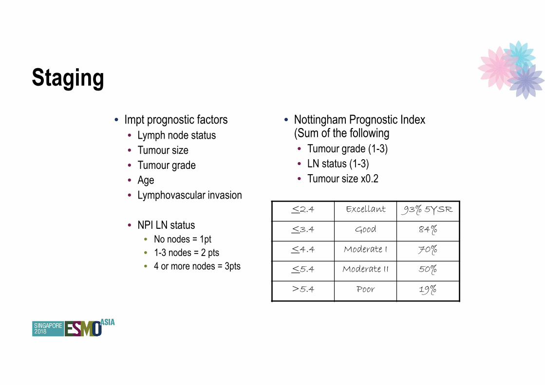
Staging
• Impt prognostic factors
• Lymph node status
• Tumour size
• Tumour grade
• Age
• Lymphovascular invasion
• NPI LN status
• No nodes = 1pt
• 1-3 nodes = 2 pts
• 4 or more nodes = 3pts
• Nottingham Prognostic Index (Sum of the following
• Tumour grade (1-3)
• LN status (1-3)
• Tumour size x0.2
<2.4 Excellant 93% 5YSR
<3.4 Good 84%
<4.4 Moderate I 70%
<5.4 Moderate II 50%
>5.4 Poor 19%

INOPERABLE CANCER
Metastatic
LABC
Neoadj chemo
� Margins
� Conservation
� Tumour biology

OPERABLE CANCER
Gradual progression towards less radical surgery
Halsted’s
Modified radical mastectomy
Breast conservation
Axillary clearance vs sentinel node biopsy

SURGERY FOR THE BREAST
BREAST SURGERY
Choice of surgery depends entirely on extent of disease
Volume of breast tissue needing resection (cancer with margins) relative to the volume of the breast
Conservation only if minimal / acceptable cosmetic impact on the operated breast achievable
� Not subject to molecular subtype, but concerns about adjacent DCIS are an important
consideration
� Radiation is mandatory (although certain subgroups suitable for de-escalating therapy)
� Aims:
� Complete excision of malignant cells
� Minimal excision of normal breast tissue
� Only what is needed for clear margins
� Minimal cosmetic impact of the affected breast
� Clear margins are the issue
� Re-operations have physical, psychological and economical repercussions
BREAST CONSERVATION SURGERY

MARGINS
Invasive breast cancer
No international consensus internationally
ASTRO / ASCO / SSO guidelines – ‘no tumour on inked margin’
USA and Netherlands: No tumour on the inked margin
UK: >2mm
Germany / Scotland / France: > 1mm
1-2mm acceptable
Moran MS, Schnitt SJ, Giuliano AE, Harris JR, Khan SA, Horton J, et al. Society of Surgical OncologyeAmerican Society for Radiation Oncology consensus guideline on margins for
breast-conserving surgery with whole-breast irra- diation in stages I and II invasive breast cancer. Int J Radiat Oncol Biol Phys 2014;88:553e64.
Kwaliteitsinstituut voor de gezondheidszorg CBO. Richtlijn mammacarci- noom. 2008. p. 76e113., http://www.oncoline.nl/uploaded/FILES/mammacarcinoom/Richtlijn Behandeling
van het Mammacarcinoom oktober 2005.pdf.
Association of Breast Surgery at B. Surgical guidelines for the management of breast cancer. Eur J Surg Oncol 2009;35(Suppl. 1):1e22.
Interdisziplin€are S3-Leitlinie für die Diagnostik, Therapie und Nachsorge des Mammakarzinoms.
http://www.awmf.org/uploads/tx_szleitlinien/032045OL_k_S3__Brustkrebs_Mammakarzinom_Diagnostik_Therapie_ Nachsorge_2012-07.pdf; 2012
Reseau Espace Sante-Cancer Rh^one-Alpes. Les Referentiels Cancer du Sein [5- 12-2013], http://www.rrc-ra.fr/Ressources/referentiels/PRA-SEI-1312SEIN. pdf; 2013.

MARGINS
DCIS
• Margins for DCIS: >2mm
• the extent of DCIS at the involved margin
• the margin which is involved
• presence of residual calcifications on mammogram
• impact of re-excision of the appearance of the breast
• life expectancy
J Clin Oncol 2016;34(33):4040e6.

MARGINS
Factors associated positive margin rate
� Lobular histology
� Adjacent DCIS to IDC
� Tumour size > 2cm
� Young age
� LVI
� Multifocality

MARGINS
Impact of close but negative margins in breast conserving surgery
� Review of patients from a prospective database, 2000-2012
� Re-excision at margins of < 2mm
� 2520 procedures, re-excision rate 12% for BCS, 2% for mastectomy
� Residual disease found in 38% and 26% respectively
Residual disease rate in positive, 0.1-0.9mm, and 1.0-1.9mm margins were 40%, 38% and 33%
� Multiple margins <2mm trended towards significance for residual disease
� Age, race, menopausal status, tumour histology, HR status, triple negative disease, LVI were not
associated with residual disease
5-year LR rates (median FU 43 mths) was 1.1% for TM, and 1.9% for BCS patients

Margins classified as
� Negative >2mm, close <2mm,
focally positive (<4mm length of
tumour touching ink), extensively
positive (>4mm length)
499 patients, Tis to T3, primary surgery (BCS)
� 43% (212) negative margins, 32%
(161) close margins, 12% (59)
focally positive, 13% (67)
extensively positive margins
MARGINS
Impact of focally positive margins
EJSO 2017; 43: 1846-1854
MARGINS
Cosmetic impact of clear margins
van den Tol et al analyzed surgical margins, and excision volumes of breast tissue following breast
conservation surgery
Central database data (PALGA – national registry in the Netherlands), 9274 reports
� Involved margins: 5.4%
� Focal involvement 11% cases
� Unsatisfactory resections – 33.8% (<1mm)
Median excised volume 46cc, calculated resection ration was 2.3 => excision was 2.3 times the optimal
resection volume
The Breast 25 (2016) 14-21

MARGINS
Methods to decrease re-excision rates
• Better definition of the target
• Determining the extent of disease
• Improve targeting of lesion
• Localisation techniques
• Immediate assessment of margins
• Cavity shave / Margin probe / Frozen section of margins
• Increase margin width *
• Oncoplastic surgery

Br J Radiol 2018; 91: 20170740.
LESION LOCALIZATION

LESION LOCALIZATION
Other techniques
Intra-operative ultrasound Annals of Surgical Oncology 2002; 9(10):994–8.
Modified ROLL – in combination with methylene blue dyeAnnals of Surgical Oncology 2011;18(1):109–13
SAVI SCOUT® localizationClin Imaging. 2018 Jul 24;52:280-286
Cryo-assisted localizationAmerican Journal of Surgery 2006;192(4):462–70.
Haematoma associated localization (post VAB)
Ann Surg Oncol. 2010 Oct;17 Suppl 3:378-83.

LESION LOCALIZATIONCochrane Database of Systematic Reviews 2015, Issue 12. Art. No.: CD009206.
DOI: 10.1002/14651858.CD009206.pub2.
11 RCTs, assessed ROLL or RSL compared to WGL
Methods were comparable
No one better than the other, ROLL / RSL are reasonable alternatives, as reliable as WGL
ROLL vs WGL: differences were seen, in favour of ROLL, but not statistically significant
Successful localization: RR 0.66, CI 0.16-2.28; 869 patients; 6 trials
Positive excision margins: RR 0.74, CI 0.42 – 1.29; 517 patients; 5 trials
Re-operation rates: RR 0.51, CI 0.21-1.23; 583 patients; 4 trials
LESION LOCALIZATION
Cochrane review
WGL vs RSL:
Successful localization: RR 3.85, CI 1.21-12.19; 402 patients, 2 trials
RSL vs WGL:
Positive margins: RR 0.67, CI 0.43-1.06; 366 patients; 2 trials
Re-operation rates: RR 0.80, CI 0.48-1.32, 305 patients, 1 trial
However for successful excisions, all 3 methods were the same, RR 1.00
WGL – fewer postoperative complications compared to both ROLL / RSL, but not significant

BREAST CONSERVATION SURGERY
Better targeting
Non-palpable tumour
• localization needed for excision
• Issues for localization
• 2D images for a 3D lesion
• Accuracy of marker placement
• Relation of the lesion to the marker
• Marker migration
• Marker transection
• Needs to be placed as a separate procedure to surgery
• Patient distress
• Knowing where the lesion is, does not increase the chance of precise excision

MARGINS
Cavity shave trials / articles
European Journal of Surgical Oncology 1999; 25: 464–469
Margin assessment by cavity shaving after breast-conserving
surgery: analysis and follow-up of 543 patients
Do additional shaved margins at the time of lumpectomy
eliminate the need for re-excision? The American Journal of Surgery (2008) 196, 556–558
Diagnostic Accuracy of Intraoperative Techniques for Margin Assessment in Breast
Cancer Surgery: A Meta-Analysis – Annals of surgery Ann Surg. 2017 Feb;265(2):300-310

MARGINS
Improving negative margin rates
RCT for margin shaves
- 1:1 comparison 235 patients, stage 0-stage 3 undergoing BCT
- Resection of routine cavity shaves vs no further resections
- Pos Margins = no ink on tumour for IDC, 1mm for DCIS
- Prior to randomization – both groups had similar positive margin rates – 36% and 34%
- After randomization for routine cavity shaves vs no further shaves, margin positive in the no shave
group remained at 34%, however margin positive rates in the routine shave group were 19%
- Re-excision rates – no shave group 21%, shave group 10 %
N Engl J Med. 2015 Aug 6;373(6):503-10.

MARGINS
Volume of Excision and Cosmesis with Routine
Cavity Shave Margins Technique
Analysed patients who had cavity shaving (CSM) vs patients treated with
standard partial mastectomy (SPM)
72 matched patients pairs-
Mean tumour size for both groups were similar 1.52 cm3 vs 1.51 cm3
Volume excised in CSM was 80.66 cm3, vs 165.1cm3 in SPM
Re-excision rates in CSM was 18.1% vs 34.6% in SPM
Cosmetic score in CSM was 2.3, vs 3.0 in SPM group
Ann Surg Oncol (2012) 19:886–891

TARGET ACQUISITION
Intraoperative margin assessment
Ann Surg. 2017 Feb;265(2):300-310
Technique Sensitivity Specificity AUROC Disadvantages
Frozen section 86% 96% 0.96Expensive, resource intensive, slow
turnaround
Cytology 91% 95% 0.98Unable to distinguish in-situ from invasive
Intraoperative US 59% 81% 0.78operator dependent, calcs not visible on US
Specimen radiography 53% 84% 0.73Unable to define non-calcified cancer, benign
calcs could be called malignant
Optical spectroscopy 85% 87% 0.88
TARGET ACQUISITION
Margin assessment
Frozen section:
Time consuming
Expensive
Subject to sampling error – 4 margins minimum, maximum 12
Snap freezing can also create compression, freezing and destructive artifacts
- However if possible, it is the most accurate way
Ann Surg Oncol (2016) 23:3290–3296
TARGET ACQUISITION
Margin assessment techniques available
Imaging:
high resolution scanners for specimen analysis
microcomputed CT, high-frequency US, MRI
Optical:
Light (of various frequencies ranging from visible to infra-red) directed on / into tissue produce spectra unique for each tissue type
Raman spectroscopy, optical coherence tomography, confocal microscopy
Bioimpedancece / Radiofrequency:Tissue exposed to radiofrequency fields and generates an electromagnetic field which is recognized as a tissue spectral signature e.g.
MarginProbe™ ClearEdge™
Mass SpectometryMeasures tissue specific ionic content linked to cellular metabolism
Rapid evaporative ionization mass spectrometry (REIMS) and Desorption electrospray Ionization (DESI)
Ann Surg. 2017 Feb;265(2):300-310
ONCOPLASTIC SURGERY
Allows the excision of larger volumes of tissue, extends option of conservation to more patients
� Larger cancers
� Multifocal disease
� EIC
Patients who could potentially omit radiation
ONCOPLASTIC SURGERY
• Oncoplastic surgery techniques
If area that requires excision (inclusive of margins) is <20% of the breast volume
• Level 1 oncoplastic technique
If volume excised is 20-50%, (50% if breast size is large) of total breast volume –
• Level 2 oncoplastic technique
• Usually entails excision of skin, and breast reduction surgery inclusive of the tumour
• Partial reconstruction also possible with the use of local pedicled flaps (TDAP, LICAP)
If >50% of total breast volume will be removed, total mastectomy with or without reconstruction

ONCOPLASTIC SURGERY
Level 2 techniques: Volume displacement
Parenchymal mobilization to fill cavity
- Tissue flaps will be somewhat ischemic
- Prone to fat necrosis
- Increased likelihood scarring / fibrosis
- Cavity sides can be clipped
BJPS 58: 889-901, 2005

ONCOPLASTIC SURGERY
Level 2 techniques: Volume displacement
• Level II
oncoplastic
techniques

ONCOPLASTIC SURGERY
Level 2 techniques: Volume replacement
Breast conservation surgery and partial reconstruction
Using L-ICAP / A-ICAP flaps
- Cavity is not re-opposed but is filled with tissue instead

ONCOPLASTIC SURGERY
Surgical approach in early breast cancer
Issues� Fat necrosis
� Tumour site� Nipple necrosis – partial / complete� Radiation –only to affected side
� Fibrosis� Positive margins
� Asymmetry� Residual volume

BREAST CONSERVATION SURGERY
The Breast 35 (2017) 32-33

BREAST CONSERVATION SURGERY
Personal reflections
1. Knowing where the target is and acquiring the target are 2 completely separate issues
2. Marker is seldom in the dead centre of the target, and where it is within or (if outside target) in relation
to the target is difficult predict
3. Assessing distances on imaging and translating it to the patient on the table is not the same
4. Although I know ‘no ink on tumour’ is acceptable, 1cm gross margins are still the aim
5. Ideal localization marker:
1. Mark the extent of disease in the patient – including DCIS
2. Can be detected just outside the margins
3. Can be visualized directly in the patient

DE-ESCALATION OF SURGERYEarly disease

EARLY STAGE BREAST CANCER
Recurrence risk for DCIS
ECOG E5194 – IBTR with omission of RT, margins >3mm1
Low-intermediate grade DCIS, <25mm, recurrence at 12 years is 14.4%
High grade DICS, <10mm, recurrence rate at 12 years is 24.6%
(5.5% low grade, 6.7% intermediate grade, 11.7% high grade recurred with invasive disease)
RTOG 9804 IBTR with RT omission2
In women aged >26 years, <25mm, >3mm margins, low-intermediate grade, not mammographically occult
7 years FU: IBTR with RT 0.9%, no RT -> 6.7%
However no impact on overall survival rates
1. J Clin Oncol, 33 (2015), pp. 3938-3944
2. J Clin Oncol, 33 (2015), pp. 709-715

EARLY STAGE BREAST CANCER
Omission of surgery in low grade DCIS
� Screen detected low / intermediate grade DCIS (HR+/-, HER2 +/-)
� <10mm, aged 70yrs or older
� Must be screen detected, diagnosed on VAB
� NOT for observation are
� Low grade DCIS in patients under 45 years of age, even with good molecular profile, <10mm in
size
Active observation
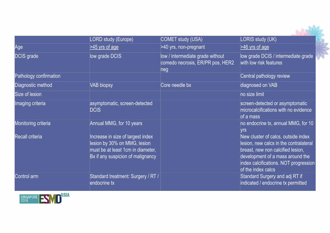
LORD study (Europe) COMET study (USA) LORIS study (UK)
Age >45 yrs of age >40 yrs, non-pregnant >46 yrs of age
DCIS grade low grade DCIS low / intermediate grade without
comedo necrosis, ER/PR pos, HER2
neg
low grade DCIS / intermediate grade
with low risk features
Pathology confirmation Central pathology review
Diagnostic method VAB biopsy Core needle bx diagnosed on VAB
Size of lesion no size limit
Imaging criteria asymptomatic, screen-detected
DCIS
screen-detected or asymptomatic
microcalcifications with no evidence
of a mass
Monitoring criteria Annual MMG, for 10 years no endocrine tx, annual MMG, for 10
yrs
Recall criteria Increase in size of largest index
lesion by 30% on MMG, lesion
must be at least 1cm in diameter,
Bx if any suspicion of malignancy
New cluster of calcs, outside index
lesion, new calcs in the contralateral
breast, new non calcified lesion,
development of a mass around the
index calcifications. NOT progression
of the index calcs
Control arm Standard treatment: Surgery / RT /
endocrine tx
Standard Surgery and adj RT if
indicated / endocrine tx permitted

DE-ESCALATION OF SURGERYAdvanced disease
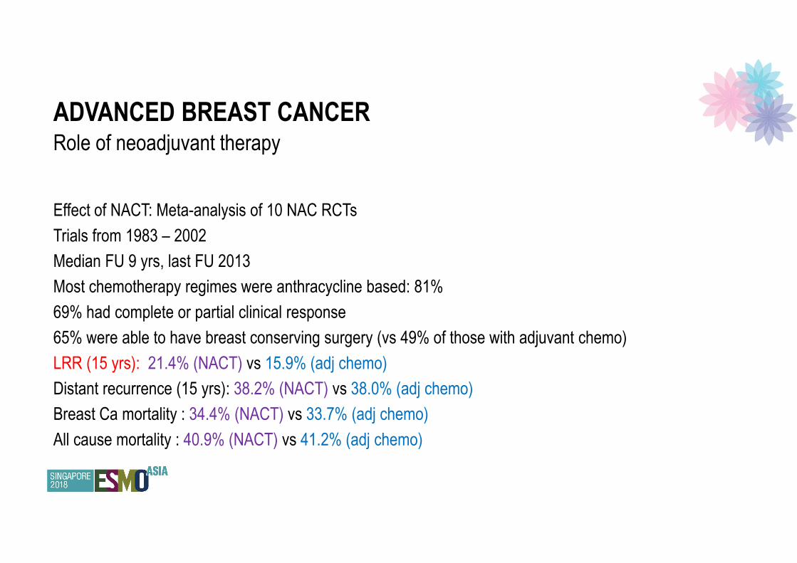
ADVANCED BREAST CANCERRole of neoadjuvant therapy
Effect of NACT: Meta-analysis of 10 NAC RCTs
Trials from 1983 – 2002
Median FU 9 yrs, last FU 2013
Most chemotherapy regimes were anthracycline based: 81%
69% had complete or partial clinical response
65% were able to have breast conserving surgery (vs 49% of those with adjuvant chemo)
LRR (15 yrs): 21.4% (NACT) vs 15.9% (adj chemo)
Distant recurrence (15 yrs): 38.2% (NACT) vs 38.0% (adj chemo)
Breast Ca mortality : 34.4% (NACT) vs 33.7% (adj chemo)
All cause mortality : 40.9% (NACT) vs 41.2% (adj chemo)

ROLE OF NEOADJUVANT THERAPY IN SURGERY
Who should receive neoadjuvant therapy?
Chemotherapy
Stage II / III HER2 positive or triple negative breast cancer
Endocrine therapy
CDK 4/6 inhibition with endocrine therapy?
Allows downsizing and downstaging of cancer
� Potential for breast conservation / makes it more feasible
� De-escalation of axillary surgery
� Elimination of micrometastatic disease
� Oligometastatic patients who are downstaged
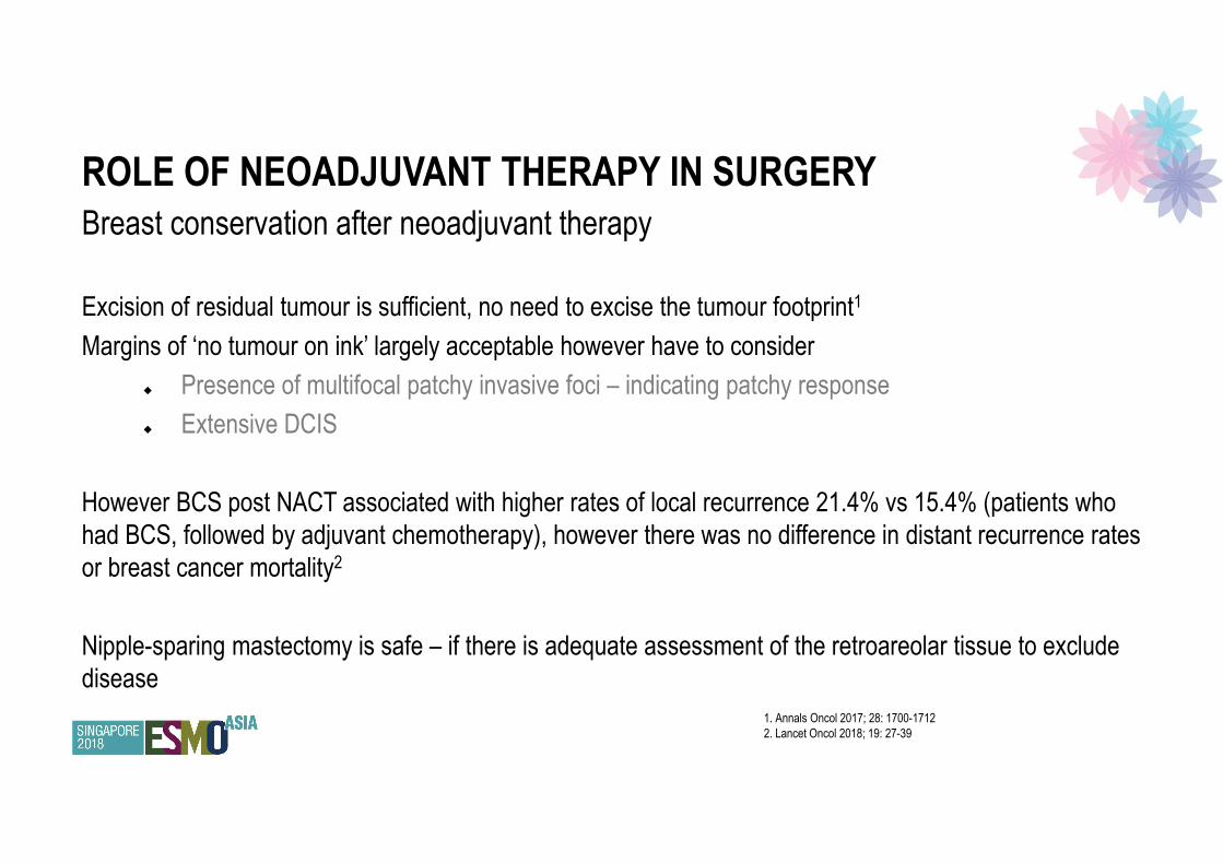
ROLE OF NEOADJUVANT THERAPY IN SURGERY
Breast conservation after neoadjuvant therapy
Excision of residual tumour is sufficient, no need to excise the tumour footprint1
Margins of ‘no tumour on ink’ largely acceptable however have to consider
� Presence of multifocal patchy invasive foci – indicating patchy response
� Extensive DCIS
However BCS post NACT associated with higher rates of local recurrence 21.4% vs 15.4% (patients who
had BCS, followed by adjuvant chemotherapy), however there was no difference in distant recurrence rates
or breast cancer mortality2
Nipple-sparing mastectomy is safe – if there is adequate assessment of the retroareolar tissue to exclude
disease
1. Annals Oncol 2017; 28: 1700-1712
2. Lancet Oncol 2018; 19: 27-39
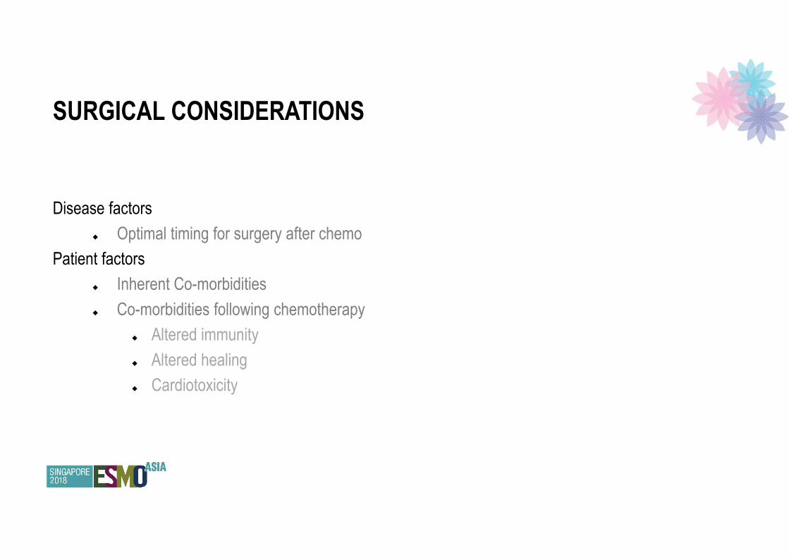
SURGICAL CONSIDERATIONS
Disease factors
� Optimal timing for surgery after chemo
Patient factors
� Inherent Co-morbidities
� Co-morbidities following chemotherapy
� Altered immunity
� Altered healing
� Cardiotoxicity

� Retrospective study assessing time to surgery (TTS)
� 319 patients, Grp A TTS <21 days, Grp B >21 days
� Grp A: 61 patients, Grp B 258 patients
� Median TTS 34 days
� No association between clinical stage, nuclear grade, chemo regime,
type or surgery with TTS was detected
� OS and RFS significantly worse for Grp B compared to Grp A, HR 3.1
(95% CI 1.1-8.6, p=0.03) and 3.1 (95% CI 1.3-7.1, p=0.008)
� Confirmed to be an independent variable on multivariate analysis
Impact of time to surgery after neoadjuvant chemotherapyin operable breast cancer patients
C. Omarini a,* , G. Guaitoli a, S. Noventa a, A. Andreotti b,A. Gambini b, E. Palma b, S. Papi b, G. Tazzioli b, S. Balduzzi c,
M. Dominici a, S. Cascinu a, F. Piacentini a
EJSO 2016

DE-ESCALATION OF SURGERYAssessment of the axilla

EARLY BREAST CANCER
Assessment of lymph nodes
Assessing for nodal involvement allows staging of the patient
provides prognostic information
also has therapeutic implications
Need for chemo / RT
But - axillary dissection does not impact overall survival (NSABP- B04)
In this age of screening and detecting more early disease, negative AC are common
Removal of normal nodes come with significant physical morbidity, risk of lymphedema, with no benefit to
the patient.
Hence SNB, omission of AC in the event of negative / low nodal burden, extending SNB to select patients
post NACT


SENTINEL NODE BIOPSYASCO guideline
7 RCTs
NSABP-B32, ALMANAC, Sentinella / GIVOM, RACS/ SNAC trial, NCT0097-983, Cambridge / East Anglia
Study grp, Canavese et al
Survival / mortality
� No difference in OS
� B32: 8 YSR 90.3% (SNB) vs 91.8% (SNB+ALND), all cause mortality 4% in each arm
DFS / EFS
� No difference is DFS / EFS
Recurrence
� No difference in rates of IBTR / Ax recurrence or DM
J Clin Oncol 2014; 32:1365-1383
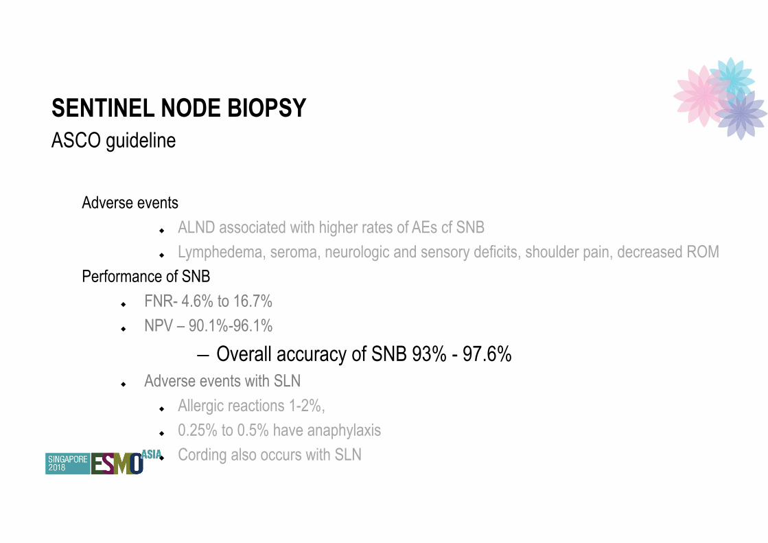
SENTINEL NODE BIOPSY
Adverse events
� ALND associated with higher rates of AEs cf SNB
� Lymphedema, seroma, neurologic and sensory deficits, shoulder pain, decreased ROM
Performance of SNB
� FNR- 4.6% to 16.7%
� NPV – 90.1%-96.1%
– Overall accuracy of SNB 93% - 97.6%
� Adverse events with SLN
� Allergic reactions 1-2%,
� 0.25% to 0.5% have anaphylaxis
� Cording also occurs with SLN
ASCO guideline

SENTINEL NODE BIOPSY
SNB: in practice for many years
- Established to reflect the state of axillary nodal involvement
- Eligibility T1 / T2, cN0.
- Dual method: Radioactive colloid (usually 99TM), and Patent V blue dye
- Rate of sentinel node detection: at least 90%
- False negative rates should be <5%
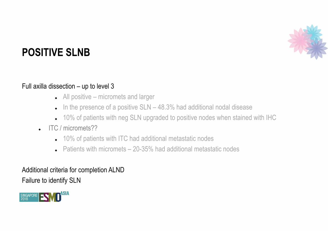
POSITIVE SLNB
Full axilla dissection – up to level 3
� All positive – micromets and larger
� In the presence of a positive SLN – 48.3% had additional nodal disease
� 10% of patients with neg SLN upgraded to positive nodes when stained with IHC
� ITC / micromets??
� 10% of patients with ITC had additional metastatic nodes
� Patients with micromets – 20-35% had additional metastatic nodes
Additional criteria for completion ALND
Failure to identify SLN

SENTINEL NODE BIOPSY
Primary surgery in early breast cancer
ASCOG Z011 trial1
Omission of full axillary dissection in patients with <2 positive nodes, undergoing breast conserving
surgery, radiation therapy and systemic therapy
AMAROS / EORTC trials
Post mastectomy patients, <2 positive nodes
Completion axillary dissection or axillary radiation offer equivalent control
JAMA 2011;305:569e75.
Ann Surg 2016;264:413e20.

Challenged need for ALND for positive SLN
� Positive SLN is often the only positive node
� NSABP- B04: upfront ALND no benefit
Criteria
� T1, T2, N0, M0 (median size 17mm), undergoing BCS with 1-2 pos SLN (H&E)
� Randomized to ALND or no Sx
� All had WBI, and most (97%) had systemic tx
� 891 pat recruited (planned 1900)
Z0011
Axillary dissection vs observation
SLND ALND
38% micromets 45% micromets
27% had additional pos
LN
LR 2% LR 4%
Axillary recurrence 5
(0.9%)
Axillary recurrence 2
(0.5%)
10 yr OS 86.3% 10 yr OS 83.6%
10 yr DFS 80.2% 10 yr DFS 78.2%
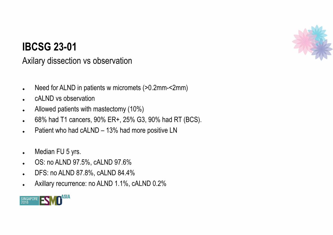
� Need for ALND in patients w micromets (>0.2mm-<2mm)
� cALND vs observation
� Allowed patients with mastectomy (10%)
� 68% had T1 cancers, 90% ER+, 25% G3, 90% had RT (BCS).
� Patient who had cALND – 13% had more positive LN
� Median FU 5 yrs.
� OS: no ALND 97.5%, cALND 97.6%
� DFS: no ALND 87.8%, cALND 84.4%
� Axillary recurrence: no ALND 1.1%, cALND 0.2%
IBCSG 23-01
Axilary dissection vs observation

� T1b-T2, N0
� BCS & TM
� Completed accrual
� 65% patients SNB neg, 29.7% patients SNB positive (1425)
� 744 – ALND, 681 had AxRT
� Median tumour size 17-18mm (13-23mm)
� 80% BCS, 90% systemic tx, 85% RT
� 1-3 LN removed in all cases, 60% macromet, 30% micromet, 10% ITC
� cALND: 32% had additional positive LN, 7.8% had > 4.
� DFS / OS similar
� 5-years axillary recurrence rate: ALND 0.43% (4 / 744 events (0.54%)) AxRT 1.19% (7 / 681 events (1.03%))
EORTC AMAROS
Axillary dissection vs RT

SNB AFTER NACT
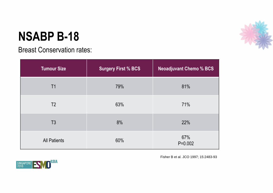
NSABP B-18 Breast Conservation rates:
Tumour Size Surgery First % BCS Neoadjuvant Chemo % BCS
T1 79% 81%
T2 63% 71%
T3 8% 22%
All Patients 60%67%
P=0.002
Fisher B et al. JCO 1997; 15:2483-93

NSABP B-18 Axillary node downstaging
Surgery First (n=743) Neoadjuvant Chemo (n=743)
1-3 nodes +ve 30% 24%
4-9 nodes +ve 17% 12%
> 10 nodes +ve 10% 4%
Overall nodes +ve 57%41%
P<0.001
Fisher B et al. JCO 1997; 15:2483-93
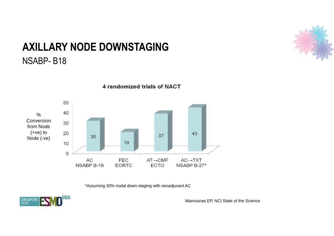
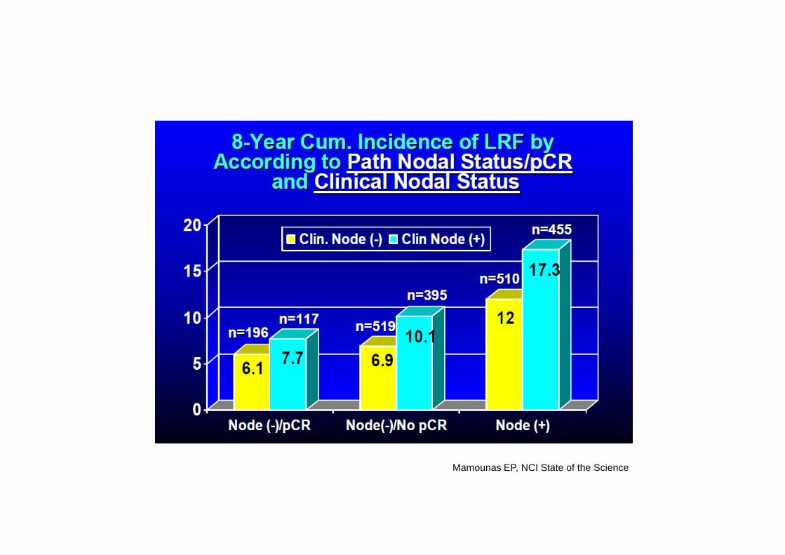
AXILLARY NODE DOWNSTAGING
NSABP- B18
% Conversion from Node
(+ve) to Node (-ve)
Mamounas EP, NCI State of the Science
*Assuming 30% nodal down-staging with neoadjuvant AC

SENTINEL NODE BIOPSY – POST NACT
After neoadjuvant therapy
cN0 at presentation, SNB recommended post NACT
cN1 at presentation, downstaged to cN0 after NACT, SNB is feasible
Nodal pCR rates are between 35-49%1,2,3
� Sentina trial
� TAD: targeted axillary dissection
Axillary dissection can be spared if 3 lymph nodes negative at the time of SNB
Fewer than 3 nodes results in unacceptably high false negative rates
1. J Clin Oncol 2015;33: 258e63.
2. JAMA 2013;310:1455e61.
3. Ann Surg Oncol 2016;23:3467e74.
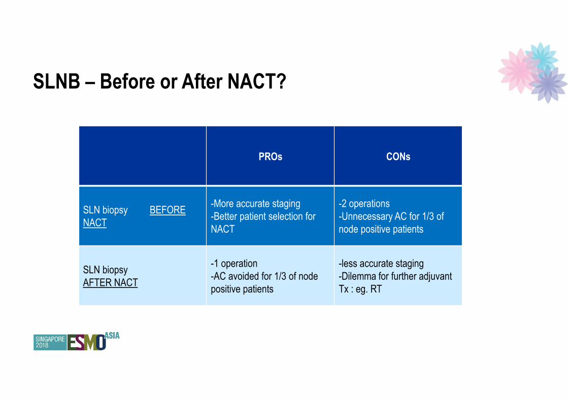
PROs CONs
SLN biopsy BEFORE
NACT
-More accurate staging
-Better patient selection for
NACT
-2 operations
-Unnecessary AC for 1/3 of
node positive patients
SLN biopsy
AFTER NACT
-1 operation
-AC avoided for 1/3 of node
positive patients
-less accurate staging
-Dilemma for further adjuvant
Tx : eg. RT
SLNB – Before or After NACT?

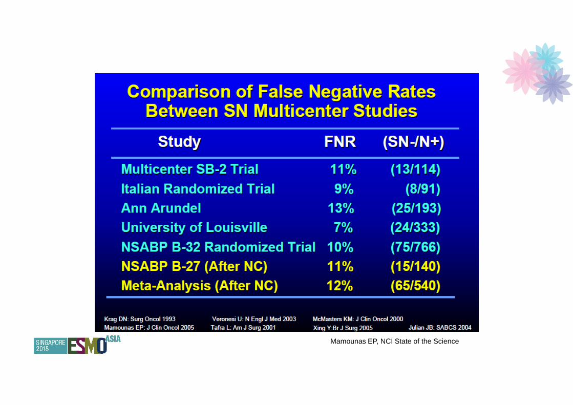
FEASIBILITY AND ACCURACY OF
SLNB POST NACT
Various studies:
� Single institution trials
� Multicenter trials
� Meta-Analyses
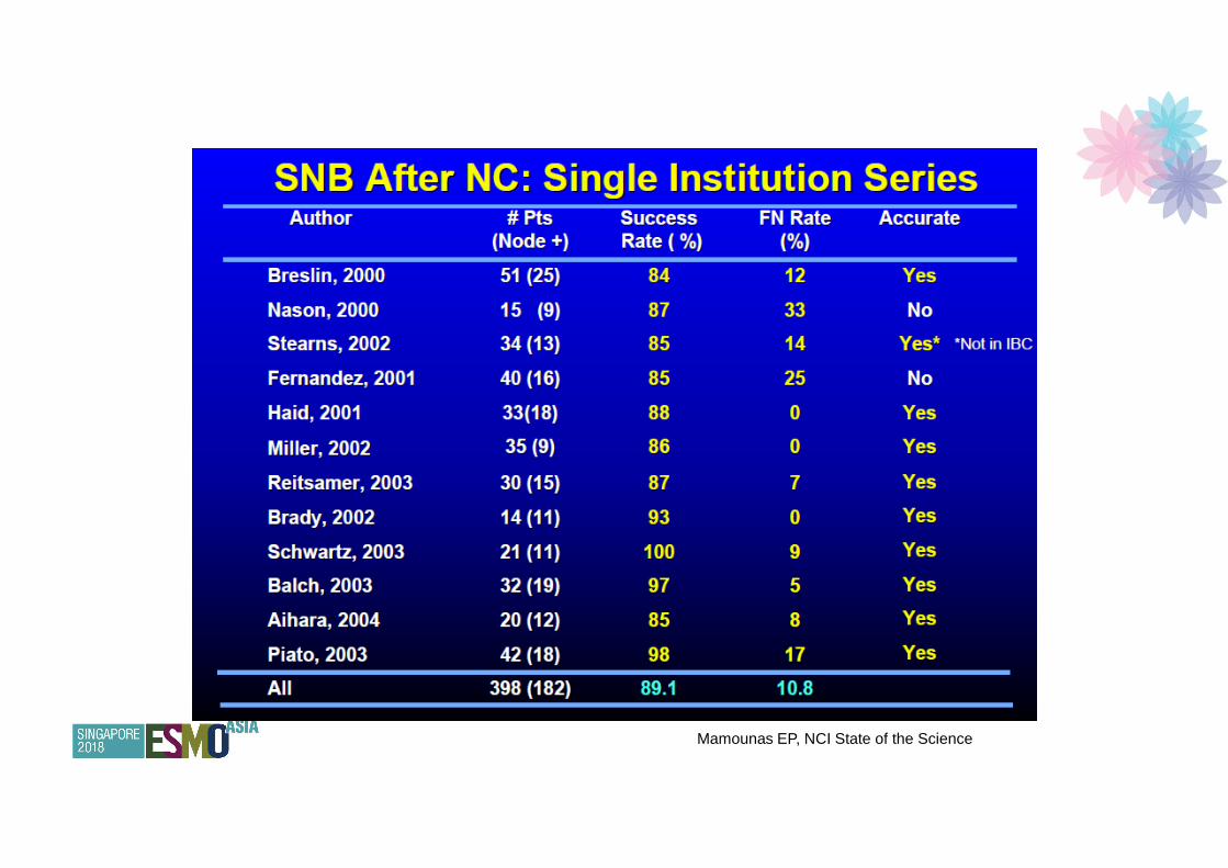
Mamounas EP, NCI State of the Science
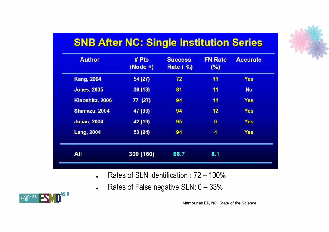
� Rates of SLN identification : 72 – 100%
� Rates of False negative SLN: 0 – 33%
Mamounas EP, NCI State of the Science
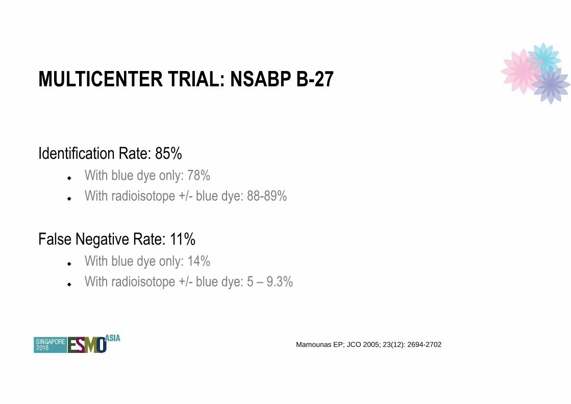
MULTICENTER TRIAL: NSABP B-27
Identification Rate: 85%
� With blue dye only: 78%
� With radioisotope +/- blue dye: 88-89%
False Negative Rate: 11%
� With blue dye only: 14%
� With radioisotope +/- blue dye: 5 – 9.3%
Mamounas EP; JCO 2005; 23(12): 2694-2702
Mamounas EP, NCI State of the Science

SLN: BEFORE OR AFTER NACT


SENTINA Trial
Outcome
1737 pts103 institutions
Arm AcN0/pN0
SLNB upfrontN=1022
Arm BcN0/SLN+ve
NACTRe-SLNB + AC
n=360
Arm CcN1-2NACT
SLNB + ACN=592
SLN Identification
rate 99% 61% 80%
False negative rate
(SLN –ve / AC +ve)
52% 14%

� False negative rate:
� By mapping technique:
� Single method (radioisotope) – 16%
� Dual method – 8.6%
� By no. of SLN removed:
� 1 SLN 24%
� 2 SLN 18%
� 3 SLN 7%
SENTINA TRIAL

LYMPH NODE POSITIVE DISEASE BEFORE NACT


� Phase 2 trial
� 701 patients (2009 – 2011)
� cT0-4, N1-2, M0 disease
� All had neoadjuvant chemotherapy (commonly AC + taxane) followed by SLNB +AC
� Clinical CR – 83%
� Pathologic CR – 41%
� SLN identification rate – 92.5%
� 79% of patients had dual method (radiocolloid + blue dye)
ACOSOG Z1071

� False negative rate:
� By mapping technique
� Single method – 20.3%
� Dual method – 10.8%
� By no. of SLN removed:
� 1 SLN 31.5%
� 2 SLN 21%
� ≥2 SLN 12.6%
� ≥3 SLN 9.1%
ACOSOG Z1071
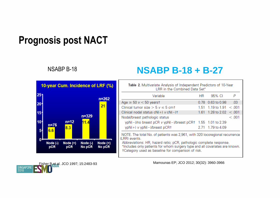
Prognosis post NACT
NSABP B-18 NSABP B-18 + B-27
Mamounas EP; JCO 2012; 30(32): 3960-3966Fisher B et al. JCO 1997; 15:2483-93
Mamounas EP, NCI State of the Science

NACT TO AVOID AXILLARY DISSECTION
Use of NAC allows avoidance of ALND in some patients
669 cN0 patients, initial BCS vs 271 patients who received NACT
In ER +, HER2 neg patients, need for ALND reduced from 34% (initial Sx/BCS by Z011 criteria) to
15% (NACT) p <0.0001
In TNBC, ALND rate was 14% for initial BCS vs 7% post NACT p=0.26
In HER2+ disease, rate was 13% for initial BCS, 8 % post NACT p=0.26
In patients undergoing mastectomy, NACT reduced need for ALND from 36% to 8%, p<0.001 in
HER2 pos, and from 25% to 7% in TNBC patients p=0.001, BUT not in ER+ cancers (37% vs 34%, p=0.62)
Optimal Treatment Plan to Avoid Axillary Lymph Node Dissection in Early-Stage Breast Cancer Patients Differs by Tumor
Subtype. In: Presented at the 2017 Society of Surgical Oncology Annual Cancer Symposium, March 15-18, 2017, Seattle,
Washington.
� Her 2 +ve / triple negative disease is responsive
� 68 - 74% axillary pCR in Her 2 +ve disease
� 57% axillary pCR in triple negative disease
� ER +ve disease is poorly responsive
� <10% axillary pCR
� Invasive Lobular Carcinoma is poorly responsive
� <5% breast / axillary pCR
� Oncotype Dx may be able to predict response to chemotherapy
PREDICTORS OF RESPONSE TO NACT
Straver ME, EJC 2009; 45(13):2284-2292Chehade HEH, Anticancer Research 2016; 36:1461-1472J Clin Oncol 2005;23:7265e77.Breast Cancer Res Treat 2015;154:299e308

AXILLARY CLEARANCE POST NACT
When it should be done
1. Clinically positive nodes after chemotherapy
2. Failure to detect lymph nodes at SLNB
3. Failure to find 3 lymph nodes
4. Lymph node positive at SLNB
Definition of positive nodes: Atypia, ITC, micro- / macro-metastases
Future: possible to avoid ALND in patients with indolent disease and low nodal burden post NACT?

Total Mastectomy
� Surgical considerations
� Clear margins
� Skin involvement
� Dermal infiltration
� Pectoralis muscle
� Closure of wound
� Reconstruction

CONSERVATIVE MASTECTOMY
Skin sparing / Areolar sparing
� Maximal excision of breast tissue
� Aesthetically not so normal
Nipple sparing
� Best results for aesthetic satisfaction
� There will be some breast tissue left in the nipple mound
� Nipple will be numb
CONSERVATIVE MASTECTOMY
Conditions for nipple sparing mastectomy:
� Early stage / Prophylactic for BRCA carriers
� Favourable biology
� IDC or DCIS at least 2 cm away from nipple
� Imaging negative for nipple involvement
� No nipple discharge
� No Paget’s disease
� Nipple base assessed and not involved with malignancy

ONCOLOGICAL SAFETYNipple sparing mastectomy
Lanitis et al 2010:
� Meta-analysis of 9 studies, 3739 patients
� LRR similar between SSM and NSM
� But SSM groups had lower proportion of distant relapse
Ann Surg 2010;251(4):632-9

ONCOLOGICAL SAFETYNipple sparing mastectomy
De La Cruz et al 2015:
Meta-analysis of 20 studies, 5594 patients
� 7 studies comparing OS
� 3.4% risk difference between NSM and SSM/MRM
� 5 Studies comparing DFS
� 9.6% risk difference between NSM and SSM/MRM
� 8 studies comparing LR
� 0.4% risk difference between the groups
Risk differences for all outcomes not statistically significant
Ann Surg Oncol 2015;22:3241-3249

ONCOLOGICAL SAFETYNipple sparing mastectomy
De La Cruz et al 2015
� At <3 yrs, 3-5 yrs, and > 5yrs
� For NSM, MRM, SSM
� OS 97.2, 97.9, 86.8%
� DFS 93.1, 92.3, 76.1%
� LR 5.4, 1.4, 11.4 %
� NAR 2.1, 1.0, 3.4%
Good biological profile – safe to undergo NSM
Age 35.6 to 61yrs, with DCIS or stage I/II IDC and TND > 2cm
Can be considered in BRCA mutation carriers however no long term FU available – so far < 5years.

COMPLICATIONS SPECIFIC TO NSM
Nipple necrosis
Flap necrosis
Headon et al: 12,358 patients pooled analysis
Overall complication rate 22.3%
Nipple necrosis rate 5.9%
However appeared to decrease over time suggesting that surgeon expertise is a factor
Arch Plast Surg 2016;43(4):328-38
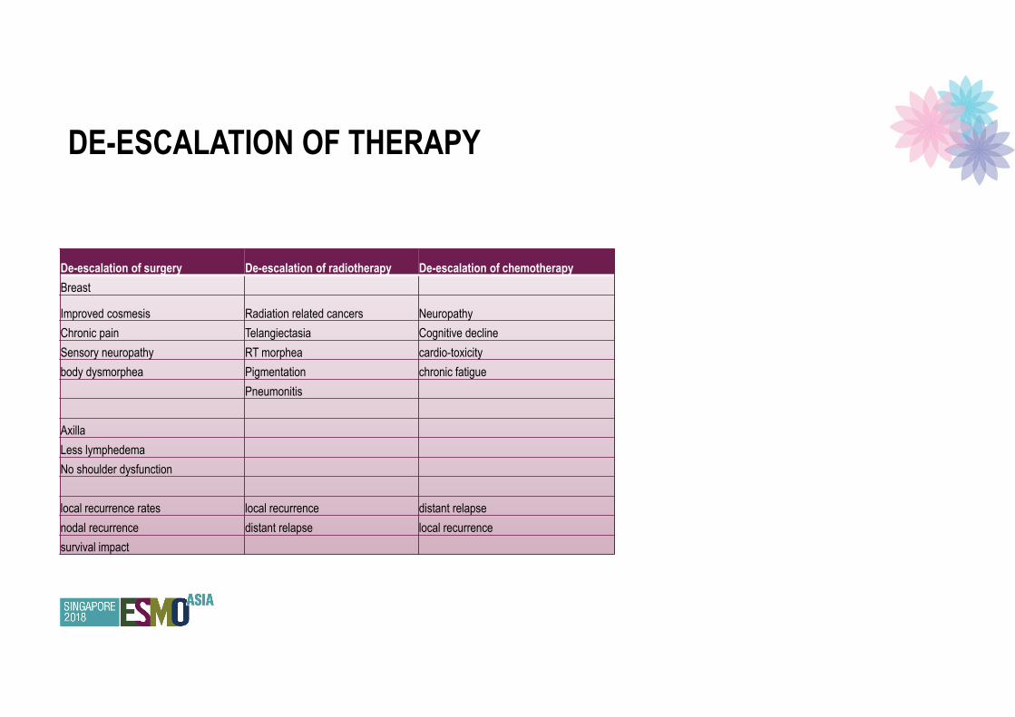
De-escalation of surgery De-escalation of radiotherapy De-escalation of chemotherapy
Breast
Improved cosmesis Radiation related cancers Neuropathy
Chronic pain Telangiectasia Cognitive decline
Sensory neuropathy RT morphea cardio-toxicity
body dysmorphea Pigmentation chronic fatigue
Pneumonitis
Axilla
Less lymphedema
No shoulder dysfunction
local recurrence rates local recurrence distant relapse
nodal recurrence distant relapse local recurrence
survival impact
DE-ESCALATION OF THERAPY

EARLY BREAST CANCER
De-escalation of treatment
With the observation of increased survival benefit and decreased local recurrence rates from long term
adjuvant radiation trials, time to question if gold standard should now be breast conservation and radiation,
over mastectomy
Patient choice?
Trade of side effects / morbidity:
Less surgery, usually means addition of RT / systemic therapy or both

DE-ESCALATION OF THERAPY
Patient discussion
Balance gain with risk
Decreased side effects vs increased recurrence risk
Need to identify patient goals
Acceptable morbidity vs relapse rates
Take into account tumour biology, anticipated lifespan, current co-morbidity

THANK YOU


Reconstruction
� Autologous vs Non-autologous

Reconstruction
� Autologous
� Free
� require microvascular anastomosis
� Increased operating time
� Flap failure rates 1.9%
� TRAM or DIEP
� Pedicled
� Failure rates 0.2%
� LD / TRAM
Reconstruction
� Autologous
� Complication rates (15-18%)
� Wound infection
� Seroma
� Wound dehiscence
� Chronic pain
Non-autologous
� Implants
� Expanders
� silicon shell with saline core that can be expanded
� Mostly silicon
� Newer ones textured
� Risk of anaplastic large cell lymphoma
� 1/1000 to 1/10,000 patients
� Presents as late, persistent seroma
� No need for prophylactic removal at present

Non-autologous
� Consequences
� Early
� Seroma
� Infection
� Late
� Tissue is stiffer – will not droop naturally
� Capsular contracture
� Implant pocket is too big
� Granuloma
� Distortion with RT
� 49% will require revision surgery

Safety of IBR
� Most guidelines recommend that IBR should be offered to all patients contemplating a mastectomy
Agrawal et al EJSO 2013;39;320-328
Local Recurrence following IBR
� Rate varies from institution to institution
� Risk factors:
� Young age
� Multiple tumours
� Larger tumours
� High grade DCIS, however most recurrences a/w invasive disease
� Higher stage disease
� Close or positive margins (<2mm)
� Median time to recurrence about 36 (7-128) months
Agrawal et al EJSO 2013;39;320-328

Prophylactic surgery

Prophylactic Surgery
� Increasing trends in the past decade
� Not just in high risk groups
� Perceived benefit
� Reduction of contralateral breast cancer risk
� ? Potential survival benefit
� Improved personal effect
� Presumed health care costs savings
� NICE guidelines
� proven genetic mutation, or
� high risk family history without a proven genetic mutation
Prophylactic Surgery
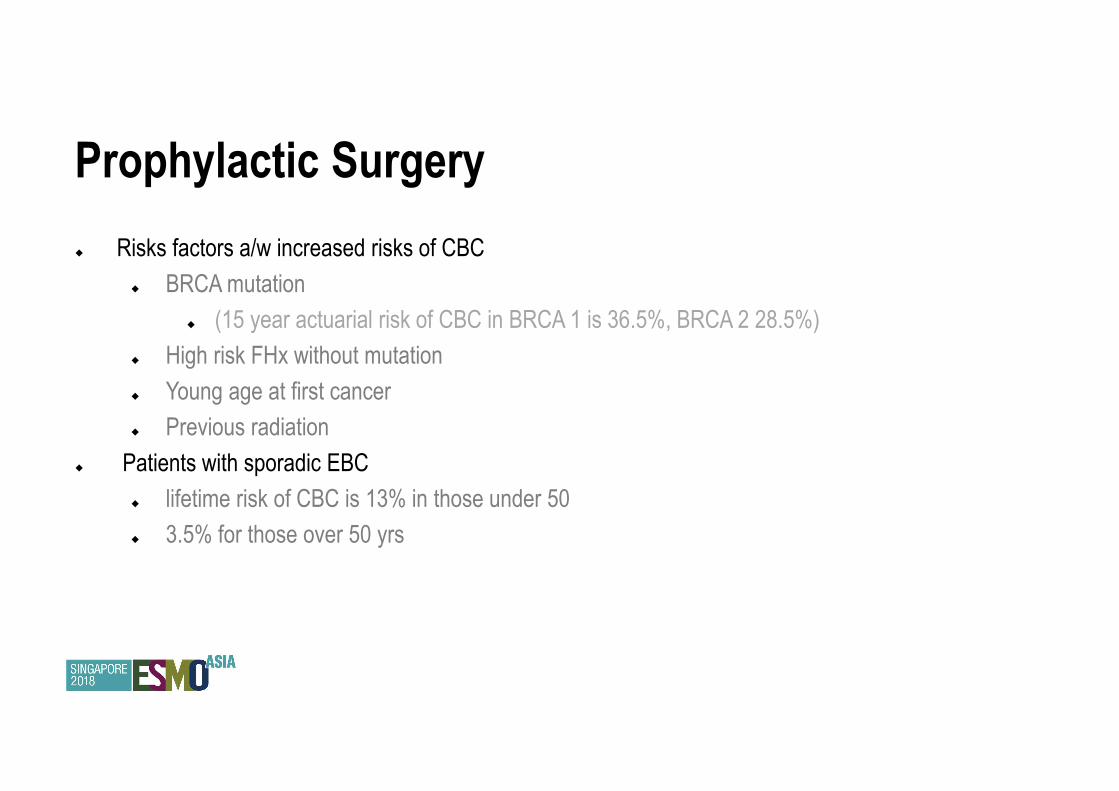
� Risks factors a/w increased risks of CBC
� BRCA mutation
� (15 year actuarial risk of CBC in BRCA 1 is 36.5%, BRCA 2 28.5%)
� High risk FHx without mutation
� Young age at first cancer
� Previous radiation
� Patients with sporadic EBC
� lifetime risk of CBC is 13% in those under 50
� 3.5% for those over 50 yrs

Prophylactic Surgery
� CBC
� Increased surveillance
� Increased awareness
� Tend to present earlier – no survival impact
� Risk of death is greater from ipsilateral metastatic disease rather than from new primary
� Even with patients with BRCA mutations, no OS benefit in patients older than 50 years
� In BRCA mutation patients > 35 years with co-morbidities, no OS benefit as well

Prophylactic Surgery
� Alternatives:
� Surveillance
� Regular CBE, MMG and MRI
� HR of death from screen detected cancers is half that of symptomatic detection
� MRI – more sensitive but lower specificity compared to MMG
� Increase risk of false positives
� Increased anxiety

Prophylactic Surgery
� Alternative:
� Chemoprevention
� STAR trial – comparing Tamoxifen vs Raloxifene in 19747 women
� 50% reduction of CBC as long as age >35 yrs, postmenopausal, or both.
� Tamoxifen slightly more effective, but higher risk of endometrial cancer and thromboembolic
events
� Similar effects noted with AIs
� ATAC trial, 2.5% CBC rates in patients on anastrozole vs 4.2% in patients on Tam at 9 years

Prophylactic Surgery
� Morbidity incurred
� Longer surgery time (can be shortened)
� Increased hospital stay
� Double the surgical risk for wound infection, dehiscence, flap necrosis
� Chronic pain
� Persistent seroma

Prophylactic Surgery
� Potential drivers of prophylactic surgery
� Psychological factors
� Perception of outcome
� Balance of risk of future cancer and effect on mortality
� vs incurred morbidity from additional surgery, psychological effect of loss of breast
� vs surveillance anxiety – biopsies etc

Palliative surgery
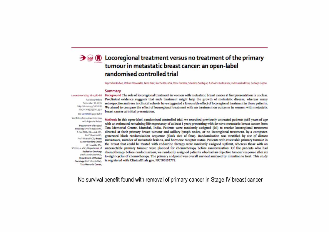
No survival benefit found with removal of primary cancer in Stage IV breast cancer

Breast cancer treatment in
mutation carriers

In BCT possible?
� Conflicting results from studies
� Recent meta-analysis of 5326 carriers, 2320 controls
� no difference in IBR rates (17.3% in carriers, 11% in controls, RR 1.45)
� However if stratify by length of FU –
� similar rates if < 7yrs,
� but IBR increases markedly after this
� Carriers – 23.9% vs 15.9% in controls

Is BCT possible?
� Comparing IBR in carriers after BCT vs mastectomy
� Cumulative risk IBR in patients with BCT is 23.5% vs 5.5% in patients who had TM at 15 years
� But BCSS with BCT was 93.5% vs 92.8%
� And OS with BCT was 91.8% vs 89.8%
� indicative of increased new primaries in patients who had BCT, unlike patients with TM who
would have had true recurrence
Pierce et al J Clin Oncol 2000;18:3360-3369

Is BCT possible?
� Factors associated with reduced risk of IBR after BCT in BRCA carriers
� Adj chemo
� OophorectomyTable 1: Risk for IBR in BRCA mutation carriers versus controls5
Cohort studies Risk Ratio (95% IC) Brekelmans 20076 0.61 [0.33, 1.11] Chappuis 20007 0.97 [0.22, 4.15] El-Tamer 20048 3.22 [1.15, 9.01] Haffty 20029 2.15 [1.13, 4.07] Robson 199810 0.46 [0.06, 3.34] Robson 200411 1.57 [0.73, 6.36] Subtotal (95% IC) 1.32 [0.70, 2.46] Case-control studies Eccles 200112 0.69 [0.30, 1.58] Garcia-Etienne 200913 4.50 [1.32, 15.35] Kirova 201014 1.90 [1.22, 2.97] Pierce 200615 1.51 [0.89, 2.56] Subtotal (95% IC) 1.60 [0.94, 2.56] Total (95% IC) 1.45 [0.98, 2.14]

Need for CPM in carriers?
� Carriers have a higher risk of CBC compared to non-carriers
Table 3: risk for CBC: BRCA-mutation carriers versus non-carriers5
Cohort studies Risk Ratio (95%
IC) Brekelmans 20076 3.54 [2.28, 5.49] Chappuis 20007 7.97 [1.39, 45.81] El-Tamer 20048 1.74 [0.98, 3.11] Haffty 20029 4.77 [1.86, 12.24] Robson 199810 4.88 [1.89, 12.58] Robson 200411 3.51 [2.05, 6.01] Stoppa-Lyonnet 200017 0.89 [0.39, 2.04] Subtotal (95% IC) 2.90 [1.85, 4.53] Case-control studies Eccles 200112 3.60 [2.15, 6.03] Garcia-Etienne 200913 15.0 [1.79, 125.57] Kirova 201014 3.67 [2.07, 6.48] Pierce 200615 8.34 [4.45, 15.63] Subtotal (95% IC) 5.0 [2.97, 8.40] Total (95% IC) 3.56 [2.50, 5.08]

� BRCA 1 carriers higher risk than BRCA 2
� Comparing BRCA 1 vs BRCA 2
� 1532 BRCA 1 vs 950 BRCA 2 carriers
� CBC rates were 21.1% and 15.1% respectively at 5 years
� Risk increases with time from diagnosis
� CPM did not did not affect OS
� but small studies, short FU
Need for CPM in carriers?
Valachis et al Breast Cancer Res Treat
2014;144:443-455
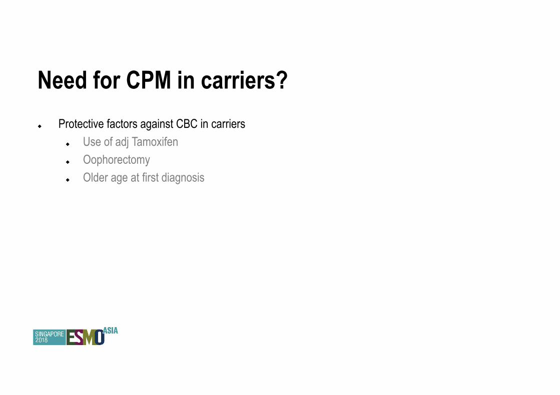
Need for CPM in carriers?
� Protective factors against CBC in carriers
� Use of adj Tamoxifen
� Oophorectomy
� Older age at first diagnosis