1. title page · web view2017-03-30 · 5department of pathology, ... pet, lung carcinoid, lung,...
TRANSCRIPT

Contents1. Title page.......................................................................................................................................... 2
2. Abstract............................................................................................................................................ 3
3. Text................................................................................................................................................... 4
Introduction........................................................................................................................................ 4
Materials and methods....................................................................................................................... 5
Results............................................................................................................................................... 7
Discussion.......................................................................................................................................... 8
4. Funding........................................................................................................................................... 12
5. Author contributions........................................................................................................................ 12
6. Acknowledgements......................................................................................................................... 12
7. Compliance with Ethical Standards................................................................................................12
8. References..................................................................................................................................... 12
9. Figures and tables..........................................................................................................................17
Table 1......................................................................................................................................... 18
Supplementary material A (Fig 1).................................................................................................19
Table 3......................................................................................................................................... 22
Supplementary material B (Table 4).............................................................................................23
Table 5......................................................................................................................................... 24
Figure 2........................................................................................................................................ 26
Supplementary material C............................................................................................................27
Table 6......................................................................................................................................... 28
Table 7......................................................................................................................................... 29
1

1. Title page
Title (80 characters):
68Gallium DOTANOC-PET imaging in Lung Carcinoids: impact on patients’ management
Short Title (46 characters incl. spaces):
68Ga-DOTANOC PET in Lung Carcinoids
Authors:
Angela Lamarca1,#, D. Mark Pritchard2,3, Thomas Westwood4, Georgios Papaxoinis1, Daisuke Nonaka5, Sobhan Vinjamuri2, Juan W Valle1,6, Prakash Manoharan4, Wasat Mansoor1
1Department of Medical Oncology, The Christie NHS Foundation Trust, ENETS Centre of
Excellence, Manchester, United Kingdom
2Liverpool ENETS Centre of Excellence, Royal Liverpool and Broadgreen University Hospitals
NHS Trust, Liverpool, United Kingdom
3 Institute of Translational Medicine, University of Liverpool, Liverpool, United Kingdom
4Department of Radiology and Nuclear Medicine, The Christie NHS Foundation Trust, ENETS
Centre of Excellence, Manchester, United Kingdom
5Department of Pathology, The Christie NHS Foundation Trust, ENETS Centre of Excellence,
Manchester, United Kingdom6Manchester Academic Health Sciences Centre, Institute of
Cancer Sciences, University of Manchester, Manchester, United Kingdom
#Part-funded by the Pancreatic Cancer Research Fund and Spanish Society of Medical
Oncology Fellowship Programme
Corresponding author: Dr Wasat Mansoor, Department of Medical Oncology, the Christie NHS
Foundation Trust, Wilmslow Road, M20 4BX, Manchester, United Kingdom. Email:
Keywords: gallium, DOTANOC, PET, lung carcinoid, lung, neuroendocrine tumour
Word count excluding references and figure/tables legends: 3716 words
2

2. Abstract
Background
68Gallium DOTA-PET imaging is preferable to standard somatostatin receptor scintigraphy where available; however, its role in the management of Lung Carcinoid tumours (LC) remains unclear.
Methods
All consecutive patients with histologically-confirmed LC from two ENETS Centres of Excellence were identified retrospectively. The primary objective was to assess the impact of 68Ga-DOTANOC-PET on clinical management in patients with LC.
Results
Of 166 patients screened, 46 were eligible: 52% female, median age 57 years (range 21-86); type of LC: DIPNECH (4%), typical (44%), atypical (35%), not reported (17%); stage: localised (63%), locally advanced (13%) and metastatic (17%) (7% unknown). A total of 47 68Ga-DOTANOCs were performed with the following rationale: LC diagnosis confirmation (4; 9%), primary tumour identification (2; 4%), post-surgical assessment (19; 40%), staging (patients with known LC present at time of 68Ga-DOTANOC) (19; 40%) and consideration of Peptide Receptor Radionuclide Therapy (PRRT) (3; 7%). Twenty-seven (57%) scans showed evidence of non-physiological uptake: median SUVmax 7.2 (range 1.42-53). 68Ga-DOTANOC provided additional information in 37% (95%CI 22-51) of patients and impacted on management in 26% (95%-CI 12-41); 9 patients (21%) were identified to have occult sites of metastases. Out of the 19 patients with post-surgical 68Ga-DOTANOC, 3 (16%) were identified to have distant metastases. There were no differences in the rate of practice changing 68Ga-DOTANOC results by type of LC (p-value 0.5).
Conclusions
Our results support the role of 68Ga-DOTANOC for optimizing the management of patients with LC, including post-surgical re-staging due to potential for identifying occult metastases.
3

3. Text
IntroductionLung neuroendocrine malignancies include a spectrum of neoplasia which share neuroendocrine
features [1]. Lung neuroendocrine tumours (NETs) are classified according to their pathological
characteristics (Table 1) [2]. Diffuse idiopathic pulmonary neuroendocrine cell hyperplasia (DIPNECH)
is considered a pre-invasive lesion with a potential toward the development of lung NETs [3, 4]. Lung
carcinoids (LC) are rare tumours [5], however incidence is increasing due to improvement in
diagnostic techniques [6]. They account for approximately 2% of all lung malignancies and around 20-
30% of all NETs. They characteristically have an indolent clinical behaviour with longer survival
intervals compared to poorly differentiated lung neuroendocrine malignancies [7]. LCare divided into
typical or atypical carcinoid tumours according to pathological characteristics, such as amount of
mitosis and necrosis (Table 1). In contrast to the well-differentiated NETs arising from the
gastrointestinal tract where the Ki-67 index is one of the parameters used in their grading
classification [8, 9], the role of Ki-67 index immunohistochemistry has not been validated as yet in
lung NETs [10, 11].
There are differences in survival between patients with typical and atypical LC, however clinical
management does not vary significantly [12, 13]. For localised stages, surgery is the treatment of
choice, performed with curative intent; there is no approved adjuvant treatment and patients are
allocated into a surveillance-only pathway; the only exception to this statement may be patients with
atypical LC with positive lymph nodes in whom adjuvant therapy could be considered on a patient by
patient basis [12]. Locally advanced inoperable or metastatic tumours are treated with palliative
approaches based on somatostatin analogues (SSAs) [14], temozolomide-based chemotherapy
combination [12]) and Peptide Receptor Radionuclide Therapy (PRRT) [15-18]. Other treatment
options such as targeted therapies (everolimus) have shown positive results and may become
available in the near future [19].
In addition to the pathological diagnosis and biochemical tumour markers which include chromogranin
and 5- Hydroxy Indol Acetic Acid (5-HIAA) (of interest in patients with carcinoid syndrome, which
accounts for around 2-5% of the LC), diagnosis, staging and assessment of response to treatment is
based on radiologic findings using computerised tomography (CT) scans and magnetic resonance
imaging (MRI) scans [12]. Nuclear medicine imaging, such as somatostatin receptor scintigraphy, has
been employed for staging of LC and patient selection for specific treatments such as PRRT.
Development of new nuclear medicine imaging techniques, including Positron Emission Tomography
(PET) combined with CT has improved diagnosis, staging and treatment of patients diagnosed with
LC [20, 21]. 18-Fluoro-deoxyglucose (18FDG), was one of the first tracers developed in oncology. Its
role in lung neuroendocrine malignancies is considered more relevant in poorly-differentiated lung
NETs compared to the LC [22-25].
4

Approximately 80% of LC express somatostatin receptors by immunohistochemistry. Based on this,
68-Gallium(68Ga)-radiolabeled PET (68Ga-DOTA-PET) tracers for functional NET imaging have
emerged as potentially useful tools. These include [68Ga-DOTA0-Tyr3]octreotate (68Ga-DOTATATE),
[68Ga-DOTA0-Tyr3]octreotide (68Ga-DOTATOC, 68Ga-EDOTREOTIDE) and [68Ga-DOTA0-
1NaI3]octreotide (68Ga-DOTANOC) [26, 27]. 68Ga-DOTA imaging, has shown superiority to
somatostatin receptor scintigraphy (such as Octreoscan), iodine-131-meta-iodobenzylguanidine
(MIBG) scintigraphy and MRI [28]. In addition, 68Ga-DOTA-PET has shown superiority to 18FDG-PET
imaging in well differentiated NETs [29]. All 68Ga-DOTA-PET tracers can bind subtypes 2 of
somatostatin receptors, while only 68Ga-DOTANOC presents a good affinity for subtypes 2,3 and 5.
However, a class effect is emerging with all of them providing similar diagnostic accuracy [30]. It is
unclear whether any of these tracers are superior to others for LC, since such patients have been
underrepresented in previously published large series on patients with NETs exploring 68Ga-DOTA-
PET imaging (accounting for 10-12% of the patients reported [29, 31]) due to their low incidence [20].
Previously published series which focused exclusively / mainly on patients diagnosed with LC were
small [20, 27], the largest including only 26 patients [32]. As per European Neuroendocrine Tumour
Society (ENETS) [12], North American Neuroendocrine Tumor Society (NANETS) [33] and National
Comprehensive Cancer Network (NCCN) [34] guidelines, the use of 68Ga-DOTA-PET scans is
preferable to standard somatostatin receptor scintigraphy whenever available, having a potential role
for selection of patients for treatment with PRRT, identification of primary tumour and pre-surgical
staging. Unfortunately, none of these guidelines specify indications for performing such imaging to LC
patients [12, 13] which remains an additional challenge for daily practice, since the real impact on
clinical management has not been specifically investigated in this patient population [31]. Therefore,
the role of these new imaging techniques in patients with LC remains unclear. This study aims to
assess the impact on clinical management of performing 68Ga-DOTANOC PET in patients with lung
carcinoids.
Materials and methodsPatients were retrospectively identified from local electronic case-note records at two ENETS Centre
of Excellence in the United Kingdom. All consecutive patients who underwent a 68Ga-DOTANOC PET
imaging scan between December 2013 and April 2016 were screened for eligibility. Patients
diagnosed with LC were eligible: typical LC, atypical LC and DIPNECH were included. Other
inclusion criteria included biopsy/cytology confirmation of LC. Patients with high grade malignancies,
non-lung primary and those whose malignancy was not confirmed pathologically at time of inclusion in
the study were considered ineligible. This study was approved in each institution by the corresponding
local audit department.
Clinical data were retrospectively collected by clinicians with an expertise in the field of LC; these data
included demographic details, together with information of stage (localised, locally advanced or
metastatic; according to American Joint Committee on Cancer (AJCC) 7th Edition [35]), treatment and
survival. Whenever feasible, samples were reviewed by experienced pathologist: type of LC, Ki-67
index (%) and mitotic account (x10 high-power fields (HPF)) were collected. The mitotic rate was
5
assessed over 10 consecutive HPF in the mitotically most active area (“hottest spot”) of each sample
at a magnification of x400 using a Nikon Eclipse microscope with a x40 objective and a x10 ocular
lens )equivalent to 2mm2 field area). In addition, all 68Ga-DOTANOC PET images were reviewed by
nuclear medicine consultants with an expertise in NETs. At the time of 68Ga-DOTANOC data
interpretation, the radiologist had access to both medical information and other imaging techniques
such as MR or CT scan whenever these had been performed in the past. Collected radiological data
included standardised uptake value (SUV) information such as maximum SUV (SUVmax). Specific
information regarding primary tumour, liver, lung, bone and lymph node metastases was collected,
including size of largest lesion and tracer uptake by site. In addition, all 68Ga-DOTANOC PET scans
were classified as “uptake” or “no-uptake” according to whether there was or not a pathological tracer
uptake, respectively. Finally, the impact of 68Ga-DOTANOC PET on patients’ management and its
addition of new information (i.e. identification of occult metastases) was assessed by clinicians, with
joint input from radiologists whenever necessary.
The primary objective of this study was to assess the role of 68Ga-DOTANOC PET in patients with
lung neuroendocrine tumours; the primary end-point was the rate at which the 68Ga-DOTANOC
results were changing practice or adding additional information. Secondary objectives included an
analysis of the role of 68Ga-DOTANOC results in different patient subgroups (i.e. resected/advanced;
typical/atypical), correlation between Ki-67 index and SUVmax and the impact of size of metastatic
deposits upon 68Ga-DOTANOC results. Impact of 68Ga-DOTANOC results (binary outcome (uptake/no
uptake) and SUVmax) on patients’ prognosis (overall survival (OS)), relapse-free survival (RFS) and
progression-free survival (PFS) was also analysed.
Statistical analysis
Statistical T-test and Chi-Squared test and Pearson-correlation test were applied as appropriate.
Receiver operating characteristic (ROC) curves were built for identification of the AUC (area under the
curve) and the most informative cut-off (as per higher sensitivity and higher specificity) of the size of
the metastatic lesions for predicting a non-physiological tracer uptake in the 68Ga-DOTANOC PET.
Logistic regression was used for identification of factors predictive of a change in patients’
management.
Relapse free-survival was measured for patients who underwent curative resection and was defined
as the time between date of surgery and date of tumour relapse. For patients who received any kind
of palliative treatment, PFS was defined as the time from starting first-line treatment for advanced LC
to the time of progression (radiological or clinical) or the date of death or last follow-up without
progression (if patient alive). OS was calculated for all patients as the time from diagnosis to the date
of death or last follow-up without death. Median RFS, PFS and OS were estimated by the Kaplan-
Meier method. The log-rank test and univariate/multivariable Cox regression models were used for
survival analysis. Those variables which showed statistically significant p-value in the univariate
analysis (defined as p-value <0.05) were included in multivariable analysis. Two-sided significance
6

test with a p-value of <0.05 was considered significant. Stata version 12.0 software was employed for
the statistical analysis.
ResultsA total of 166 patients were screened and reviewed for eligibility: 46 patients were eligible for inclusion
into the study (Figure 1; Supplementary Material A), who accounted for a total of 47 68Ga-
DOTANOC PET scans (one patient was scanned twice).
Baseline characteristics
Baseline characteristics for the 46 eligible patients are summarised in Table 2. Subtype of lung
carcinoid was as follows: 44% typical carcinoid, 35% atypical carcinoid, 4% DIPNECH and 17% not
specified (however subtype was not specified, tumours were confirmed to be LC). All patients
diagnosed with DIPNECH were based on pathological findings only and were asymptomatic (not
meeting criteria for DIPNECH syndrome). Mean Ki-67 index was higher in atypical LC (7.2% (95% CI
5.1-9.3) compared to typical LC (2.3% (95% CI 0.8-3.9)); p-value <0.001. Similar findings were shown
with the mitotic count: atypical LC (4.6 per 10 HPF (95% CI 1.8-7.7) compared to typical LC (0.4 per
10 HPF (95% CI 0.1-0.7)); p-value 0.002.
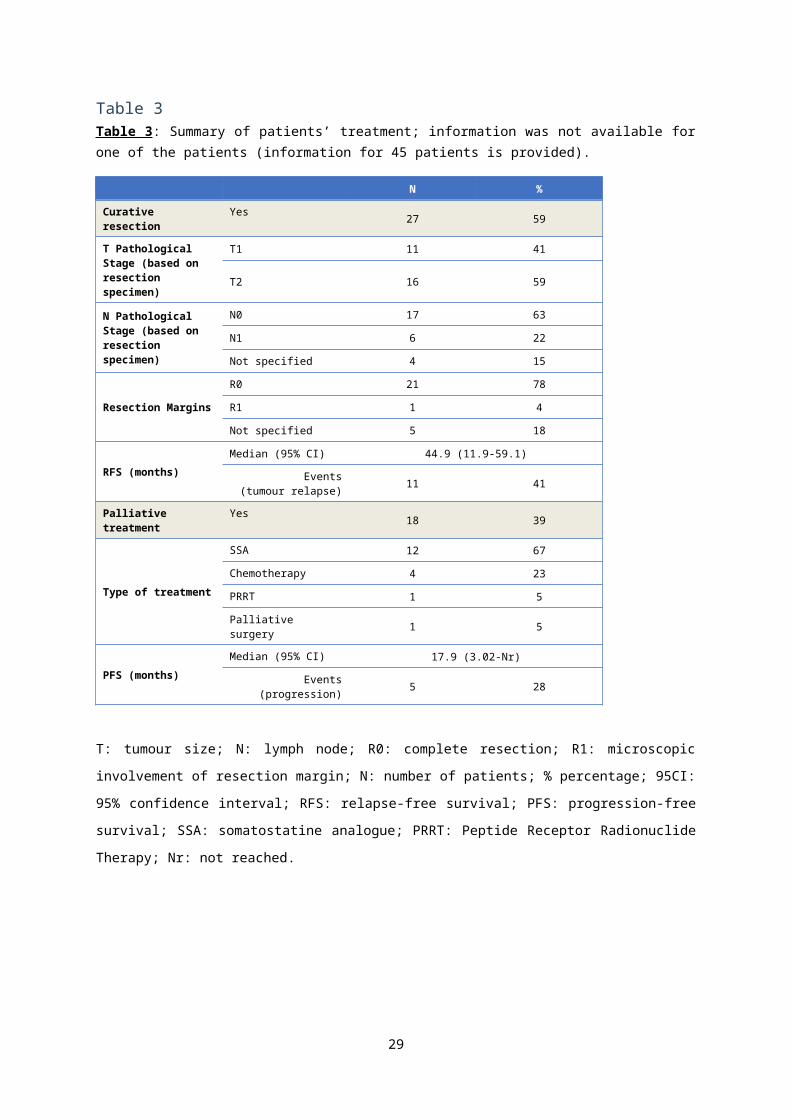
Most patients were diagnosed at localised stages (63%). Median time of follow-up for this study was
11.3 months (range 3.2-266.1). Treatment characteristics are specified in Table 3; for one of the
patients, information about treatment was not available. The majority of patients were treated with
curative resection (59%); achieving a median RFS was 44.9 months (95% CI 11.9-59.1). Only 39%
received any form of palliative treatment during the follow-up period, such as somatostatin analogues
(67%) or chemotherapy (23%); median PFS to first-line palliative treatment was 17.9 months (95% CI
3.02-not reached). Median overall survival was not reached (Table 2).
68Ga-DOTANOC PET imaging
A total of 47 68Ga-DOTANOC imaging scans were performed. In total, 27 scans (57%) showed
pathological uptake, while 20 (43%) were classified as “no-uptake” (no uptake or physiological uptake
only identified). Out of the 28 patients who had primary or metastatic LC in situ at the time of 68Ga-
DOTANOC-PET, 24 showed pathological uptake (sensitivity of 86%). Median SUVmax was 7.2 (95%
CI 6.1-19.9). There was neither significant correlation between Ki-67 index index and SUVmax (16
observations; Spearman rho=0.004; p-value 0.9) nor differences in SUVmax between typical and
atypical LC (mean SUVmax 9.7 (95% CI 1.3-18.1) vs 23.6 (95% CI 11.1-36.1), respectively; p-value
0.07).
The maximum size and tracer uptake of primary tumour and metastatic sites are summarised in Table 4 (Supplementary Material B). Overall, the biggest lesions were identified within the liver (median of
7 cm), however all tumour sites explored showed a high rate of tracer uptake (between 80-100%): 68Ga-DOTANOC showed high sensitivity regardless of the organ been explored. Regarding the most
suitable size cut-off of the largest metastatic lesion per cancer site for predicting tracer uptake in 68Ga-
7

DOTANOC imaging, ROC curves identified 1.4 cm and 2 cm as the most informative cut-offs for
primary lung tumour and lung metastases, respectively (Table 4; Supplementary Material B).
Rationale for performing 68Ga-DOTANOC imaging
Regarding the rationale behind performing the 68Ga-DOTANOC imaging, most scans were performed
as a post-surgical assessment of patients assumed to be cancer-free (post-surgical re-staging) (40%)
or for completing staging of patients with localised/advanced lung carcinoid tumours (Table 5). Other
reasons included confirmation of diagnosis of NET (9%), consideration of treatment with PRRT (7%)
and identification of primary tumour site (4%). Rate of positivity in the 68Ga-DOTANOC PET changed
according to rationale behind performing such imaging (Chi square p-value < 0.001). As expected, the
lowest uptake rate (16%) was shown in the post-surgical re-staging group, followed by patients in
whom 68Ga-DOTANOC was performed in order to confirm NET diagnosis (75%).
Impact of Ga-DOTANOC results on patients’ management
A total of 17 (37%) 68Ga-DOTANOC-PET imaging scans added additional information to the previous
imaging techniques (including CT and MR imaging), and 10 (26%) impacted patients’ management.
As detailed in Table 5, lower rate of additional information and change in patients’ management was
shown in the post-surgical re-staging group (21% and 12%, respectively), followed by patients in
whom 68Ga-DOTANOC-PET was performed as staging (42% and 29%, respectively). The rationale
behind the PET imaging did not have a statistically significant impact on whether the 68Ga-DOTANOC
added any extra information or not (Chi square p-value 0.065).
Type of additional information provided by the 68Ga-DOTANOC scan was analysed further. Out of the
47 scans performed, 9 (21%) identified new sites of disease, three of which were performed in
patients who were expected to be “cancer free” after curative resection (Figure 2). These accounted
for 16% (3 out of 19) scans performed in this population; none of these patients were early stage
(pT1aN0 completely resected (R0)) typical LC or DIPNECH. In addition, one scan changed options of
treatment, identifying a patient suitable for PRRT. See Table 5 for full information.
Subtype of LC did not change the rate of the 68Ga-DOTANOC-PET scan impacting patients’
management: DIPNECH (0%), typical LC (16%) and atypical LC (29%) (Chi square p-value 0.541);
however our data showed a trend for more significant benefit in patients diagnosed with typical or
atypical LC compared to DIPNECH.
Factors predictive of change in patients’ management and Survival analyses
Our data were unable to specify which population of patients may benefit more from performing a 68Ga-DOTANOC-PET scan (Supplementary Material C). Baseline factors and 68Ga-DOTANOC
results (such as SUVmax) were explored as potential prognostic factors impacting OS, RFS or PFS.
No prognostic factors were identified (all p-values >0.05; full data not shown) (Supplementary Material C).
8

DiscussionPreviously published series which focused exclusively / mainly on patients diagnosed with LC were
small [20, 27], the largest including only 26 patients [32] (Table 6). To our knowledge, our series is
one of the largest series exploring “real” role of 68Ga-DOTANOC PET exclusively in patients
diagnosed with LC. Our results showed that 68Ga-DOTANOC PET often changes management in
patients with LC tumours and should, therefore, be part of the baseline staging imaging (for both
metastatic and post-resected patients) regardless of site of metastases and grade (Table 7).
Our results confirmed similar sensitivity to that previously reported with 68Ga-DOTATATE [40, 41] and
was agreement with previous literature regarding none of the patients diagnosed with DIPNECH
showed any uptake [36]. Based on this, we would question whether performing such imaging in
patients with DIPNECH is of any value? 68Ga-DOTANOC PET could be beneficial to exclude
presence of small foci of typical LC in patients with DIPNECH, but its role is, otherwise, limited. On the
other hand, we did not identify any differences in 68Ga-DOTANOC uptake (in terms of SUVmax)
between typical and atypical LC; however this has been previously suggested by other researchers
[36]. Finally, all organs explored (lung, liver bone and lymph node) showed a good sensitivity and
therefore capacity of 68Ga-DOTANOC for identification of LC primary or metastases seems
independent of the organ being investigated. We would therefore suggest that 68Ga-DOTA-PET
imaging is performed regardless of type of LC (with the exception of DIPNECH) and site (organ) and
size of metastases.
The proportion of 68Ga-DOTANOC PET scans providing the clinician with additional information was
clinically relevant (37%), which translated into an impact on management in 26% of patients. These
results are in agreement with previous experience [39]. Interestingly, this benefit was not limited to a
specific sub-population, since no factors predictive of impact in patients’ management were identified
in our series: all patients seemed to benefit from performing the 68Ga-DOTA-PET, regardless of the
rationale behind such investigation or other baseline characteristics (including type of LC).
The predominant ways in which the 68Ga-DOTANOC PET scan impacted management were by the
identification of occult sites of metastases, assessment of disease distribution and tumour burden and
selection of patients suitable for PRRT.
Identification of new metastatic sites was retrospectively evaluated by Ambrosini et al. who assessed
the capacity of 68Ga-DOTANOC PET to identify occult bone metastases in 223 patients diagnosed
with NET (any site): 68Ga-DOTANOC PET was more accurate than CT for the identification of bone
lesions and led to a change in clinical management in nine patients who had a previous negative CT
scan [42]. Our results concur with these findings by identifying new occult bone and lung metastases
in patients who had the PET performed for accurate staging of metastatic disease (20%) and also in
patients in whom the PET scan was performed as a post-surgical re-staging test and who were
thought to be tumour-free (16%). This is therefore a novel indication for 68Ga-DOTANOC PET imaging
in LC; based on our results we would suggest incorporating a baseline 68Ga-DOTA-PET scan in all
patients following curative resection of LC due to the risk of identification of occult metastases as has
9

been previously suggested by other colleagues [38]. The potential role of 68Ga-DOTA-PET as a pre-
surgical staging evaluation could not be addressed by this study, since none of the patients had this
imaging performed pre-operatively. This aspect should be explored in further studies; in the mean-
time, pre-surgical 68Ga-DOTA-PET would be worth considering due to the same rationale for
recommending performing post-surgical re-staging.
Regarding the assessment of disease distribution and tumour burden in patients with LC, it is worth
highlighting that 68Ga-DOTANOC PET impacted on clinical management in 29% of patients with
known metastatic disease. This is comparable to rates previously described by other colleagues in
smaller series (33%) [39]. In fact, an accurate staging for assessment of tumour burden may be useful
for selection of treatment approaches, such as 1) assessment of liver remnant for patients due to
have liver embolisation; 2) assessment of extrahepatic disease in order to select patients for liver
directed therapies, such as liver embolisation, debulking surgery, liver radioembolisation; 3) selection
of systemic approaches such as SSA or chemotherapy based on tumour burden (SSA for patients
with low tumour burden; chemotherapy in scenarios with high tumour burden when rapid tumour
volume reduction is desirable) [13]. In summary, assessment of tumour distribution and tumour
burden may be helpful for the decision making process in the context of multidisciplinary case
discussion, when planning the treatment pathway for patients with advanced disease; 68Ga-DOTA-
PET should be part of the baseline assessment of patients with advanced LC and part of the
multidisciplinary discussion.
Finally, the role of 68Ga-DOTA-PET as a selection tool for patients with PRRT is widely accepted by
the NET community [12]; our results support such a use, since findings will help with the selection of
patients likely to respond to treatment and also to assess tumour burden and predict toxicity [13, 43-
45].
Limitations of all retrospective studies apply, such as reporting bias, since clinical data available was
restricted to previously collected information. Although all consecutive patients were included in order
to reduce to a minimum patient selection bias, it is impossible to completely avoid such a problem.
Patients who were referred for 68Ga-DOTANOC PET in the first instance could be somewhat selected
due to the clinical picture. Finally, the sample size was small, which is not surprising taking into
account the extremely selected patient population that this study was targeting. The limited sample
size, reduced the power of our study which should be considered a “proof of concept” study and which
conclusions should be confirmed in a larger cohort. In order to attenuate the above mention
limitations, it is worth highlighting the following. First, our population was as homogeneous as
possible, by exclusion of patients without pathological confirmation of LC and with high-grade lung
NETs. Second, all 68Ga-DOTA-PET reported in this study employed the same tracer (68Ga-
DOTANOC) providing consistency. Third, both centres involved in the study are recognised as high
volume and high expertise centres, not only in NETs, but also in nuclear medicine. Finally, all patients
had a pathology review by pathologist with an expertise in NETs; reflection of this is the fact that the
mitotic count was significantly higher in atypical LC compared to typical and that such differences
were significant, as expected according to international classifications of LC.
10

In summary (See Table 7), 68Ga-DOTANOC PET often changes management in patients with LC
tumours and should, therefore, be part of the baseline staging imaging (for both metastatic and post-
resected patients) regardless of site of metastases and grade. Results should be discussed in a
multidisciplinary meeting for planning patients’ management according to disease distribution and
tumour burden, including consideration of suitability for PRRT.
11
4. FundingThis study did not receive any external funding.
5. Author contributions Formulating the research question: AL, GP, WM
Designing the study: AL, GP, WM
Data collection: AL, MP, SV, PM
Analysing the data: AL
Result interpretation: All authors
Manuscript writing: All authors
6. AcknowledgementsAngela Lamarca was part-funded by the Pancreatic Cancer Research Fund and Spanish Society of Medical Oncology (SEOM) Fellowship Programme
George Papaxoinis received a grant from the Hellenic Society of Medical Oncology (HeSMO)
This work has been partially presented in ESMO (European Society of Medical Oncology) Annual Conference 2016 (Copenhagen, Denmark) and UKINETS (UK and Ireland Neuroendocrine Tumour Society) Annual Conference 2016 (London, United Kingdom).
7. Compliance with Ethical StandardsCompliance with Ethical Standards: All authors declares that there is no conflict of interest.
8. References
Reference List
1. Travis WBEM-HHH. The concept of pulmonary neuroendocrine tumours.. In: Pathology & Genetics: Tumours of the Lung, Pleura, Thymus, and Heart., IARC Press, Lyon 2004. p.19., 2004
2. Pelosi G, Fabbri A, Cossa M, Sonzogni A, Valeri B, Righi L, Papotti M. What clinicians are asking pathologists when dealing with lung neuroendocrine neoplasms? Semin.Diagn.Pathol. 2015; 32:469-479
3. Chauhan A, Ramirez RA. Diffuse Idiopathic Pulmonary Neuroendocrine Cell Hyperplasia (DIPNECH) and the Role of Somatostatin analogs: A Case Series. Lung 2015; 193:653-657
4. Marchevsky AM, Walts AE. Diffuse idiopathic pulmonary neuroendocrine cell hyperplasia (DIPNECH). Semin.Diagn.Pathol. 2015; 32:438-444
12

5. Yao JC, Hassan M, Phan A, Dagohoy C, Leary C, Mares JE, Abdalla EK, Fleming JB, Vauthey JN, Rashid A, Evans DB. One hundred years after "carcinoid": epidemiology of and prognostic factors for neuroendocrine tumors in 35,825 cases in the United States. J Clin Oncol 2008; 26:3063-3072
6. Hemminki K, Li X. Incidence trends and risk factors of carcinoid tumors: a nationwide epidemiologic study from Sweden. Cancer 2001; 92:2204-2210
7. Modlin IM, Lye KD, Kidd M. A 5-decade analysis of 13,715 carcinoid tumors. Cancer. 2003; 97:934-959
8. Tang LH, Basturk O, Sue JJ, Klimstra DS. A Practical Approach to the Classification of WHO Grade 3 (G3) Well-differentiated Neuroendocrine Tumor (WD-NET) and Poorly Differentiated Neuroendocrine Carcinoma (PD-NEC) of the Pancreas. Am.J Surg.Pathol. 2016;
9. Heetfeld M, Chougnet CN, Olsen IH, Rinke A, Borbath I, Crespo G, Barriuso J, Pavel M, O'Toole D, Walter T. Characteristics and treatment of patients with G3 gastroenteropancreatic neuroendocrine neoplasms. Endocr.Relat Cancer. 2015; 22:657-664
10. Rindi G, Klersy C, Inzani F, Fellegara G, Ampollini L, Ardizzoni A, Campanini N, Carbognani P, De Pas TM, Galetta D, Granone PL, Righi L, Rusca M, Spaggiari L, Tiseo M, Viale G, Volante M, Papotti M, Pelosi G. Grading the neuroendocrine tumors of the lung: an evidence-based proposal. Endocr.Relat Cancer. 2013; 21:1-16
11. Pelosi G, Papotti M, Rindi G, Scarpa A. Unraveling tumor grading and genomic landscape in lung neuroendocrine tumors. Endocr.Pathol. 2014; 25:151-164
12. Caplin ME, Baudin E, Ferolla P, Filosso P, Garcia-Yuste M, Lim E, Oberg K, Pelosi G, Perren A, Rossi RE, Travis WD. Pulmonary neuroendocrine (carcinoid) tumors: European Neuroendocrine Tumor Society expert consensus and recommendations for best practice for typical and atypical pulmonary carcinoids. Ann.Oncol 2015; 26:1604-1620
13. Horsch D, Schmid KW, Anlauf M, Darwiche K, Denecke T, Baum RP, Spitzweg C, Grohe C, Presselt N, Stremmel C, Heigener DF, Serke M, Kegel T, Pavel M, Waller CF, Deppermann KM, Arnold R, Huber RM, Weber MM, Hoffmann H. Neuroendocrine tumors of the bronchopulmonary system (typical and atypical carcinoid tumors): current strategies in diagnosis and treatment. Conclusions of an expert meeting February 2011 in Weimar, Germany. Oncol Res.Treat. 2014; 37:266-276
14. Aparicio T, Ducreux M, Baudin E, Sabourin JC, de BT, Mitry E, Schlumberger M, Rougier P. Antitumour activity of somatostatin analogues in progressive metastatic neuroendocrine tumours. Eur.J Cancer. 2001; 37:1014-1019
15. Waldherr C, Pless M, Maecke HR, Haldemann A, Mueller-Brand J. The clinical value of [90Y-DOTA]-D-Phe1-Tyr3-octreotide (90Y-DOTATOC) in the treatment of neuroendocrine tumours: a clinical phase II study. Ann Oncol. 2001; 12:941-945
16. Imhof A, Brunner P, Marincek N, Briel M, Schindler C, Rasch H, Macke HR, Rochlitz C, Muller-Brand J, Walter MA. Response, survival, and long-term toxicity after therapy with the radiolabeled somatostatin analogue [90Y-DOTA]-TOC in metastasized neuroendocrine cancers. J Clin Oncol. 2011; 29:2416-2423
17. Kwekkeboom DJ, de Herder WW, Kam BL, van Eijck CH, van EM, Kooij PP, Feelders RA, van Aken MO, Krenning EP. Treatment with the radiolabeled somatostatin analog [177 Lu-DOTA 0,Tyr3]octreotate: toxicity, efficacy, and survival. J Clin Oncol. 2008; 26:2124-2130
13
18. van EM, Krenning EP, Bakker WH, de Herder WW, van Aken MO, Kwekkeboom DJ. Peptide receptor radionuclide therapy with 177Lu-octreotate in patients with foregut carcinoid tumours of bronchial, gastric and thymic origin. Eur.J Nucl.Med Mol.Imaging. 2007; 34:1219-1227
19. Yao JC, Fazio N, Singh S, Buzzoni R, Carnaghi C, Wolin E, Tomasek J, Raderer M, Lahner H, Voi M, Pacaud LB, Rouyrre N, Sachs C, Valle JW, Delle FG, Van CE, Tesselaar M, Shimada Y, Oh DY, Strosberg J, Kulke MH, Pavel ME. Everolimus for the treatment of advanced, non-functional neuroendocrine tumours of the lung or gastrointestinal tract (RADIANT-4): a randomised, placebo-controlled, phase 3 study. Lancet. 2016; 387:968-977
20. Lococo F, Cesario A, Paci M, Filice A, Versari A, Rapicetta C, Ricchetti T, Sgarbi G, Alifano M, Cavazza A, Treglia G. PET/CT assessment of neuroendocrine tumors of the lung with special emphasis on bronchial carcinoids. Tumour.Biol. 2014; 35:8369-8377
21. Lococo F, Treglia G, Cesario A, Paci M, Filice A, Versari A, Filosso PL. Functional imaging evaluation in the detection, diagnosis, and histologic differentiation of pulmonary neuroendocrine tumors. Thorac.Surg.Clin 2014; 24:285-292
22. Abgral R, Leboulleux S, Deandreis D, Auperin A, Lumbroso J, Dromain C, Duvillard P, Elias D, de BT, Guigay J, Ducreux M, Schlumberger M, Baudin E. Performance of (18)fluorodeoxyglucose-positron emission tomography and somatostatin receptor scintigraphy for high Ki67 (>/=10%) well-differentiated endocrine carcinoma staging. J Clin Endocrinol.Metab. 2011; 96:665-671
23. Park CM, Goo JM, Lee HJ, Kim MA, Lee CH, Kang MJ. Tumors in the tracheobronchial tree: CT and FDG PET features. Radiographics. 2009; 29:55-71
24. Daniels CE, Lowe VJ, Aubry MC, Allen MS, Jett JR. The utility of fluorodeoxyglucose positron emission tomography in the evaluation of carcinoid tumors presenting as pulmonary nodules. Chest. 2007; 131:255-260
25. Pattenden HA, Leung M, Beddow E, Dusmet M, Nicholson AG, Shackcloth M, Mohamed S, Darr A, Naidu B, Iyer S, Marchbank A, Greenwood A, West D, Granato F, Kirk A, Ariyaratnam P, Loubani M, Lim E. Test performance of PET-CT for mediastinal lymph node staging of pulmonary carcinoid tumours. Thorax. 2015; 70:379-381
26. Virgolini I, Ambrosini V, Bomanji JB, Baum RP, Fanti S, Gabriel M, Papathanasiou ND, Pepe G, Oyen W, De CC, Chiti A. Procedure guidelines for PET/CT tumour imaging with 68Ga-DOTA-conjugated peptides: 68Ga-DOTA-TOC, 68Ga-DOTA-NOC, 68Ga-DOTA-TATE. Eur.J Nucl.Med Mol.Imaging. 2010; 37:2004-2010
27. Sollini M, Erba PA, Fraternali A, Casali M, Di Paolo ML, Froio A, Frasoldati A, Versari A. PET and PET/CT with 68gallium-labeled somatostatin analogues in Non GEP-NETs Tumors. ScientificWorldJournal. 2014; 2014:194123. doi: 10.1155/2014/194123. eCollection;%2014.:194123
28. Mojtahedi A, Thamake S, Tworowska I, Ranganathan D, Delpassand ES. The value of (68)Ga-DOTATATE PET/CT in diagnosis and management of neuroendocrine tumors compared to current FDA approved imaging modalities: a review of literature. Am.J Nucl.Med Mol.Imaging. 2014; 4:426-434
29. Panagiotidis E, Alshammari A, Michopoulou S, Skoura E, Naik K, Maragkoudakis E, Mohmaduvesh M, Al-Harbi M, Belda M, Caplin ME, Toumpanakis C, Bomanji J. Comparison of the impact of 68Ga-DOTATATE and 18F-FDG PET/CT on clinical management in patients with neuroendocrine tumors. J Nucl.Med. 2016; jnumed
14
30. Ambrosini V, Nicolini S, Caroli P, Nanni C, Massaro A, Marzola MC, Rubello D, Fanti S. PET/CT imaging in different types of lung cancer: an overview. Eur.J Radiol. 2012; 81:988-1001
31. Skoura E, Michopoulou S, Mohmaduvesh M, Panagiotidis E, Al HM, Toumpanakis C, Almukhailed O, Kayani I, Syed R, Navalkissoor S, Ell PJ, Caplin ME, Bomanji J. The Impact of 68Ga-DOTATATE PET/CT Imaging on Management of Patients with Neuroendocrine Tumors: Experience from a National Referral Center in the United Kingdom. J Nucl.Med. 2016; 57:34-40
32. Venkitaraman B, Karunanithi S, Kumar A, Khilnani GC, Kumar R. Role of 68Ga-DOTATOC PET/CT in initial evaluation of patients with suspected bronchopulmonary carcinoid. Eur.J Nucl.Med Mol.Imaging. 2014; 41:856-864
33. Phan AT, Oberg K, Choi J, Harrison LH, Jr., Hassan MM, Strosberg JR, Krenning EP, Kocha W, Woltering EA, Maples WJ. NANETS consensus guideline for the diagnosis and management of neuroendocrine tumors: well-differentiated neuroendocrine tumors of the thorax (includes lung and thymus). Pancreas 2010; 39:784-798
34. NCCN. National Comprehensive Cancer Network (NCCN) guidelines. 2016.
35. Edge SBDRCCCFAGGFLTAE. AJCC Cancer Staging Manual. 7th ed. 2010.ISBN 978-0-387-88440-0. In: Anonymous2010
36. Kayani I, Conry BG, Groves AM, Win T, Dickson J, Caplin M, Bomanji JB. A comparison of 68Ga-DOTATATE and 18F-FDG PET/CT in pulmonary neuroendocrine tumors. J Nucl.Med 2009; 50:1927-1932
37. Jindal T, Kumar A, Venkitaraman B, Meena M, Kumar R, Malhotra A, Dutta R. Evaluation of the role of [18F]FDG-PET/CT and [68Ga]DOTATOC-PET/CT in differentiating typical and atypical pulmonary carcinoids. Cancer Imaging. 2011; 11:70-5. doi: 10.1102/1470-7330.2011.0010.:70-75
38. Jindal T, Kumar A, Venkitaraman B, Dutta R, Kumar R. Role of (68)Ga-DOTATOC PET/CT in the evaluation of primary pulmonary carcinoids. Korean J Intern.Med. 2010; 25:386-391
39. Ambrosini V, Castellucci P, Rubello D, Nanni C, Musto A, Allegri V, Montini GC, Mattioli S, Grassetto G, Al-Nahhas A, Franchi R, Fanti S. 68Ga-DOTA-NOC: a new PET tracer for evaluating patients with bronchial carcinoid. Nucl.Med Commun. 2009; 30:281-286
40. Haug AR, Cindea-Drimus R, Auernhammer CJ, Reincke M, Wangler B, Uebleis C, Schmidt GP, Goke B, Bartenstein P, Hacker M. The role of 68Ga-DOTATATE PET/CT in suspected neuroendocrine tumors. J Nucl.Med 2012; 53:1686-1692
41. Souvatzoglou M, Ziegler SI, Martinez MJ, Busch R, Dzewas G, Schwaiger M, Bengel F. Standardised uptake values from PET/CT images: comparison with conventional attenuation-corrected PET. Eur.J Nucl.Med Mol.Imaging. 2007; 34:405-412
42. Ambrosini V, Nanni C, Zompatori M, Campana D, Tomassetti P, Castellucci P, Allegri V, Rubello D, Montini G, Franchi R, Fanti S. (68)Ga-DOTA-NOC PET/CT in comparison with CT for the detection of bone metastasis in patients with neuroendocrine tumours. Eur.J Nucl.Med Mol.Imaging. 2010; 37:722-727
43. Oksuz MO, Winter L, Pfannenberg C, Reischl G, Mussig K, Bares R, Dittmann H. Peptide receptor radionuclide therapy of neuroendocrine tumors with (90)Y-DOTATOC: is treatment response predictable by pre-therapeutic uptake of (68)Ga-DOTATOC? Diagn.Interv.Imaging. 2014; 95:289-300
15

44. Bodei L, Kidd M, Paganelli G, Grana CM, Drozdov I, Cremonesi M, Lepensky C, Kwekkeboom DJ, Baum RP, Krenning EP, Modlin IM. Long-term tolerability of PRRT in 807 patients with neuroendocrine tumours: the value and limitations of clinical factors. Eur.J Nucl.Med Mol.Imaging. 2015; 42:5-19
45. Mariniello A, Bodei L, Tinelli C, Baio SM, Gilardi L, Colandrea M, Papi S, Valmadre G, Fazio N, Galetta D, Paganelli G, Grana CM. Long-term results of PRRT in advanced bronchopulmonary carcinoid. Eur.J Nucl.Med Mol.Imaging. 2016; 43:441-452
16

9. Figures and tables
17

Table 1Table 1: Classification of lung neuroendocrine tumours (adapted from features [1, 2, 4]).
Premalignant Lung carcinoids Lung NECsDIPNECH Typical LC Atypical LC Large-cell NEC Small-cell NEC
Grade Low Low Intermediate High HighMitotic rate (x10 HPF)
<2 2-10 >10 >10
Necrosis None Often (focal) Often (diffuse) Often (diffuse)Morphology Well-differentiated Well-differentiated Well-
differentiatedPoorly-differentiated Poorly-differentiated
Pathological characteristics
Characterised by widespread
neuroendocrine cell hyperplasia and
tumourlets
Cells are bland, polygonal in shape with round nuclei and finely dispersed
chromatin. Cells are arranged in distinct organoid, trabecular, or insular growth
patterns with a delicate vascular stroma.Immunohistochemical staining for
neuropeptides (i.e. CD56 chromogranin, synaptophysin) is usually present.
Cells are arranged in organoid, trabecular, or
palisading patterns.Cells are large and present
abundant eosinophilic cytoplasm. Nuclear
chromatin tends to be granular. Cells present
immunoreactivity for chromogranin and
synaptophysin.
Round, oval and angulated cells, small amounts of cytoplasm, nuclei with
dispersed "salt and pepper" chromatin
Tumour cells are usually positive for one or more of
chromogranin or synaptophysin, although
around 10 percent may be unreactive for neuroendocrine
markers.
LC: lung carcinoid; HPF: high-power fields: HPF; NEC: neuroendocrine carcinoma. DIPNECH: Diffuse idiopathic pulmonary neuroendocrine cell hyperplasia.
The mitotic rate was assessed over 10 consecutive high-power fields (HPF) in the mitotically most active area (“hottest spot”) of each sample at a
magnification of x400 using a Nikon Eclipse microscope with a x40 objective and a x10 ocular lens (equivalent to 2mm2 field area).
18
Supplementary material A (Fig 1)
Figure 1 (supplementary material): Patients’ flow.
Out of the 166 patients screened, 46 were eligible. All patients included had pathological diagnosis of lung carcinoid and had a gallium DOTANOC-PET scan performed. LC: lung carcinoid.
19
Table 2
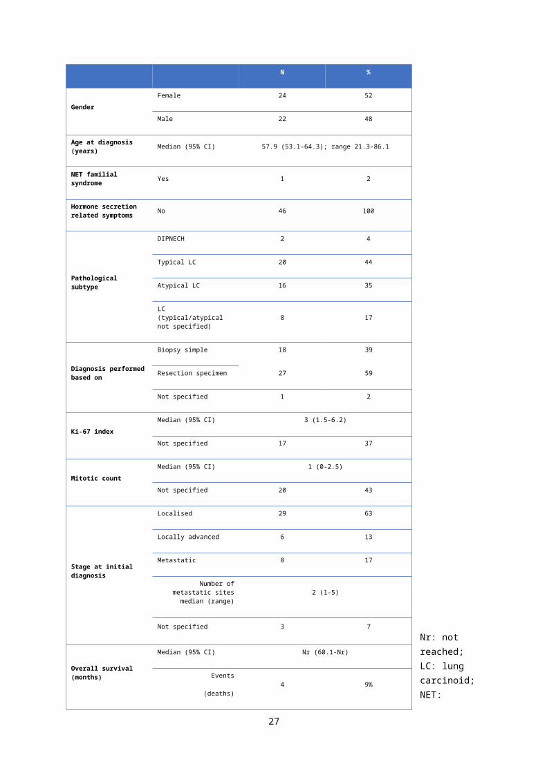
Table 2: Summary of patients’ characteristics (46 patients).
20
N %
GenderFemale 24 52
Male 22 48
Age at diagnosis (years) Median (95% CI) 57.9 (53.1-64.3); range 21.3-86.1
NET familial syndrome Yes 1 2
Hormone secretion related symptoms No 46 100
Pathological subtype
DIPNECH 2 4
Typical LC 20 44
Atypical LC 16 35
LC (typical/atypical not specified) 8 17
Diagnosis performed based on
Biopsy simple 18 39
Resection specimen 27 59
Not specified 1 2
Ki-67 indexMedian (95% CI) 3 (1.5-6.2)
Not specified 17 37
Mitotic countMedian (95% CI) 1 (0-2.5)
Not specified 20 43
Stage at initial diagnosis
Localised 29 63
Locally advanced 6 13
Metastatic 8 17
Number of metastatic sites median (range) 2 (1-5)
Not specified 3 7
Overall survival (months)
Median (95% CI) Nr (60.1-Nr)
Events
(deaths)4 9%

Nr: not reached; LC: lung carcinoid; NET: neuroendocrine tumour; N: number; % percentage; 95CI: 95% confidence interval; DIPNECH: Diffuse idiopathic pulmonary neuroendocrine cell hyperplasia; Nr: not reached.
21

Table 3Table 3: Summary of patients’ treatment; information was not available for one of the patients (information for 45 patients is provided).
N %
Curative resection Yes 27 59
T Pathological Stage (based on resection specimen)
T1 11 41
T2 16 59
N Pathological Stage (based on resection specimen)
N0 17 63
N1 6 22
Not specified 4 15
Resection Margins
R0 21 78
R1 1 4
Not specified 5 18
RFS (months)Median (95% CI) 44.9 (11.9-59.1)
Events(tumour relapse) 11 41
Palliative treatment Yes 18 39
Type of treatment
SSA 12 67
Chemotherapy 4 23
PRRT 1 5
Palliative surgery 1 5
PFS (months)Median (95% CI) 17.9 (3.02-Nr)
Events(progression) 5 28
T: tumour size; N: lymph node; R0: complete resection; R1: microscopic involvement of resection
margin; N: number of patients; % percentage; 95CI: 95% confidence interval; RFS: relapse-free
survival; PFS: progression-free survival; SSA: somatostatine analogue; PRRT: Peptide Receptor
Radionuclide Therapy; Nr: not reached.
22

Supplementary material B (Table 4) Table 4 (Supplementary material): Size of largest metastatic deposit and tracer uptake by metastatic site: primary lung tumour, liver, lung, bone and lymph node metastases.
Primary tumour in situ Liver
metastases
Lung metastases
Bone metastases
Lymph node metastases
Patients with these lesions present at the time of performing 68Ga-DOTANOC scan (N (%))
16 (24%)10 (21%)
10 (21%) 13 (28%) 12 (26%)
If yes, maximum size of metastatic deposit within this site (cm; median (95% CI))
4.3 (1.9-21.1)7.5 (1.8-7.8) 2.7 (1.2-13)
2.15 (1.5-2.5) 2.6 (1.9-3.6)
If present, did this tumour site show tracer uptake in the 68Ga-DOTANOC? (Sensitivity) (N (%; 95% CI))
13/16 (81%; 95% CI 67.2-
100)8/10 (80%; 95%
CI 49.8-100)
8/10 (80%; 95% CI 49.8-100)
13/13 (100%; 95% CI 100-
100)
11/12 (91.7%; 95% CI 73.3-
100)
ROC curve (AUC), prediction of tracer uptake according to size of metastases
0.9231 Cannot calculate*
1Cannot
calculate*Cannot
calculate*
Most informative size cut-off to predict tracer uptake (cm) (Se/Sp)
1.4(Se 92% / Sp
100%)
Cannot calculate*
2(Se 100% / Sp
100%)
Cannot calculate*
Cannot calculate*
N: number of patients; % percentage; 95% CI: 95% confidence interval; cm: centimeter; ROC: receiver operating characteristic; AUC: area under the curve; Se: sensitivity; Sp: specificity. *ROC curve cannot be drawn due to not having enough information for the analysis (i.e. all lesions with maximum size available showed tracer uptake).
23
Table 5Table 5: Rationale and outcome (total of 47 scans) and clinical impact (information available for 42 scans) of 68Ga-DOTANOC PET imaging.
Outcome of the 68Ga-DOTANOC Impact of the 68Ga-DOTANOC on patients’ management
How many showed uptake?
(N=47)
How many added extra information?
(N=46)
How many changed
management? (N=38)
Identified new sites of disease (N=42)
Changed options of treatment (in terms of
PRRT) (N=42)
TOTAL 27/47 (57%) 17/46 (37%) 10/38 (26%) 9/42 (21%) 1/42 (2%)
Rat
iona
le fo
r per
form
ing
68G
a-D
OTA
NO
C
Confirmation of NET (N=4) 3/4 (75%) 2/4 (50%) 1/3 (33%) 1/4 (25%) 0/4 (0%)
Identification of primary tumour (N=2) 2/2 (100%) 0/1 (0%) 0/1 (0%) 0/2 (0%) 0/2 (0%)
Post-surgical re-staging (N=19) 3/19 (16%) 4*/19 (21%) 2&/17 (12%) 3/19 (16%) 0/19 (0%)
Staging (patients with cancer) (N=19) 16/19 (84%) 8/19 (42%) 4/14 (29%) 3/15 (20%) 0/15 (0%)
Consideration of PRRT (N=3) 3/3 (100%) 3/3 (100%) 3$/3 (100%) 2/3 (67%) 1/3 (33%)
Four reasons for performing 68Ga-DOTANOC imaging were evaluated: confirmation of diagnosis of NET (4 scans; 9%), identification of primary tumour in patients originally diagnosed with unknown primary malignancy (2 scans; 4%), post-surgical re-staging in patients who were thought to be “cancer free” after curative resection (19 scans; 40%), completion of staging (for patients known to have localised, locally advanced or metastatic cancer in situ) (19 scans; 40%) and pre-PRRT assessment (3 scans; 7%). Outcome was measured as per the rate of pathological tracer uptake, whether the imaging added or not additional information and whether that additional information impacted patients’ management or not. Data information regarding the additional information provided by the 68Ga-DOTANOC PET scan (if any) was collected whenever available. *3 pts were identified to have metastatic disease; 1 patient showed suspicious uptake within lungs which did not change the management (patient continued follow-up); &for one of the patients in whom metastatic disease was identified,
24

information regarding impact on management was not available; $two patients were found to have too much tumour burden for PRRT. NET: neuroendocrine tumour, PRRT: Peptide Receptor Radionuclide Therapy; N: number of scans; %: percentage.
25

Figure 2
Figure 2: Bone metastasis identified in patient who underwent a post-surgical re-staging 68Ga-DOTANOC PET imaging.
26
Supplementary material C
Factors predictive of change in patients’ management
Our data were unable to specify which population of patients may benefit more from performing a 68Ga-DOTANOC-PET scan. Logistic regression was performed to explore selected baseline
characteristics which may be able to identify patient subgroups with more benefit from performing 68Ga-DOTANOC PET in terms of impact on patients’ management. Univariate analysis did not identify
any significant factors: type of LC (OR (odds ratio) 2.3 (95% CI 0.5-11.6); p-value 0.308), stage (OR
1.4 (95% CI 0.5-3.7); p-value 0.474), previous curative resection (OR 1.3 (95% CI 0.3-6.2); p-value
0.744), aim of the 68Ga-DOTANOC (all p-values > 0.05). Other factors such as Ki-67 index, mitotic
count and sites of metastases were also not significant (all p-values > 0.05; full data not shown).
Survival analyses
Baseline factors and 68Ga-DOTANOC results (such as SUVmax) were explored as potential
prognostic factors impacting OS, RFS or PFS. Tracer uptake and SUVmax did not show impact on
OS (Hazard Ratio (HR) 0.2 (95% CI 0.01-3.3; p-value 0.233) and HR 0.8 (95% CI 0.6-1.2; p-value
0.385), respectively), RFS (HR 2.5 (95% CI 0.2-27.6; p-value 0.457) and HR 1.01 (95% CI 0.9-1.05;
p-value 0.840), respectively) or PFS (HR 1.8 (95% CI 0-not estimated; p-value 1) and HR 1.04 (95%
CI 0.9-1.1; p-value 0.249), respectively). No prognostic factors were identified (all p-values >0.05; full
data not shown).
27

Table 6 Table 6: Summary of the largest LC series exploring the role of 68Ga-DOTA-PET imaging.
Author, reference
Number of LC patients
Total number of patients
TracerFindings
Kayani et al. [36]
18 18 68Ga-
DOTATAT
E
Typical and atypical LC showed higher and lower uptake of 68Ga-DOTATATE, respectively.
DIPNECH showed no uptake.
Jindal et al. [37] 20 20 68Ga-
DOTATOC
Typical and atypical LC showed higher and lower uptake of 68Ga-DOTATOC, respectively.
Ratios of SUVmax on 68Ga-DOTATOC to that 18F-FDG were significantly higher in typical
LC compared with atypical LC.
Jindal et al. [38] 19 19 68Ga-
DOTATOC
Tumour detection rate of 95%. SUVmax ranged from 1.1 to 66, with a median value of
21.6. In one patient (out of 20), 68Ga-DOTATOC PET revealed additional lesions:
recommended to be included in the diagnostic work-up of these patients.
Ambrosini et al. [39]
11 11 68Ga-
DOTANOC
68Ga-DOTANOC was useful in LC patients because it led to a better evaluation of the
extent of the disease, detecting higher number of lesions in 5 patients (out of 11) and
providing additional information in nine of 11 patients leading to the changes in the clinical
management of three of nine patients (33%).
Venkitaraman et al. [32]
26 32 68Ga-
DOTATOC
The sensitivity, specificity and accuracy of 68Ga-DOTATOC in the diagnosis of LC were
96%, 100% and 97%, respectively; whereas those of 18F-FDG were 78%, 11% and 59%,
respectively. Authors concluded that 68Ga-DOTATOC was useful for the evaluation of LC,
while 18F-FDG PET suffered from low sensitivity and specificity.
Lamarca et al. (This series)
46 46 68Ga-
DOTANOC
68Ga-DOTANOC PET often changes management in patients with LC tumours (26%) and
should, therefore, be part of the staging imaging (for both metastatic and resected
patients) regardless of site of metastases and grade.
28
68Ga-DOTA-PET: 68-Gallium-radiolabeled positron emission tomography; LC: lung carcinoid.
29

Table 7Table 7: Summary of recommendations (applicable whenever 68Ga-DOTA-PET is available).
Recommendations (applicable whenever 68Ga-DOTA-PET is available)#1 68Ga-DOTA-PET imaging should be performed regardless of type of LC (typical/atypical),
with the exception of DIPNECH. 68Ga-DOTA-PET could be beneficial to exclude presence of small foci of typical LC in patients with DIPNECH, but its role in DIPNECH seems, otherwise, limited.
#2 68Ga-DOTA-PET imaging should be performed regardless of site (organ) and size of metastases.
#3 68Ga-DOTA-PET should be considered and incorporate to the baseline imaging to be performed to all patients following curative resection of LC due to the potential for identification of occult metastases; exceptions may include pT1aN0R0 typical LC and DIPNECH. Its role pre-surgery could not be explored in the current study but would be worth considering due to the same rational.
#4 68Ga-DOTA-PET should be part of the baseline assessment of patients with advanced LC and part of the multidisciplinary discussion, in order to provide information regarding tumour distribution and tumour burden.
#5 All patients to be considered for PRRT should require previous assessment with 68Ga-DOTA-PET whenever available.
68Ga-DOTA-PET: 68-Gallium-radiolabeled positron emission tomography; DIPNECH: diffuse idiopathic pulmonary neuroendocrine cell hyperplasia; LC: lung carcinoid; PRRT: peptide receptor radionuclide therapy; R0: complete resection.
30