1 chapter 4 viruses monera structure and shape classification reproduction retrovirus origin of...
TRANSCRIPT

1
CHAPTER 4VIRUSES MONERA
• STRUCTURE AND SHAPE• CLASSIFICATION• REPRODUCTION• RETROVIRUS• ORIGIN OF
VIRSUSES
• CLASSIFICATION• STRUCTURE• ECOLOGY/ ADAPTATION• REPRODUCTION• ECONOMIC
IMPORTANCE
• Bacteria Testing
2
Structure and Shape of viruses
• Don’t possess life functions
• Composed of Protein coat and Genetic materials ( DNA or RNA)
• Most are spherical or other geometric form
3
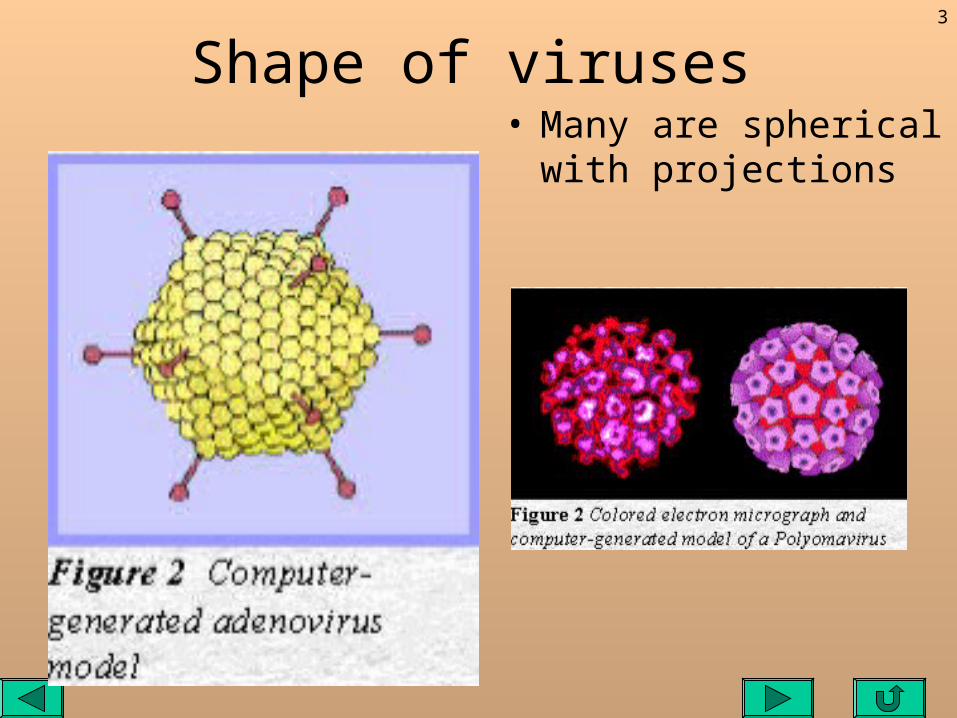
Shape of viruses• Many are spherical with
projections
4
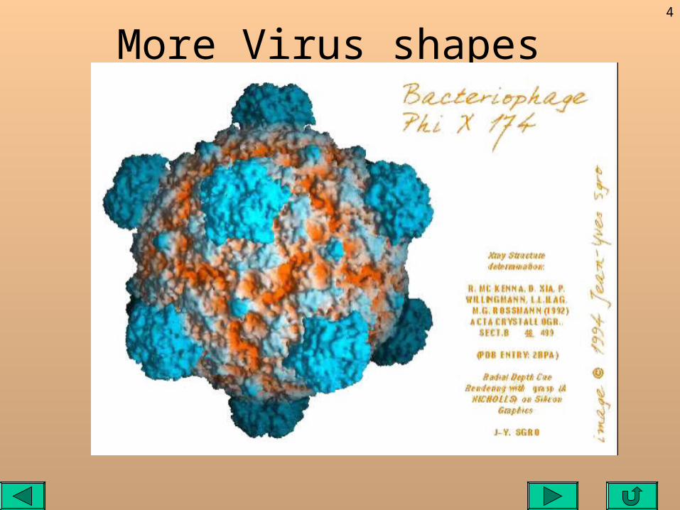
More Virus shapes

5
Filamentous virusEbola

6
Complex Virus Structures

7
HIV MODEL
RNA
Enzymes
Core Protein Coat
Inner protein

8
Virus slides
1- Influenza virus
2- Polio virus
3- Tobacco Mosaic virus

9
Virus Sizes• With electron microscopy the level of
resolution is 5nm (1nm = 10-9 meters). To put this into some kind of perspective:
• an atom is about 0.2-0.3 nm in diameter• DNA is about 2nm in diameter. A small
virus• parvovirus has a diameter of about
25nm. A large virus (e.g.• poxviruses) have a diameter of up to
300nm.

10
Classification of Viruses
• Grouped by the type of genetic material they have
– Single strand of DNA
– Double strands of DNA
– Single strand of RNA
– Double Strands of RNA
• Shape and size

11Virus Families• Poxviridae (pox viruses)• Parvoviridae• Reoviridae• Picornaviridae (Hepatitis A virus, foot-and-
mouth disease virus)• Togaviridae (Rubella virus)• Flaviviridae (Hepatitis C virus, yellow fever
virus)• Rhabdoviridae• Bunyaviridae (Hantaan virus)• Herpesviridae (Human Herpes Simplex
Viruses 1&2, VZV, Human
12
Virus Families continued• Adenoviridae• Papovaviridae (Papillomaviruses)• Hepadnaviridae (Hepatitis B virus)• Caliciviridae• Arenaviridae• * Paramyxoviridae (Measles virus)• * Orthomyxoviridae (Influenza viruses
A-C)• * Filoviridae (Ebola virus)• * Retroviridae (HIV-1&2, HTLV-1)• * Astroviridae

13
Viral Reproductions
• Since viruses are nonliving they must use a host for reproduction. The host provided all the material and energy to replicate itself.

14
Viral Reproductions-2
• Viruses are very specific in which types of cell they require as host. This is why it is very difficult (but not impossible)to get a virus infection from an animal.
15
Viral Reproductions-3
• Two types of reproductive cycles
–Lytic cycle
–Lysogenic cycle
16
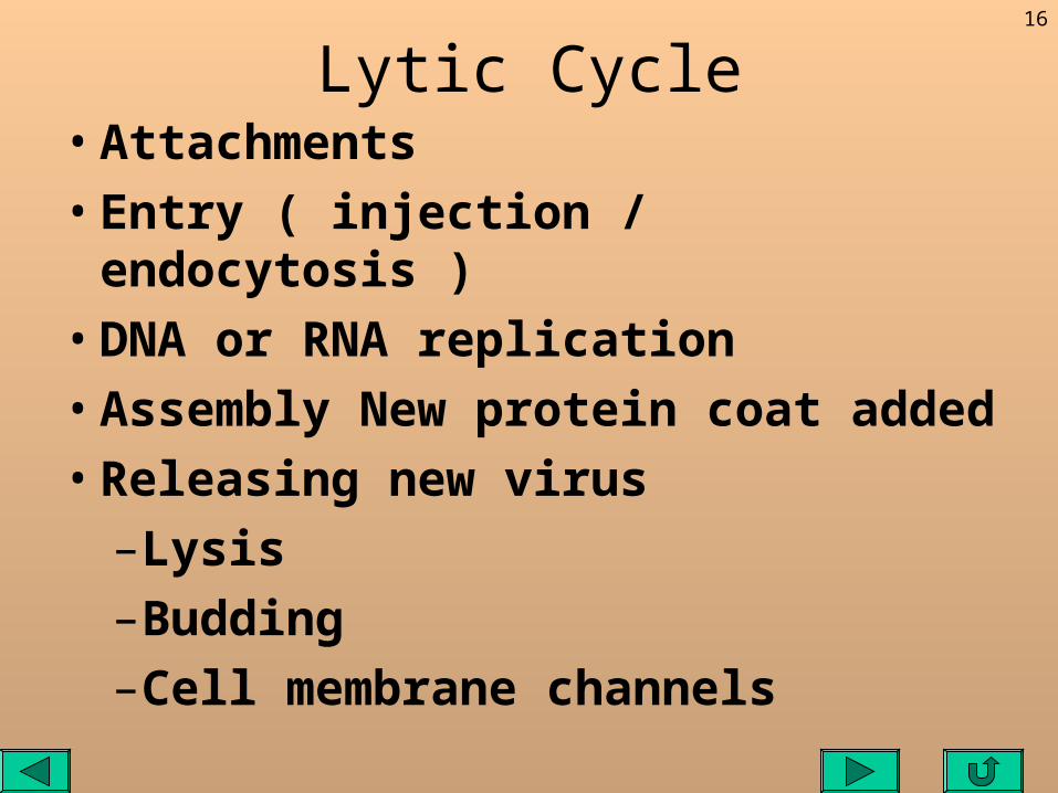
Lytic Cycle• Attachments
• Entry ( injection / endocytosis )
• DNA or RNA replication
• Assembly New protein coat added
• Releasing new virus
– Lysis
– Budding
– Cell membrane channels

17
Replication steps

18
Movie clip showing ReplicationQuickTime™ and aVideo decompressor
are needed to see this picture.
19
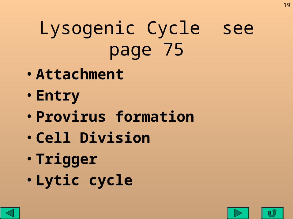
Lysogenic Cycle see page 75
• Attachment
• Entry
• Provirus formation
• Cell Division
• Trigger
• Lytic cycle
20
Retrovirus
• The most complex RNA viruses
• During injection of their RNA they also inject a special enzyme that help in the reverse transcriptase
• HIV is such a virus
21
ORIGIN OF VIRUSES
• The theory is that viruses originated from cells who DNA or RNA some how escaped a developed a way to reproduce as parasites.
• This would indicate that new viruses could be continually being made.
22
Monera (Bacteria)
• Archaebacteria - ancient bacteria that live in extreme enviroments.
– Oxygen free environments
– salt water environments
– hot acidic waters

23
Life on Mars
24
Eubacteria- Heterotrophs
• Heterotrophs- decomposers
• Eubacteria - Chemosynthetic
• Eubacteria- Photosynthetic

25
Bacteria structures
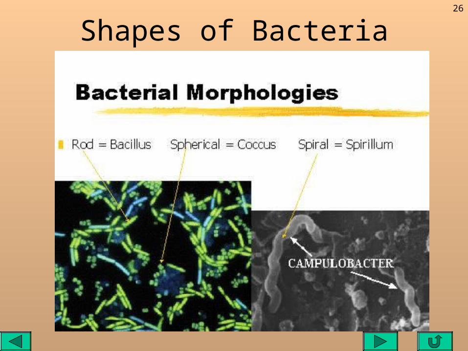
• Shapes
– coccus- round
– bacillus- rod shape
– spirallus- cork screw shape
• Arrangements
– diplo-
– staphylo-
– strepto-
26
Shapes of Bacteria

27
Coccus - Round shape

28
Bacillus- Rod Shape
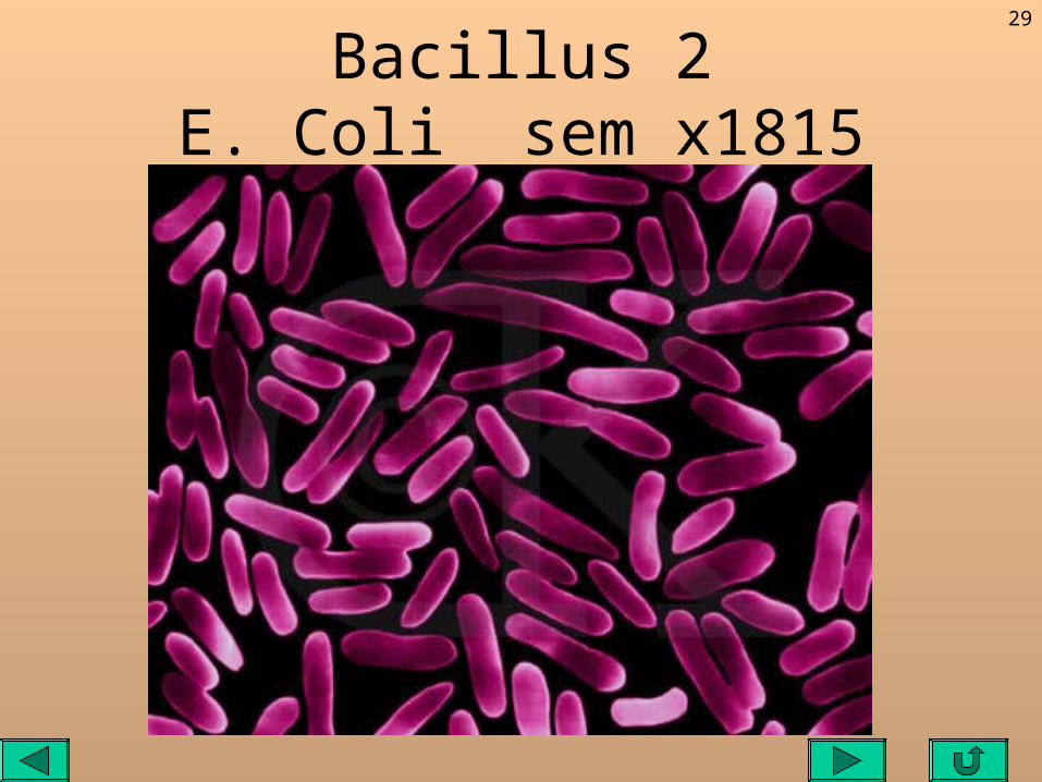
29
Bacillus 2E. Coli sem x1815

30
E.coli Sem x49,440

31
Streptomyces sem x 5,510
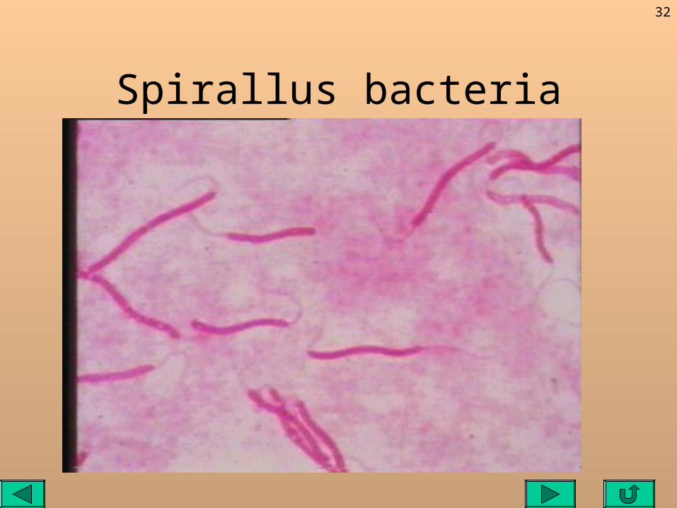
32
Spirallus bacteria

33
Naming of Bacteria
• Names are a combinations of the shape and the cluster arrangements.
– Example
–diplococcus
–streptococcus
–staphylbaccillus

34
A typical bacteria structure 1
• Prokaryotes- Lack a membrane bound nucleus.
• Cell wall- Different chemical composition than plants- complex polysaccharide (not found in eukaryotes)
• Plant cell walls contain cellulose.
• See Transparency # 44
35
Structures-2
• Capsule- slimy material that covers the cell wall. Protects the bacteria.
EX. Capsule protects the cell from the white blood cells and antibodies produced by animal cells.

36
Structures-3
• Cell membrane- located just inside the cell wall. Prokaryotes lack organelles. All reactions take place in the folds of the cell membrane.

37
Structure continued 2
• Cytoplasm- contains ribosome (synthesize proteins). If bacteria carry out photosynthesis chlorophyll is contained here.
• Hereditary material (DNA)- Lack a "true" nucleus. DNA is circular. Found in the nucleolid. Plamids are smaller segments of DNA.
38
Structures -3• 6.Endospores- Formed within the
cytoplasm. Contain DNA and a small amount of cytoplasm. Form when conditions are unfavorable. Allows the bacteria to remain dormant. When conditions become favorable the bacteria will grow again. Developed this trait for survival
39
Protection from Osmotic rupture
• Like most living things the concentration of water and other liquids is higher outside the organism then inside
• Most bacteria have a thick cell wall composed of sugar molecules linked with amino acids.
40
Penicillin- Bacteria Killer?
• Penicillin kills bacteria by interfering with the amino acids that link the sugars together in the cell wall
• This rupturing of the wall allows water to rush in lysing the cell
41
Ecology and Adaptations
• Obligate aerobes bacteria require oxygen
• Obligate anaerobes live in an oxygen free environment. - oxygen will kill them.
• Endospores are formed by some bacteria when conditions become harmful to them

42
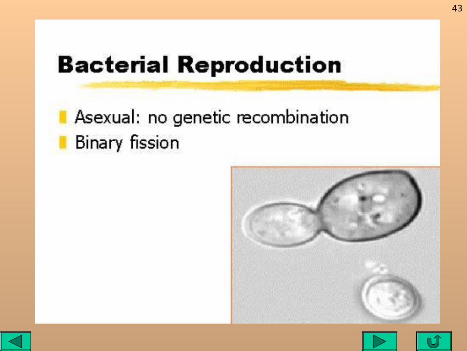
Reproductions
• Binary fission- asexual process - bacteria will simply undergo mitosis
• Sexual reproduction - Chromosomes are exchanged from one bacteria to another through the Pili
43
Reproduction

44
Rod shape bacteria with Pilus

45
SEM of Pili

46
Economic Importance
• Nitrogen fixation - all organism need nitrogen to construct things like protein, DNA, RNA and ATP.
• Nitrogen fixation - occurs in some bacteria that are able to get nitrogen from the air (N2) and convert it to NH3 or NO2, NO3

47
Nitrogen Fixing nodules

48
Economic Importance 2
• Bacteria cause organic material to decay. This allows for the recycling of nutrients.
• Some bacteria use fermentation which makes a variety of molecules with distinctive flavors and aromas- Yogurt, cheese, vinegar.

49
Why to we culture bacteria?
• to study them in more detail
• to study or improve strains of bacteria.
• to identify which bacterium has infected you and therefore what treatment to begin.

50
How Bacteria are cultured?
• Life forms require certain foods, water and temperatures to exist bacteria are no exception.
• Each type of bacteria prefers either sugars, starch, fats or proteins. So by providing a certain nutrient you will encourage a specific type of bacteria to grow.
51
How Bacteria are cultured? -2
• Temperature should be 20 C to 37 C

52
Inoculation
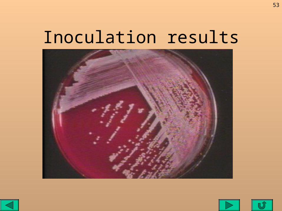
• Adding bacteria to a culture dish is called inoculation
53
Inoculation results

54
Inoculation results 2

55
Inoculation results 3
56
Culture Results
• By Studying pure culture plates of a bacterial species, and observing the texture, aroma, color, growth pattern, height of the growth, and other physical characteristics of the colonies, you can learn a lot about the specimen.

57
Testing Bacteria
• One way to determine how to treat a bacteria is to determine the type of cell wall it has.
– Thick wall usually indicate a Gram positive type
– Thin wall usually indicate a Gram negative type

58
Gram Negative test
• It was found that thick wall bacteria will stain differently than those that have thin wall.
• The Gram negative test uses a process to stain bacteria.

59
Gram Positive • They are usually Coccus and Bacillus
in shape
• Most are harmless to people and are used for their fermentation process to make foods.
• Examples of common Gram-positive cells include Staphylococcus aureusand Streptococcus cremoris, a bacterium used in dairy production.

60
Gram Negative
• These bacteria are more harmful then helpful
• Afflicted individuals are usually treated with streptomycin or erythromycin.