1 6 bones and skeletal tissues part a. 2 skeletal cartilage contains no blood vessels or nerves ...
TRANSCRIPT
1
6Bones and Skeletal Tissues
Part A
2
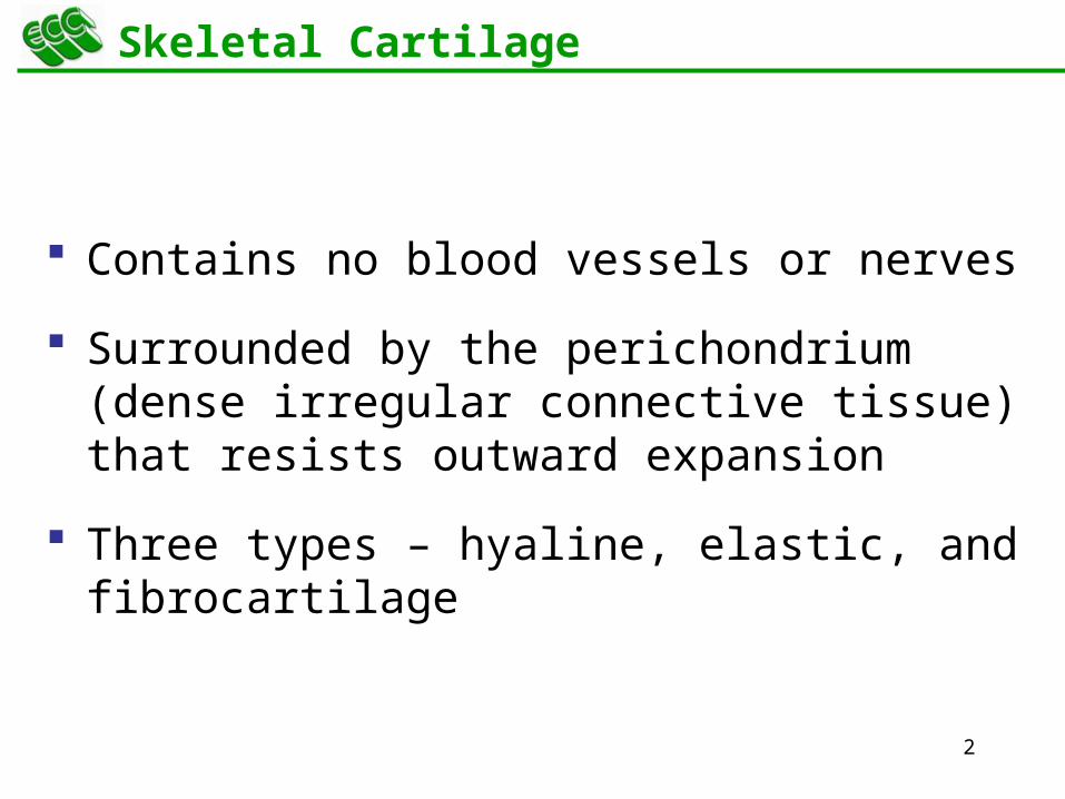
Skeletal Cartilage
Contains no blood vessels or nerves
Surrounded by the perichondrium (dense irregular connective tissue) that resists outward expansion
Three types – hyaline, elastic, and fibrocartilage
3
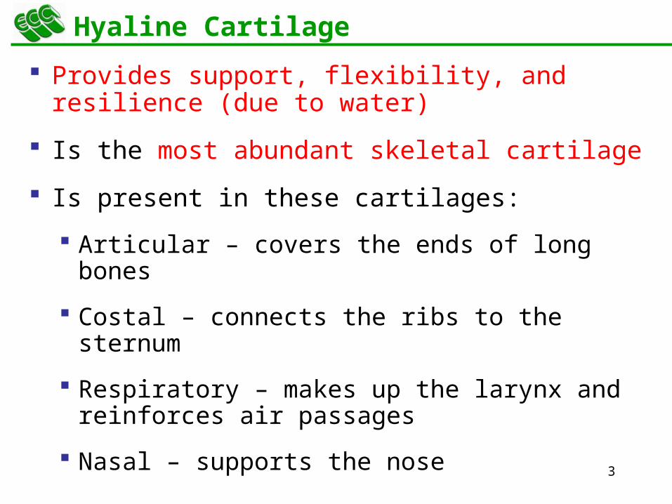
Hyaline Cartilage
Provides support, flexibility, and resilience (due to water)
Is the most abundant skeletal cartilage
Is present in these cartilages:
Articular – covers the ends of long bones
Costal – connects the ribs to the sternum
Respiratory – makes up the larynx and reinforces air passages
Nasal – supports the nose
4
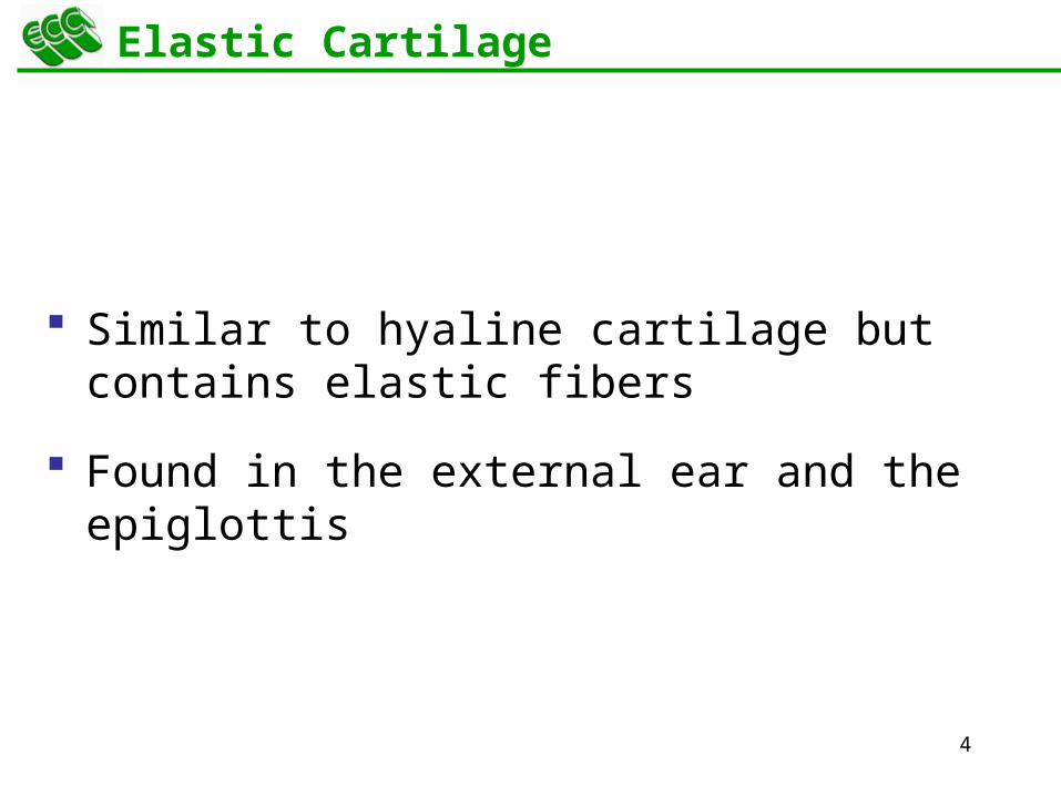
Elastic Cartilage
Similar to hyaline cartilage but contains elastic fibers
Found in the external ear and the epiglottis
5
Fibrocartilage
Highly compressed with great tensile strength
Contains collagen fibers
Found in menisci of the knee and in intervertebral discs
6
Growth of Cartilage
Appositional – cells in the perichondrium secrete matrix against the external face of existing cartilage
Interstitial – lacunae-bound chondrocytes inside the cartilage divide and secrete new matrix, expanding the cartilage from within
Calcification of cartilage occurs
During normal bone growth
During old age
7
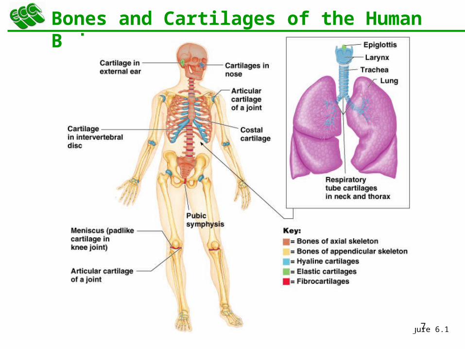
Bones and Cartilages of the Human Body
Figure 6.1

8
Classification of Bones
Axial skeleton – bones of the skull, vertebral column, and rib cage
Appendicular skeleton – bones of the upper and lower limbs, shoulder, and hip

9
Classification of Bones: By Shape
Long bones – longer than they are wide (e.g., humerus)
Figure 6.2a
10
Classification of Bones: By Shape
Figure 6.2b
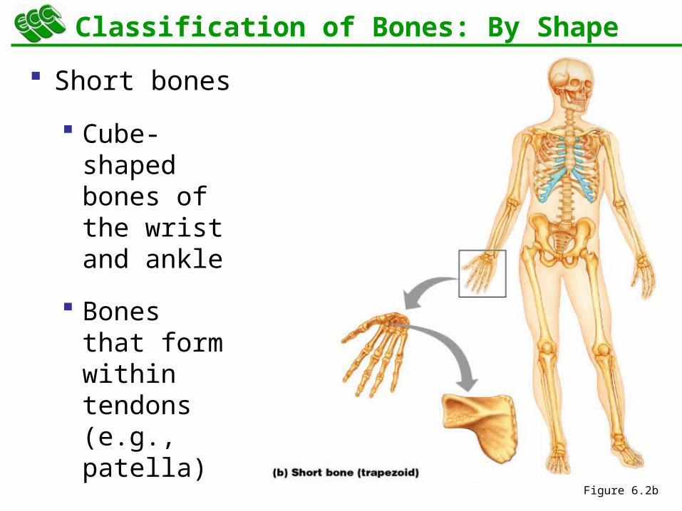
Short bones
Cube-shaped bones of the wrist and ankle
Bones that form within tendons (e.g., patella)
11
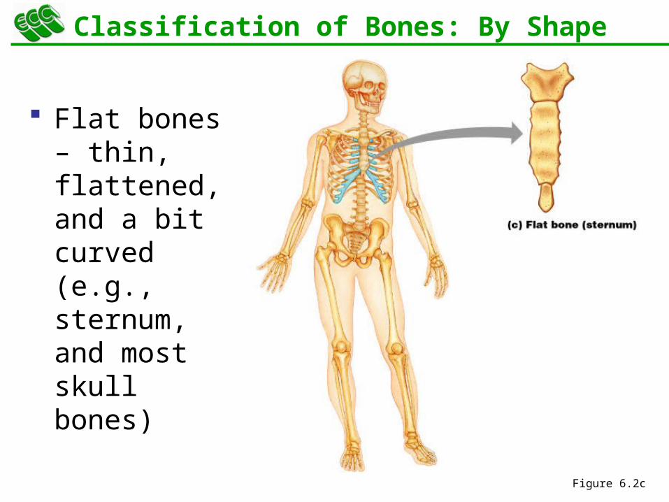
Classification of Bones: By Shape
Flat bones – thin, flattened, and a bit curved (e.g., sternum, and most skull bones)
Figure 6.2c
12
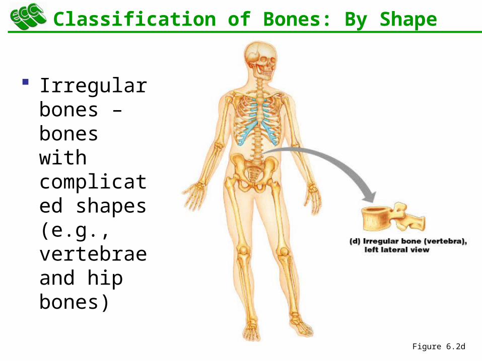
Classification of Bones: By Shape
Irregular bones – bones with complicated shapes (e.g., vertebrae and hip bones)
Figure 6.2d
13
Function of Bones
Support – form the framework that supports the body and cradles soft organs
Protection – provide a protective case for the brain, spinal cord, and vital organs
Movement – provide levers for muscles
Mineral storage – reservoir for minerals, especially calcium and phosphorus
Blood cell formation – hematopoiesis occurs within the marrow cavities of bones
14
Bone Markings
Bulges, depressions, and holes that serve as:
Sites of attachment for muscles, ligaments, and tendons
Joint surfaces
Conduits for blood vessels and nerves

15
Tuberosity – rounded projection
Crest – narrow, prominent ridge of bone
Trochanter – large, blunt, irregular surface
Line – narrow ridge of bone
Bone Markings: Projections – Sites of Muscle and Ligament Attachment
16
Tubercle – small rounded projection
Epicondyle – raised area above a condyle
Spine – sharp, slender projection
Process – any bony prominence
Bone Markings: Projections – Sites of Muscle and Ligament Attachment
17
Head – bony expansion carried on a narrow neck
Facet – smooth, nearly flat articular surface
Condyle – rounded articular projection
Ramus – armlike bar of bone
Bone Markings: Projections – Projections That Help to Form Joints
18
Bone Markings: Depressions and Openings
Meatus – canal-like passageway
Sinus – cavity within a bone
Fossa – shallow, basinlike depression
Groove – furrow
Fissure – narrow, slitlike opening
Foramen – round or oval opening through a bone
19
Gross Anatomy of Bones: Bone Textures
Compact bone – dense outer layer
Spongy bone – honeycomb of trabeculae filled with yellow bone marrow
20
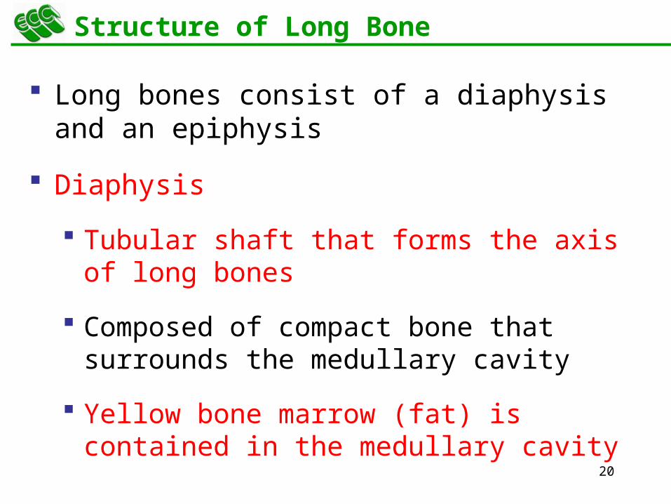
Structure of Long Bone
Long bones consist of a diaphysis and an epiphysis
Diaphysis
Tubular shaft that forms the axis of long bones
Composed of compact bone that surrounds the medullary cavity
Yellow bone marrow (fat) is contained in the medullary cavity
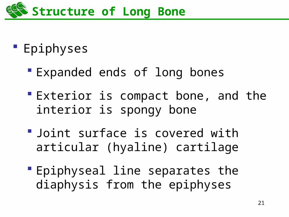
21
Structure of Long Bone
Epiphyses
Expanded ends of long bones
Exterior is compact bone, and the interior is spongy bone
Joint surface is covered with articular (hyaline) cartilage
Epiphyseal line separates the diaphysis from the epiphyses
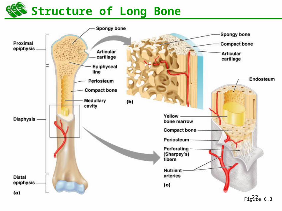
22
Structure of Long Bone
Figure 6.3
23
Bone Membranes
Periosteum – double-layered protective membrane
Outer fibrous layer is dense regular connective tissue
Inner osteogenic layer is composed of osteoblasts and osteoclasts
Richly supplied with nerve fibers, blood, and lymphatic vessels, which enter the bone via nutrient foramina
Secured to underlying bone by Sharpey’s fibers
Endosteum – delicate membrane covering internal surfaces of bone
24
Structure of Short, Irregular, and Flat Bones
Thin plates of periosteum-covered compact bone on the outside with endosteum-covered spongy bone (diploë) on the inside
Have no diaphysis or epiphyses
Contain bone marrow between the trabeculae
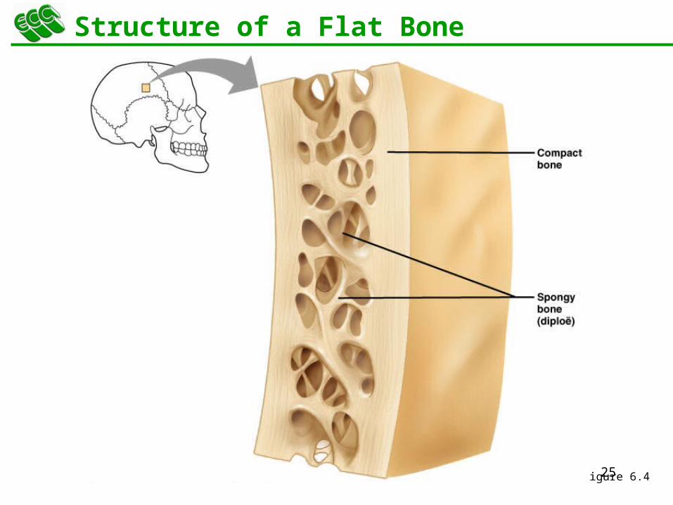
25
Structure of a Flat Bone
Figure 6.4
26
Location of Hematopoietic Tissue (Red Marrow)
In infants
Found in the medullary cavity and all areas of spongy bone
In adults
Found in the diploë of flat bones, and the head of the femur and humerus
27
Microscopic Structure of Bone: Compact Bone
Haversian system, or osteon – the structural unit of compact bone
Lamella – weight-bearing, column-like matrix tubes composed mainly of collagen
Haversian, or central canal – central channel containing blood vessels and nerves
Volkmann’s canals – channels lying at right angles to the central canal, connecting blood and nerve supply of the periosteum to that of the Haversian canal
28
Microscopic Structure of Bone: Compact Bone
Osteocytes – mature bone cells
Lacunae – small cavities in bone that contain osteocytes
Canaliculi – hairlike canals that connect lacunae to each other and the central canal

29
Microscopic Structure of Bone: Compact Bone
Figure 6.6a, b
30
Chemical Composition of Bone: Organic
Osteoblasts – bone-forming cells and secretes matrix
Osteocytes – mature bone cells
Osteoclasts – large cells that resorb or break down bone matrix
Osteoid – unmineralized bone matrix composed of proteoglycans, glycoproteins, and collagen
31
Chemical Composition of Bone: Inorganic
Hydroxyapatites, or mineral salts
Sixty-five percent of bone by mass
Mainly calcium phosphates
Responsible for bone hardness and its resistance to compression
32
Bone Development
Osteogenesis and ossification – the process of bone tissue formation, which leads to:
The formation of the bony skeleton in embryos
Bone growth until early adulthood
Bone thickness, remodeling, and repair
33
Formation of the Bony Skeleton
Begins at week 8 of embryo development
Intramembranous ossification – bone develops from a fibrous membrane
Endochondral ossification – bone forms by replacing hyaline cartilage
34
Intramembranous Ossification
Formation of most of the flat bones of the skull and the clavicles
Fibrous connective tissue membranes are formed by mesenchymal cells
35
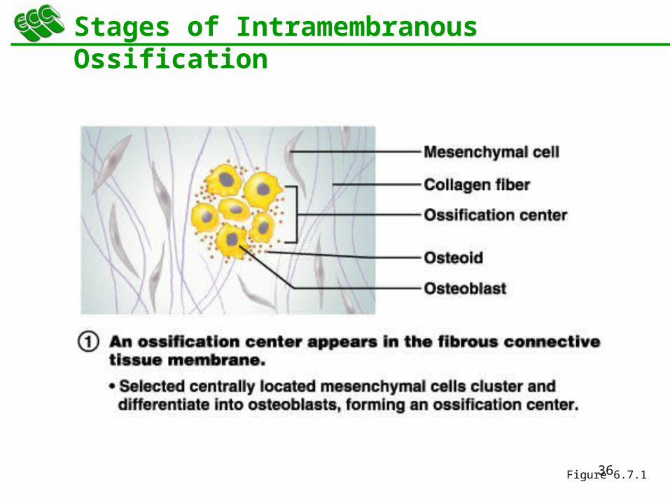
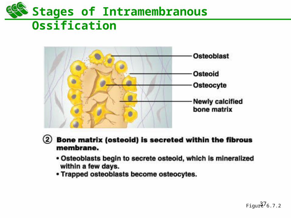
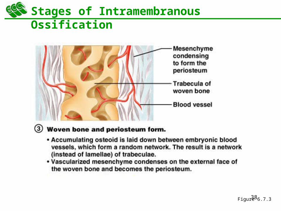
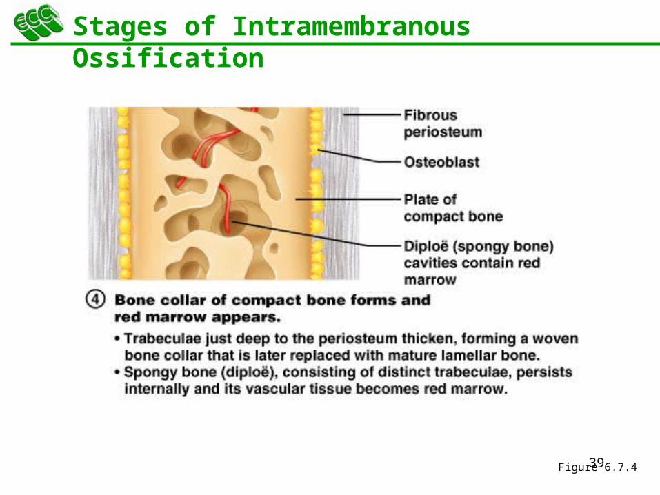
Stages of Intramembranous Ossification
An ossification center appears in the fibrous connective tissue membrane
Bone matrix is secreted within the fibrous membrane
Woven bone and periosteum form
Bone collar of compact bone forms, and red marrow appears
36
Stages of Intramembranous Ossification
Figure 6.7.1
37
Stages of Intramembranous Ossification
Figure 6.7.2
38
Stages of Intramembranous Ossification
Figure 6.7.3
39
Stages of Intramembranous Ossification
Figure 6.7.4

40
Endochondral Ossification
Begins in the second month of development
Uses hyaline cartilage “bones” as models for bone construction
Requires breakdown of hyaline cartilage prior to ossification
41
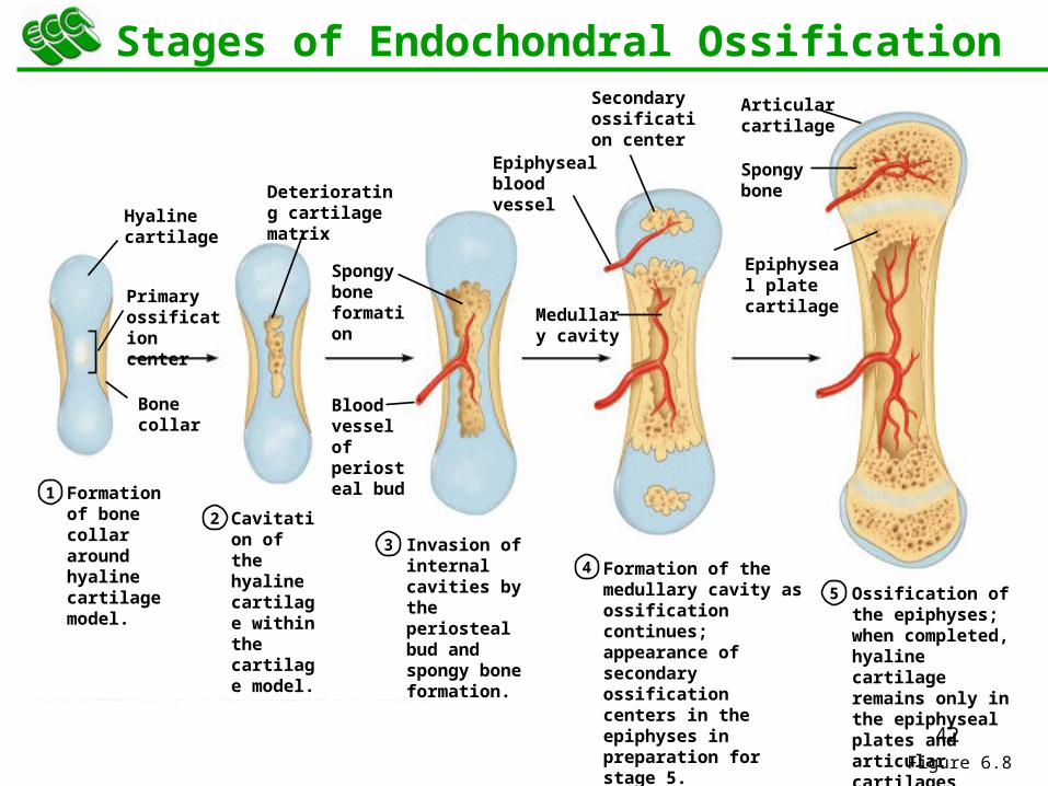
Stages of Endochondral Ossification
Formation of bone collar
Cavitation of the hyaline cartilage
Invasion of internal cavities by the periosteal bud, and spongy bone formation
Formation of the medullary cavity; appearance of secondary ossification centers in the epiphyses
Ossification of the epiphyses, with hyaline cartilage remaining only in the epiphyseal plates
42
Formation of bone collar around hyaline cartilage model.
1
2
3
4
Cavitation of the hyaline cartilage within the cartilage model.
Invasion of internal cavities by the periosteal bud and spongy bone formation.
5 Ossification of the epiphyses; when completed, hyaline cartilage remains only in the epiphyseal plates and articular cartilages
Formation of the medullary cavity as ossification continues; appearance of secondary ossification centers in the epiphyses in preparation for stage 5.
Hyaline cartilage
Primary ossification center
Bone collar
Deteriorating cartilage matrix
Spongy bone formation
Blood vessel of periosteal bud
Secondary ossification center
Epiphyseal blood vessel
Medullary cavity
Epiphyseal plate cartilage
Spongy bone
Articular cartilage
Stages of Endochondral Ossification
Figure 6.8
43
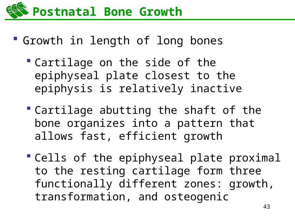
Postnatal Bone Growth
Growth in length of long bones
Cartilage on the side of the epiphyseal plate closest to the epiphysis is relatively inactive
Cartilage abutting the shaft of the bone organizes into a pattern that allows fast, efficient growth
Cells of the epiphyseal plate proximal to the resting cartilage form three functionally different zones: growth, transformation, and osteogenic
44
Functional Zones in Long Bone Growth
Growth zone – cartilage cells undergo mitosis, pushing the epiphysis away from the diaphysis
Transformation zone – older cells enlarge, the matrix becomes calcified, cartilage cells die, and the matrix begins to deteriorate
Osteogenic zone – new bone formation occurs
45
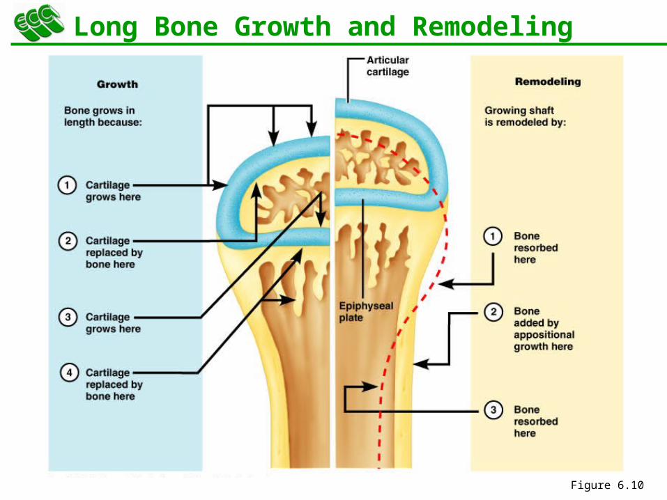
Long Bone Growth and Remodeling
Growth in length – cartilage continually grows and is replaced by bone as shown
Growth in width (Remodeling) – bone is resorbed and added by appositional growth as shown
46
Long Bone Growth and Remodeling
Figure 6.10
47
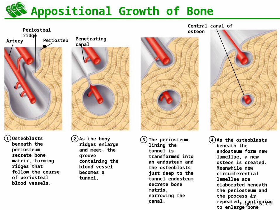
Osteoblasts beneath the periosteum secrete bone matrix, forming ridges that follow the course of periosteal blood vessels.
1 2 3 4As the bony ridges enlarge and meet, the groove containing the blood vessel becomes a tunnel.
The periosteum lining the tunnel is transformed into an endosteum and the osteoblasts just deep to the tunnel endosteum secrete bone matrix, narrowing the canal.
As the osteoblasts beneath the endosteum form new lamellae, a new osteon is created. Meanwhile new circumferential lamellae are elaborated beneath the periosteum and the process is repeated, continuing to enlarge bone diameter.
Artery Periosteum Penetrating canal
Central canal of osteonPeriosteal ridge
Appositional Growth of Bone
Figure 6.11
48
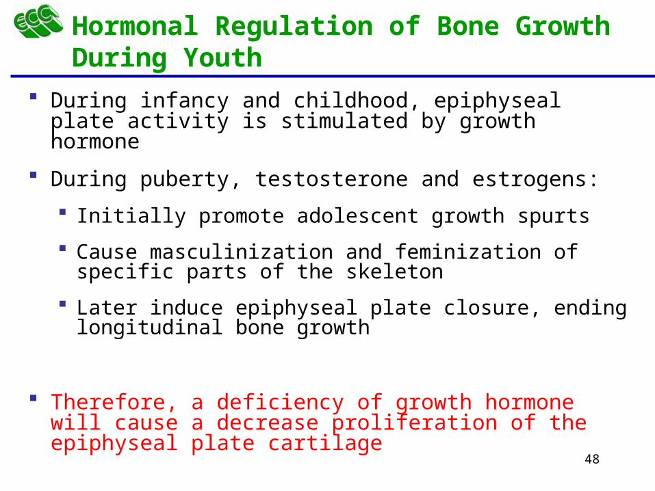
During infancy and childhood, epiphyseal plate activity is stimulated by growth hormone
During puberty, testosterone and estrogens:
Initially promote adolescent growth spurts
Cause masculinization and feminization of specific parts of the skeleton
Later induce epiphyseal plate closure, ending longitudinal bone growth
Therefore, a deficiency of growth hormone will cause a decrease proliferation of the epiphyseal plate cartilage
Hormonal Regulation of Bone Growth During Youth
49
Bone Remodeling
Remodeling units – adjacent osteoblasts and osteoclasts deposit and resorb bone at periosteal and endosteal surfaces
50
Bone Deposition
Occurs where bone is injured or added strength is needed
Requires a diet rich in protein, vitamins C, D, and A, calcium, phosphorus, magnesium, and manganese
Alkaline phosphatase is essential for mineralization of bone
Sites of new matrix deposition are revealed by the:
Osteoid seam – unmineralized band of bone matrix
Calcification front – abrupt transition zone between the osteoid seam and the older mineralized bone
51
Bone Resorption
Accomplished by osteoclasts
Resorption bays – grooves formed by osteoclasts as they break down bone matrix
Resorption involves osteoclast secretion of:
Lysosomal enzymes that digest organic matrix
Acids that convert calcium salts into soluble forms
Dissolved matrix is transcytosed across the osteoclast’s cell where it is secreted into the interstitial fluid and then into the blood
52
Importance of Ionic Calcium in the Body
Calcium is necessary for:
Transmission of nerve impulses
Muscle contraction
Blood coagulation
Secretion by glands and nerve cells
Cell division
53
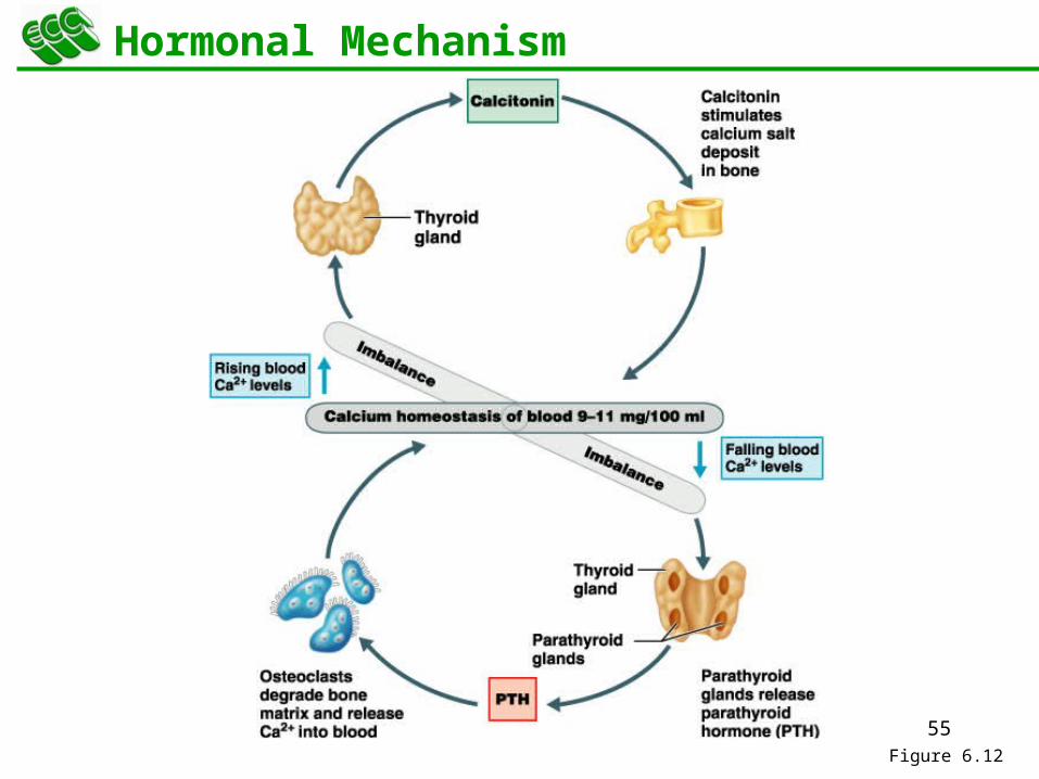
Control of Remodeling
Two control loops regulate bone remodeling
Hormonal mechanism maintains calcium homeostasis in the blood
Mechanical and gravitational forces acting on the skeleton
54
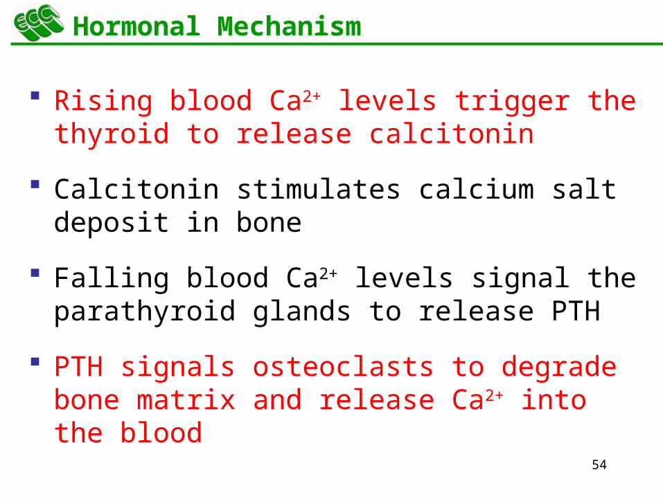
Hormonal Mechanism
Rising blood Ca2+ levels trigger the thyroid to release calcitonin
Calcitonin stimulates calcium salt deposit in bone
Falling blood Ca2+ levels signal the parathyroid glands to release PTH
PTH signals osteoclasts to degrade bone matrix and release Ca2+ into the blood
55
Hormonal Mechanism
Figure 6.12
56
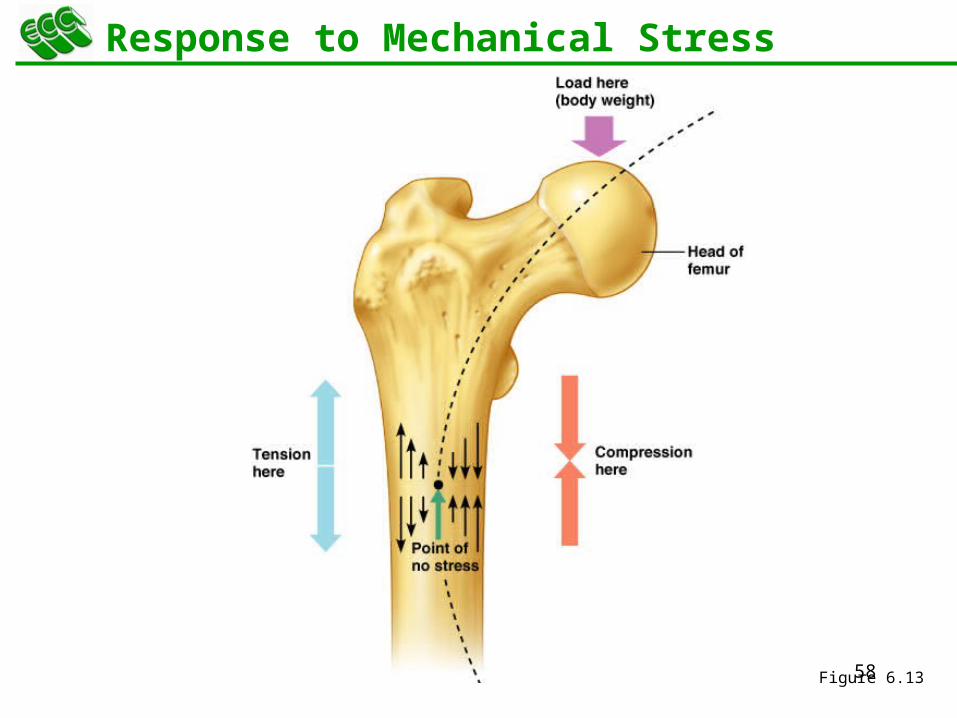
Response to Mechanical Stress
Wolff’s law – a bone grows or remodels in response to the forces or demands placed upon it
Observations supporting Wolff’s law include
Long bones are thickest midway along the shaft (where bending stress is greatest)
Curved bones are thickest where they are most likely to buckle
57
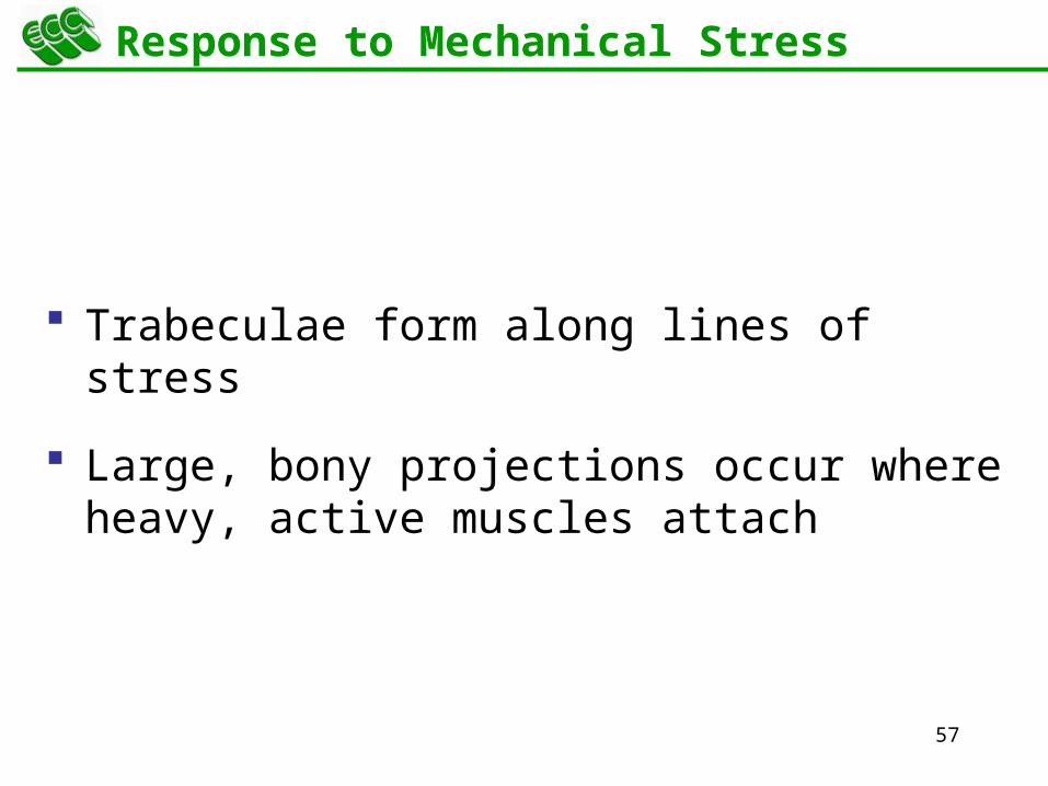
Response to Mechanical Stress
Trabeculae form along lines of stress
Large, bony projections occur where heavy, active muscles attach
58
Response to Mechanical Stress
Figure 6.13
59
Bone Fractures (Breaks)
Bone fractures are classified by:
The position of the bone ends after fracture
The completeness of the break
The orientation of the bone to the long axis
Whether or not the bones ends penetrate the skin
60
Types of Bone Fractures
Nondisplaced – bone ends retain their normal position
Displaced – bone ends are out of normal alignment
Complete – bone is broken all the way through
Incomplete – bone is not broken all the way through
Linear – the fracture is parallel to the long axis of the bone
61
Types of Bone Fractures
Transverse – the fracture is perpendicular to the long axis of the bone
Compound (open) – bone ends penetrate the skin
Simple (closed) – bone ends do not penetrate the skin
62
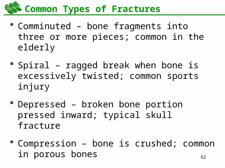
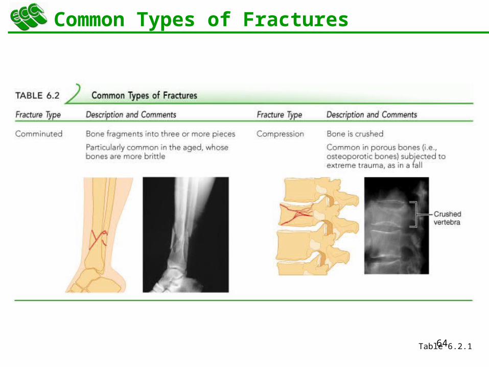
Common Types of Fractures
Comminuted – bone fragments into three or more pieces; common in the elderly
Spiral – ragged break when bone is excessively twisted; common sports injury
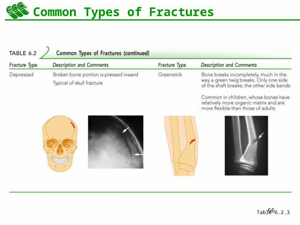
Depressed – broken bone portion pressed inward; typical skull fracture
Compression – bone is crushed; common in porous bones
63
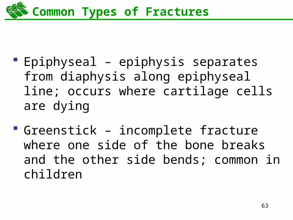
Common Types of Fractures
Epiphyseal – epiphysis separates from diaphysis along epiphyseal line; occurs where cartilage cells are dying
Greenstick – incomplete fracture where one side of the bone breaks and the other side bends; common in children
64
Common Types of Fractures
Table 6.2.1

65
Common Types of Fractures
Table 6.2.2
66
Common Types of Fractures
Table 6.2.3
67
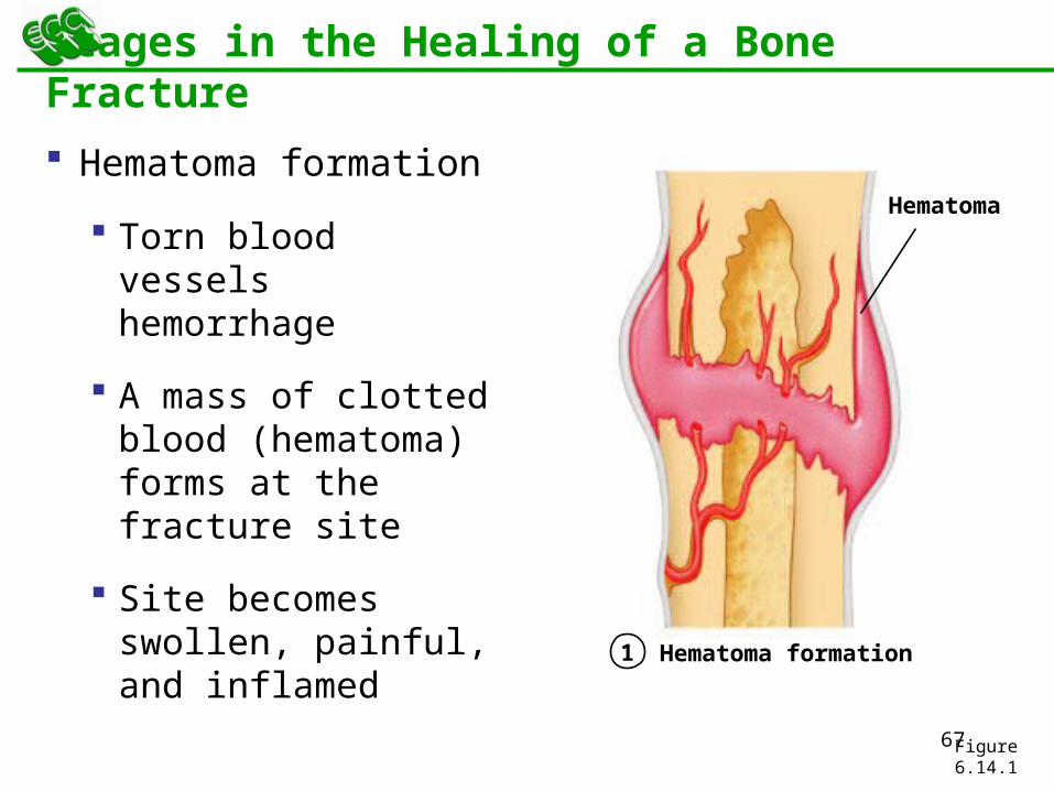
Stages in the Healing of a Bone Fracture
Hematoma formation
Torn blood vessels hemorrhage
A mass of clotted blood (hematoma) forms at the fracture site
Site becomes swollen, painful, and inflamed
Figure 6.14.1
1
Hematoma
Hematoma formation

68
Stages in the Healing of a Bone Fracture
Fibrocartilaginous callus forms
Granulation tissue (soft callus) forms a few days after the fracture
Capillaries grow into the tissue and phagocytic cells begin cleaning debris
Figure 6.14.2
2 Fibrocartilaginous callus formation
External callus
New blood vessels
Spongy bone trabeculae
Internal callus (fibrous tissue and cartilage)
69
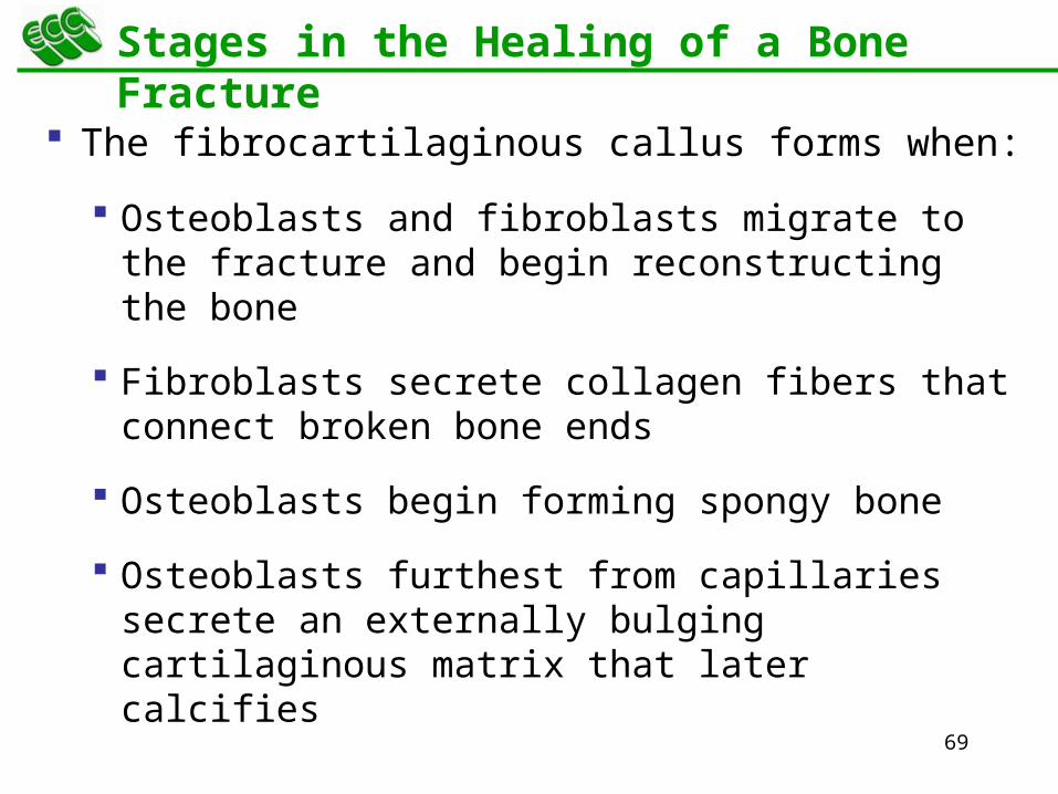
Stages in the Healing of a Bone Fracture
The fibrocartilaginous callus forms when:
Osteoblasts and fibroblasts migrate to the fracture and begin reconstructing the bone
Fibroblasts secrete collagen fibers that connect broken bone ends
Osteoblasts begin forming spongy bone
Osteoblasts furthest from capillaries secrete an externally bulging cartilaginous matrix that later calcifies
70
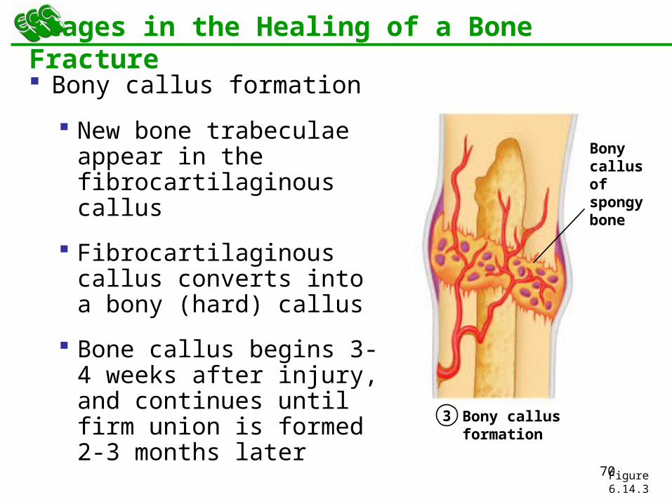
Stages in the Healing of a Bone Fracture
Bony callus formation
New bone trabeculae appear in the fibrocartilaginous callus
Fibrocartilaginous callus converts into a bony (hard) callus
Bone callus begins 3-4 weeks after injury, and continues until firm union is formed 2-3 months later
Figure 6.14.3
3 Bony callus formation
Bony callus of spongy bone
71
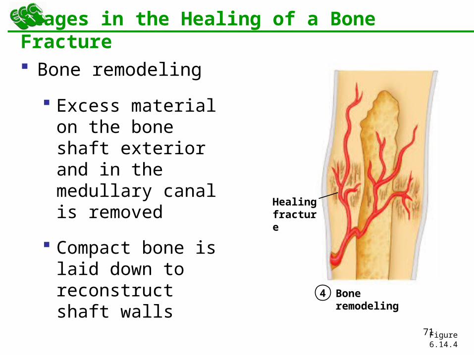
Stages in the Healing of a Bone Fracture
Bone remodeling
Excess material on the bone shaft exterior and in the medullary canal is removed
Compact bone is laid down to reconstruct shaft walls
Figure 6.14.4
4 Bone remodeling
Healing fracture
72
Homeostatic Imbalances
Osteomalacia
Bones are inadequately mineralized causing softened, weakened bones
Bone formed is poorly mineralized and soft.
Deforms on weight-bearing
Main symptom is pain when weight is put on the affected bone
Caused by insufficient calcium in the diet, or by vitamin D deficiency
73
Homeostatic Imbalances
Rickets
Bones of children are inadequately mineralized causing softened, weakened bones
Bowed legs and deformities of the pelvis, skull, and rib cage are common
Caused by insufficient calcium in the diet, or by vitamin D deficiency

74
Homeostatic Imbalances
Osteoporosis
Group of diseases in which bone reabsorption outpaces bone deposit
Spongy bone of the spine is most vulnerable
Bones are porous and thin but bone composition is normal
Occurs most often in postmenopausal women
Bones become so fragile that sneezing or stepping off a curb can cause fractures
75
Osteoporosis: Treatment
Calcium and vitamin D supplements
Increased weight-bearing exercise
Hormone (estrogen) replacement therapy (HRT) slows bone loss
Natural progesterone cream prompts new bone growth
Statins increase bone mineral density
76
Paget’s Disease
Characterized by excessive bone formation and breakdown
Abnormal bone formation and reabsorption
Pagetic bone with an excessively high ratio of woven to compact bone is formed
Pagetic bone, along with reduced mineralization, causes spotty weakening of bone
Osteoclast activity wanes, but osteoblast activity continues to work
May be prevented by increasing dietary vitamin C
77
Paget’s Disease
Usually localized in the spine, pelvis, femur, and skull
Unknown cause (possibly viral)
Treatment includes the drugs Didronate and Fosamax
78
Developmental Aspects of Bones
Mesoderm gives rise to embryonic mesenchymal cells, which produce membranes and cartilages that form the embryonic skeleton
The embryonic skeleton ossifies in a predictable timetable that allows fetal age to be easily determined from sonograms
At birth, most long bones are well ossified (except for their epiphyses)
79
Developmental Aspects of Bones
By age 25, nearly all bones are completely ossified
In old age, bone resorption predominates
A single gene that codes for vitamin D docking determines both the tendency to accumulate bone mass early in life, and the risk for osteoporosis later in life