03 cleavage and implantation complete - tulane …embryo/lectures/exam 1/03 cleavage... · cleavage...
TRANSCRIPT
Cleavage
Cell Division – Cell Cycle Control Morula – CompactionBlastocyst – HatchingImplantation – Decidual ReactionEarly Cell Lineages
Inner Cell MassTrophoblasts (Extra-embryonic)
Anomalies

Cleavage

Cleavage
Blastomere
Equal
Asynchronous
40 hours – 4 cells
72 hours – 6-12 cells
96 hours – 16-32 cells
Cleavage – Molecular Events
In mammals – no large maternal stores of RNA and ribosomes
Zygotic transcription begins by 2-4 cell stageOct-3 – Transcription factor expressed in egg
KO in mouse – arrest at 1 cell stageExpressed in blastomeres up to morula stageExpressed in germ cells
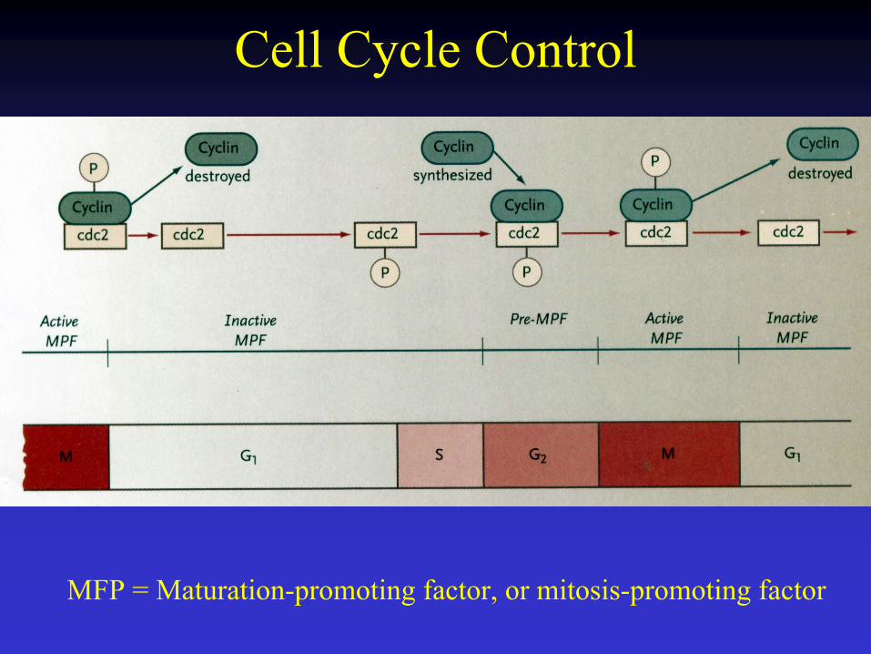
Cell Cycle Control
MFP = Maturation-promoting factor, or mitosis-promoting factor
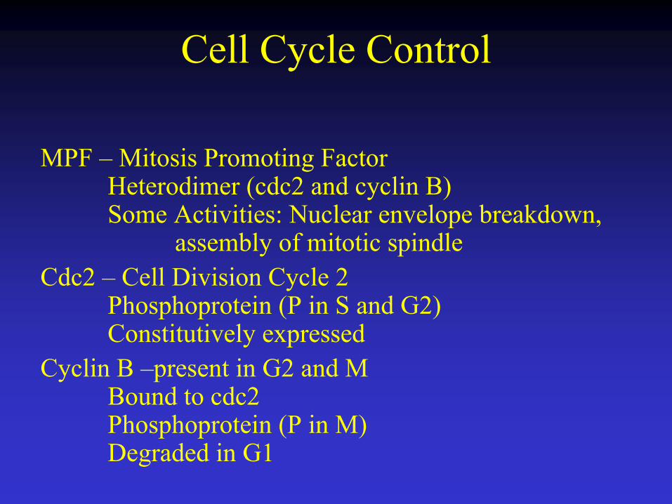
Cell Cycle Control
MPF – Mitosis Promoting FactorHeterodimer (cdc2 and cyclin B)Some Activities: Nuclear envelope breakdown,
assembly of mitotic spindleCdc2 – Cell Division Cycle 2
Phosphoprotein (P in S and G2)Constitutively expressed
Cyclin B –present in G2 and MBound to cdc2Phosphoprotein (P in M)Degraded in G1

Morula32 cell stage ‘Berry’ - appearance
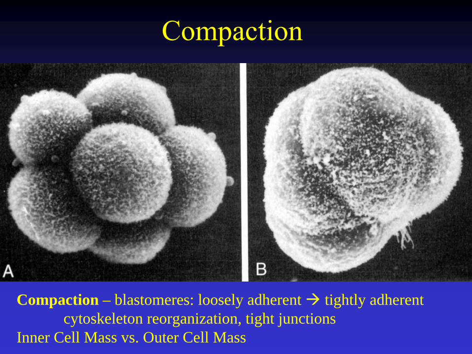
Compaction
Compaction – blastomeres: loosely adherent tightly adherent cytoskeleton reorganization, tight junctions
Inner Cell Mass vs. Outer Cell Mass
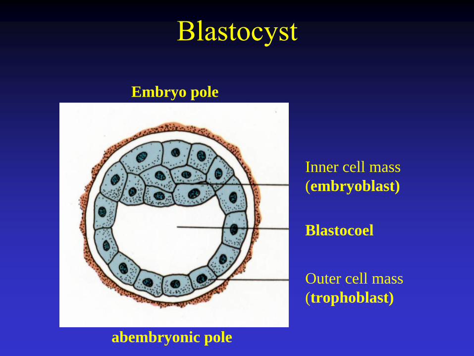
Blastocyst
Inner cell mass (embryoblast)
Outer cell mass (trophoblast)
Blastocoel
Embryo pole
abembryonic pole

Hatching(from zona pellucida)
Hatching: Enzymatic production by Trophoblasts - digestion of the Zona Pellucida

Zona Pellucida - Functions
Species-specific sperm penetrationPermanent block to polyspermyActs as a porous selective filter - uterine tube
signalsImmunological barrier -
no HLA (histocompatibility antigens)Keeps blastomeres together (loosely adherent)Prevents premature implantation
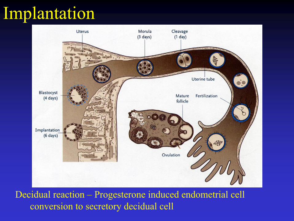
Implantation
Decidual reaction – Progesterone induced endometrial cell conversion to secretory decidual cell

Implantation
Days 6 –12
Adhesion, blastocyst to endometrium
Trophoblastproliferation
Syncytiotrophoblast
Secretion of hydrolytic enzymes
Breakdown of endometrium
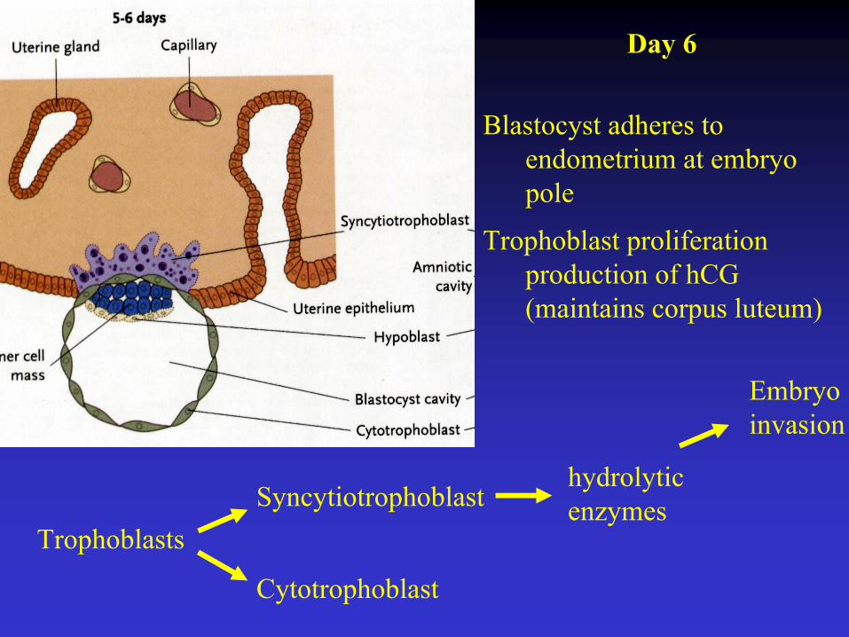
Day 6
Blastocyst adheres to endometrium at embryo pole
Trophoblast proliferation production of hCG(maintains corpus luteum)
TrophoblastsSyncytiotrophoblast
Cytotrophoblast
hydrolytic enzymes
Embryo invasion
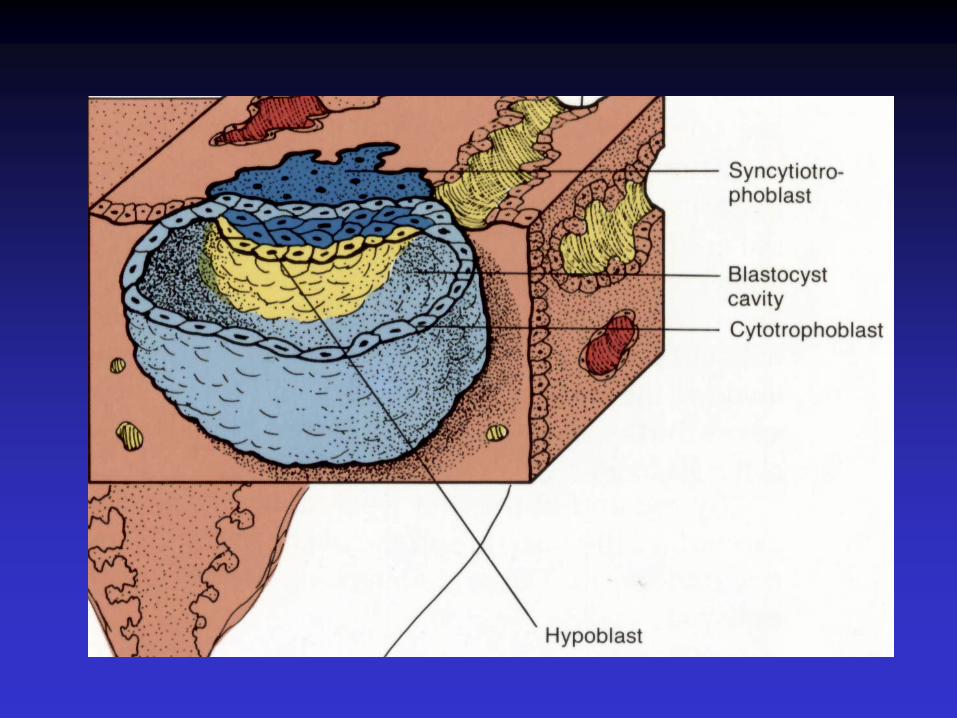

Day 7-8
Syncytiotrophoblastexpansion
Lacunae form – filled with fluid (embryotroph)
Embryotroph provides nutrients to the embryo. Derived from maternal blood.
Embryo - Bilaminar germ disc:Epiblast layer – cavitates to form the amnionic cavity. Hypoblast layer form the exocoelomic cavity / primary yolk sac

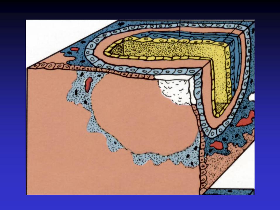
Day 9-10
Lacunae enlarge
Syncytiotrophoblastexpands around entire blastocyst
Cytotrophoblasts form primary villus –initiation of placenta formation
Implantation Complete
Coagulation Plug forms
Embryo: hypoblast exocoelomic membrane = Hauser’s membraneExtraembryonic mesoderm from yolk sac
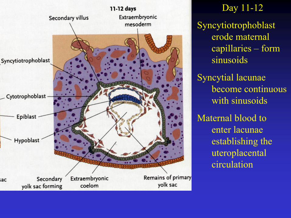
Day 11-12
Syncytiotrophoblasterode maternal capillaries – form sinusoids
Syncytial lacunae become continuous with sinusoids
Maternal blood to enter lacunae establishing the uteroplacentalcirculation
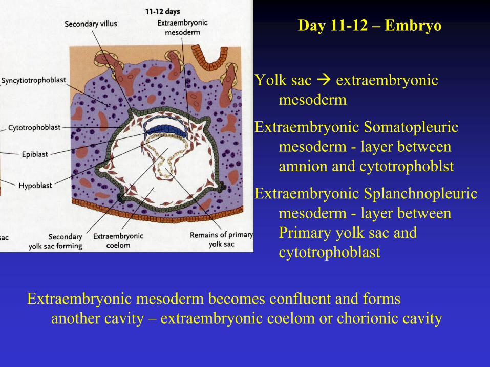
Day 11-12 – Embryo
Yolk sac extraembryonicmesoderm
Extraembryonic Somatopleuricmesoderm - layer between amnion and cytotrophoblst
Extraembryonic Splanchnopleuricmesoderm - layer between Primary yolk sac and cytotrophoblast
Extraembryonic mesoderm becomes confluent and forms another cavity – extraembryonic coelom or chorionic cavity
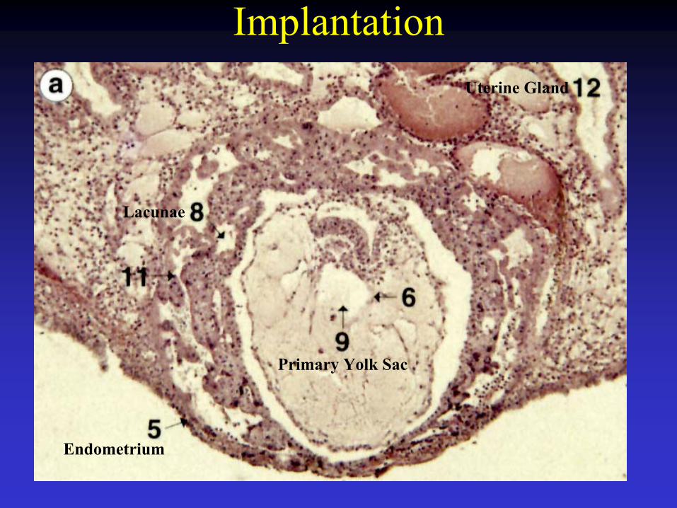
Implantation
Primary Yolk Sac
Lacunae
Endometrium
Uterine Gland

Cytotrophoblast
Amniotic Cavity
Ectoderm
Endoderm
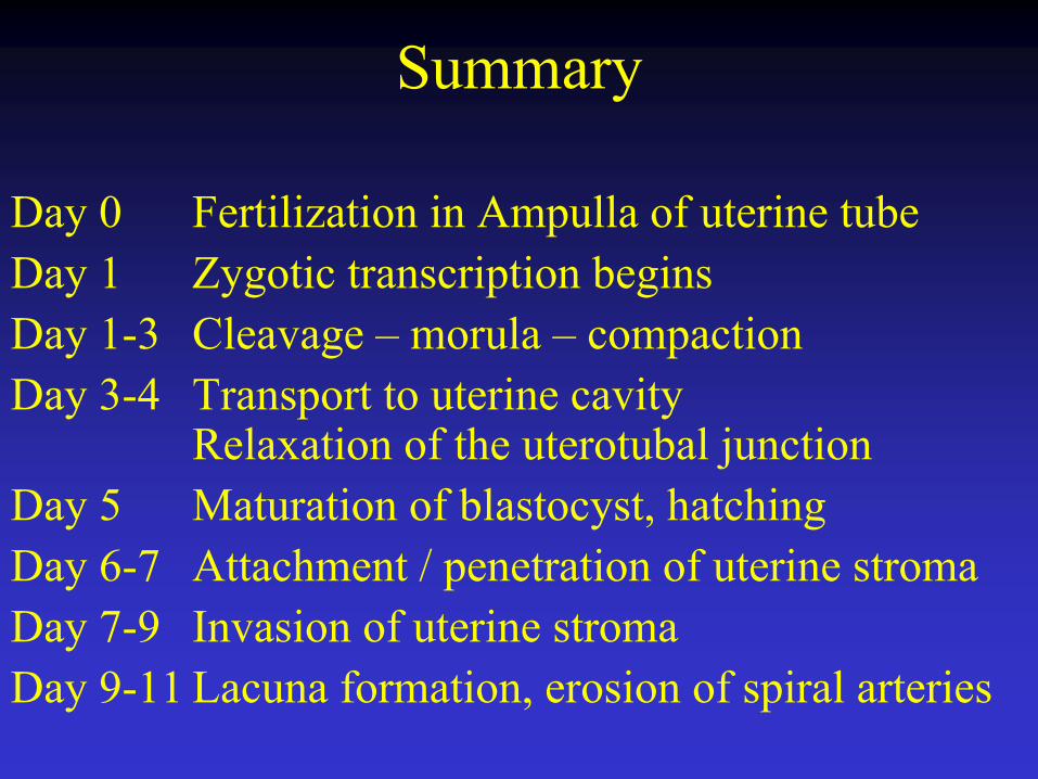
Summary
Day 0 Fertilization in Ampulla of uterine tubeDay 1 Zygotic transcription beginsDay 1-3 Cleavage – morula – compactionDay 3-4 Transport to uterine cavity
Relaxation of the uterotubal junctionDay 5 Maturation of blastocyst, hatchingDay 6-7 Attachment / penetration of uterine stromaDay 7-9 Invasion of uterine stromaDay 9-11 Lacuna formation, erosion of spiral arteries

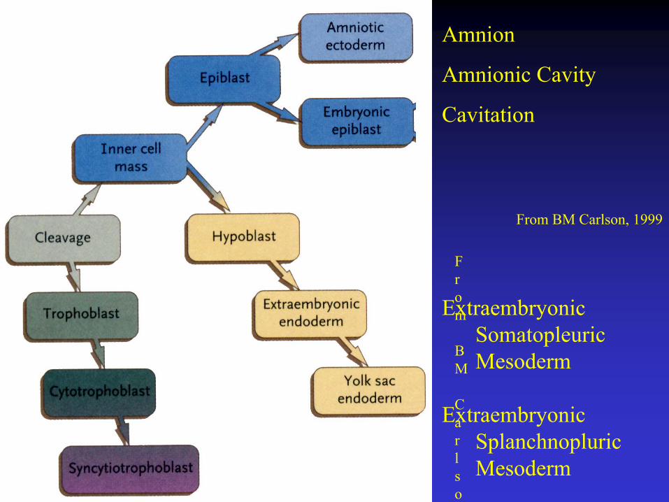
Early Cell Lineages
ExtraembryonicSomatopleuricMesoderm
ExtraembryonicSplanchnopluricMesoderm
From BM Carlson, 1999
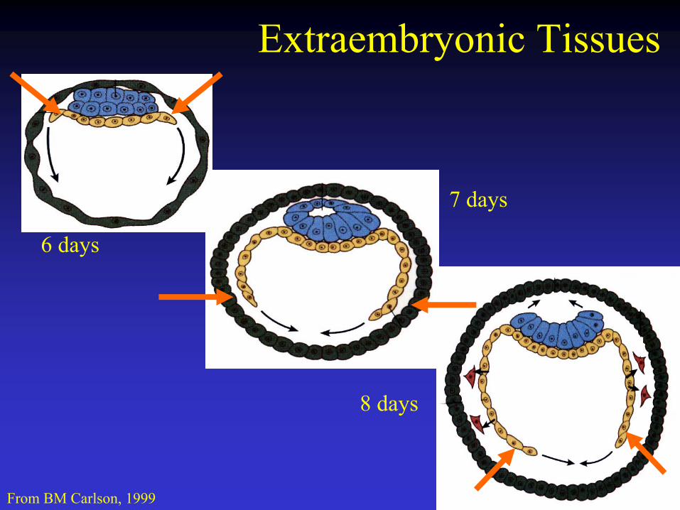
Extraembryonic Tissues
6 days
7 days
8 days
From BM Carlson, 1999

Extraembryonic Tissues
8 days
9 days
14 days
From BM Carlson, 1999
From
BM
Carlso
Amnion
Amnionic Cavity
Cavitation
ExtraembryonicSomatopleuricMesoderm
ExtraembryonicSplanchnopluricMesoderm
From BM Carlson, 1999

Extraembryonic Tissues
8 days
9 days
14 days
From BM Carlson, 1999

Implantation Sites
Typical - Mid-Uterus
Distribution of atypical implantation sites
From BM Carlson, 1999

Dizygotic Twins
From TW Sadler, 2000
Dichorial
Monochorial
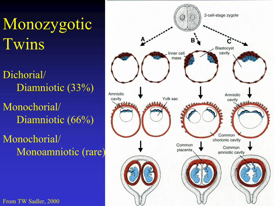
Monozygotic Twins
From TW Sadler, 2000
Dichorial/Diamniotic (33%)
Monochorial/Diamniotic (66%)
Monochorial/Monoamniotic (rare)
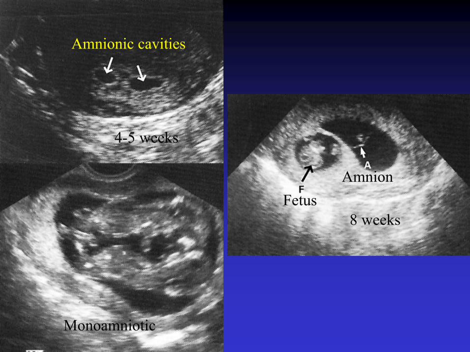
Amnionic cavities
FetusAmnion
Monoamniotic
4-5 weeks
8 weeks
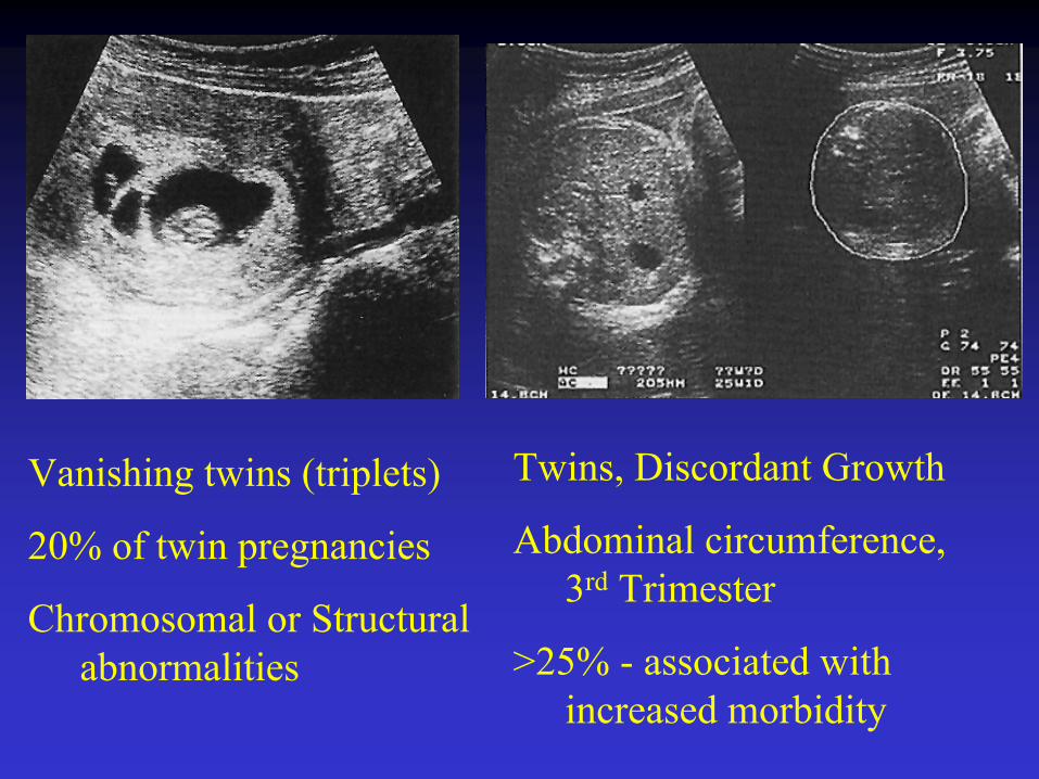
Vanishing twins (triplets)
20% of twin pregnancies
Chromosomal or Structural abnormalities
Twins, Discordant Growth
Abdominal circumference, 3rd Trimester
>25% - associated with increased morbidity
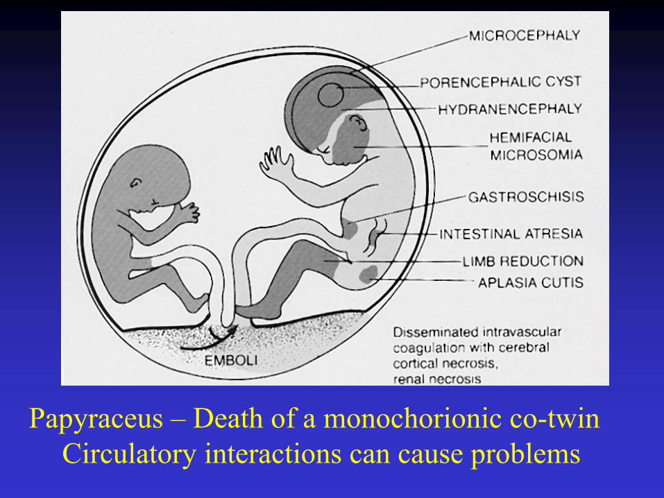
Papyraceus – Death of a monochorionic co-twin Circulatory interactions can cause problems

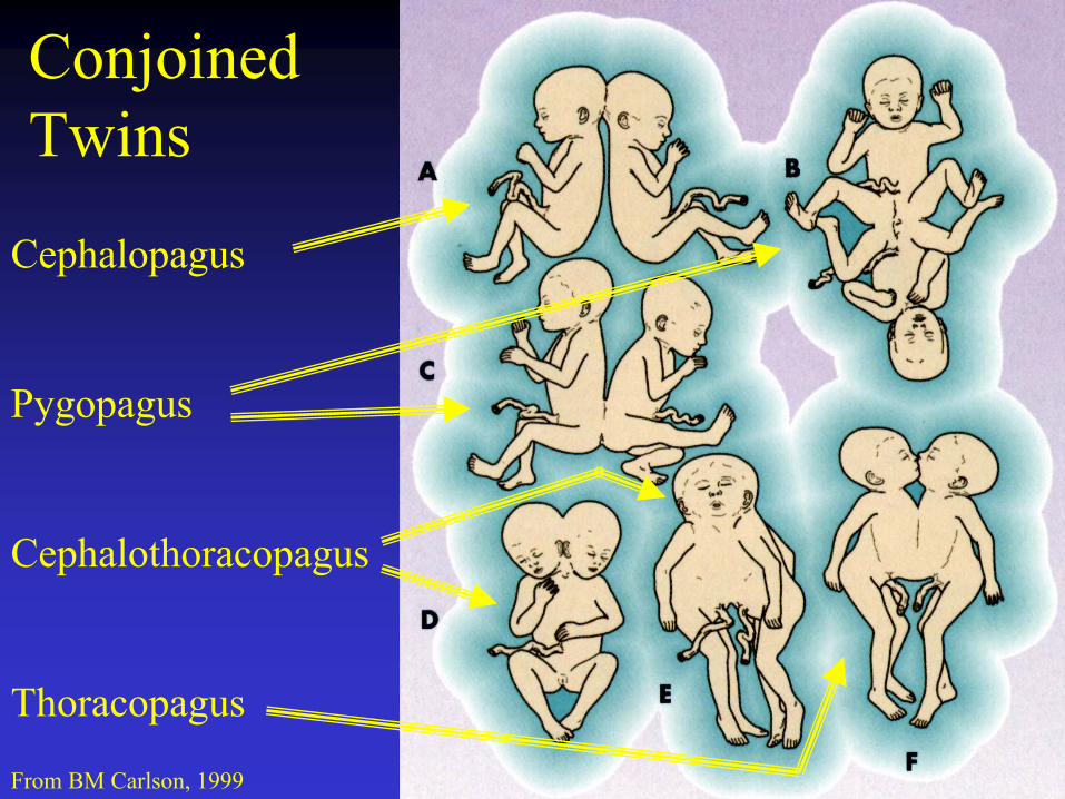
ConjoinedTwins
From BM Carlson, 1999
Cephalopagus
Pygopagus
Cephalothoracopagus
Thoracopagus
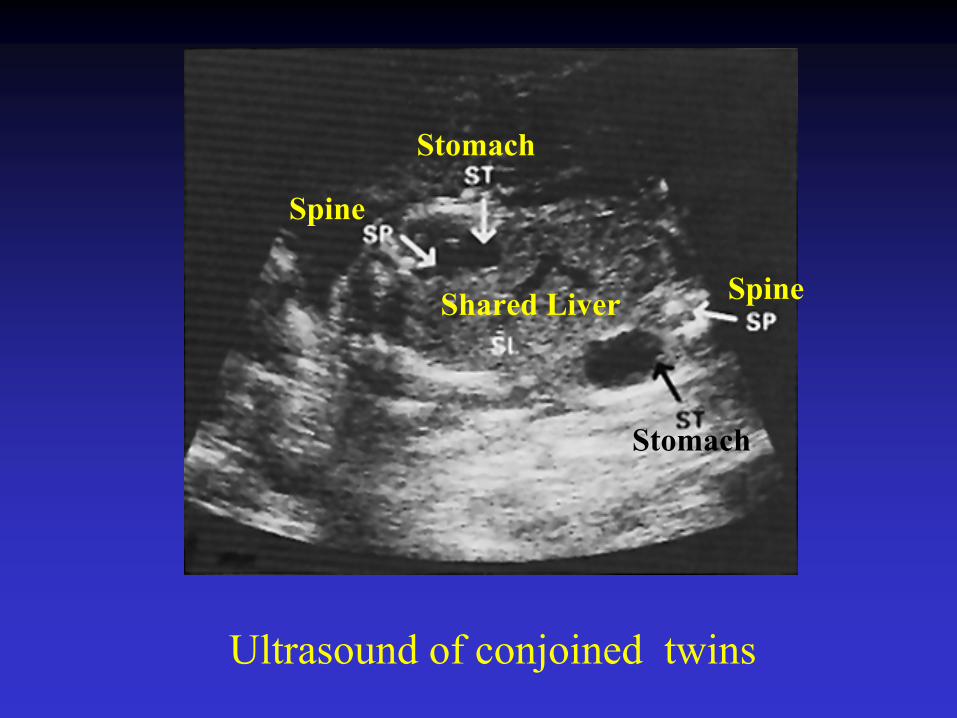
Ultrasound of conjoined twins
Shared Liver Spine
Stomach
Spine
Stomach

Pygopagus
Posterior union of the rump19% of all conjoined twins.
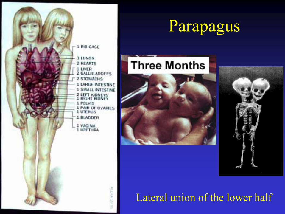
Parapagus
Lateral union of the lower half
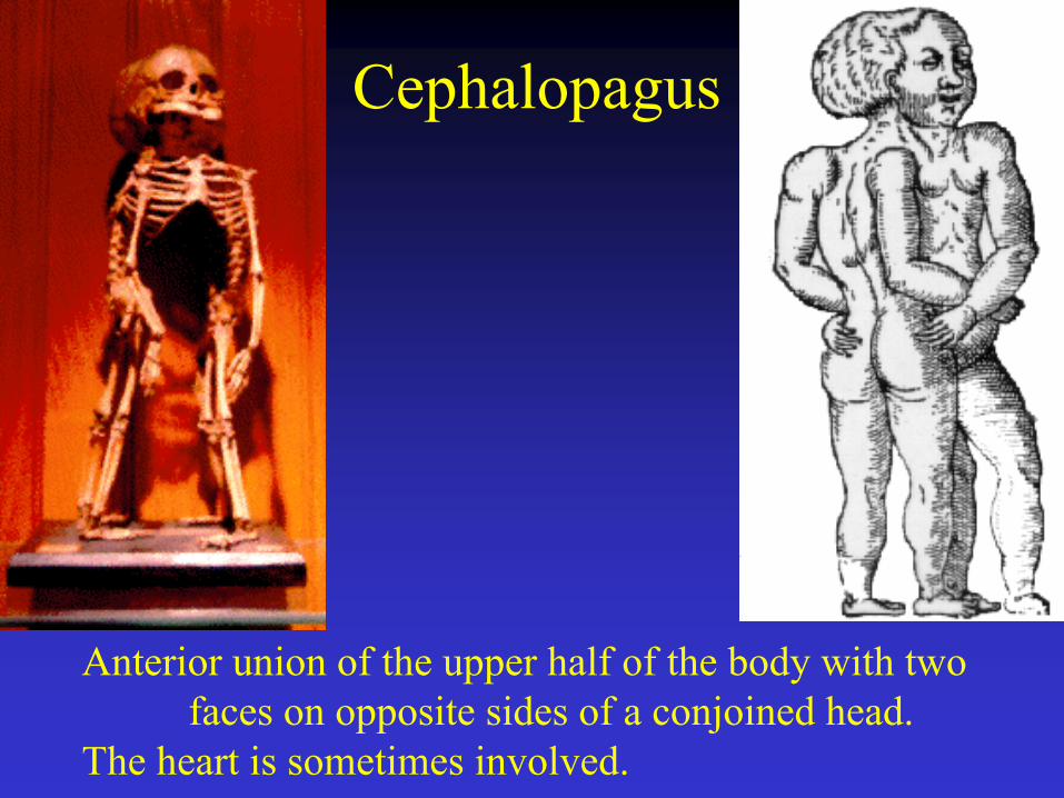
Cephalopagus
Anterior union of the upper half of the body with two faces on opposite sides of a conjoined head.
The heart is sometimes involved.
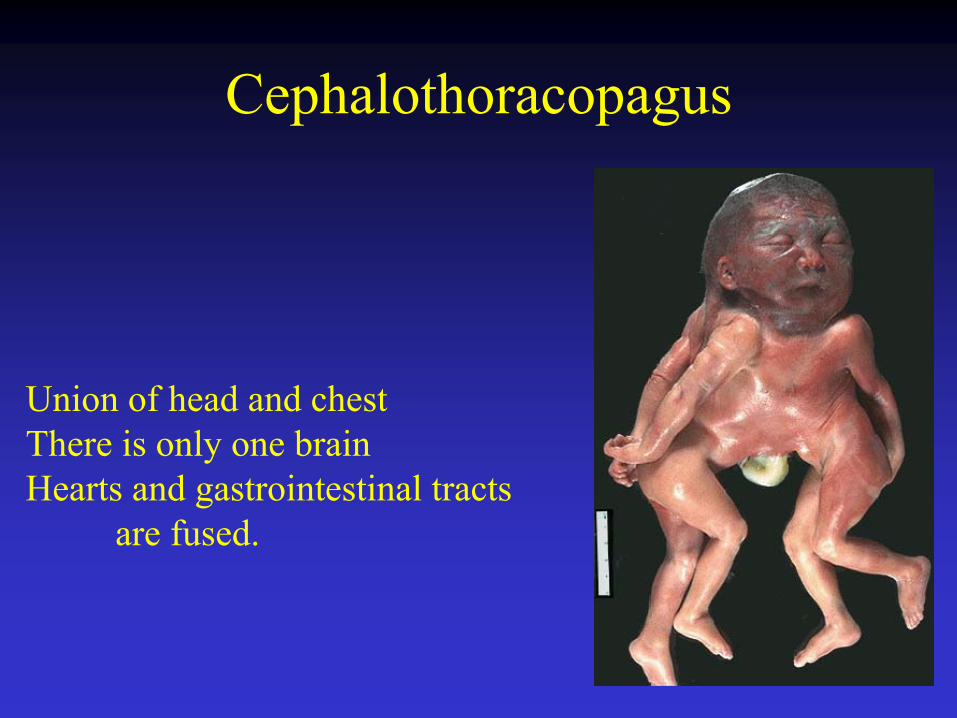
Cephalothoracopagus
Union of head and chestThere is only one brainHearts and gastrointestinal tracts
are fused.
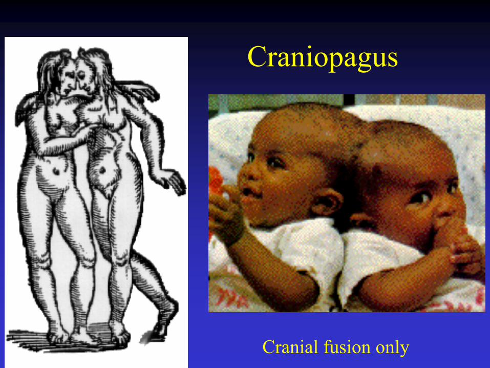
Craniopagus
Cranial fusion only

Parasitic Conjoined Twins
One twin without brain or heart
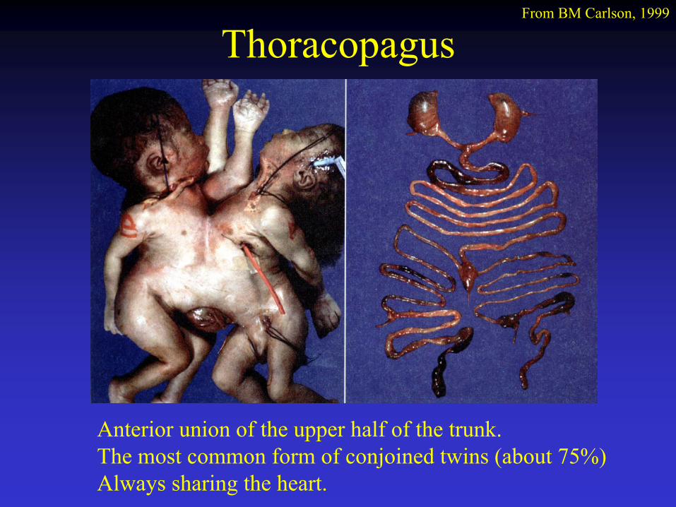
ThoracopagusFrom BM Carlson, 1999
Anterior union of the upper half of the trunk. The most common form of conjoined twins (about 75%)Always sharing the heart.
Hydatidiform Mole
Pregnancy without an embryo (complete or partial mole)
Complete Mole = Only a placenta / No fetus – Diploid but with 2 sets of paternal chromosomes, no maternal contribution
Partial Mole = Triploid (Maternal, 1N; Paternal, 2N)
Diagnosis – high hCG levels; ploidy analysis (flow cytometry)
1:1200 pregnancies in US; 1:200 pregnancies in Latin America/Asia
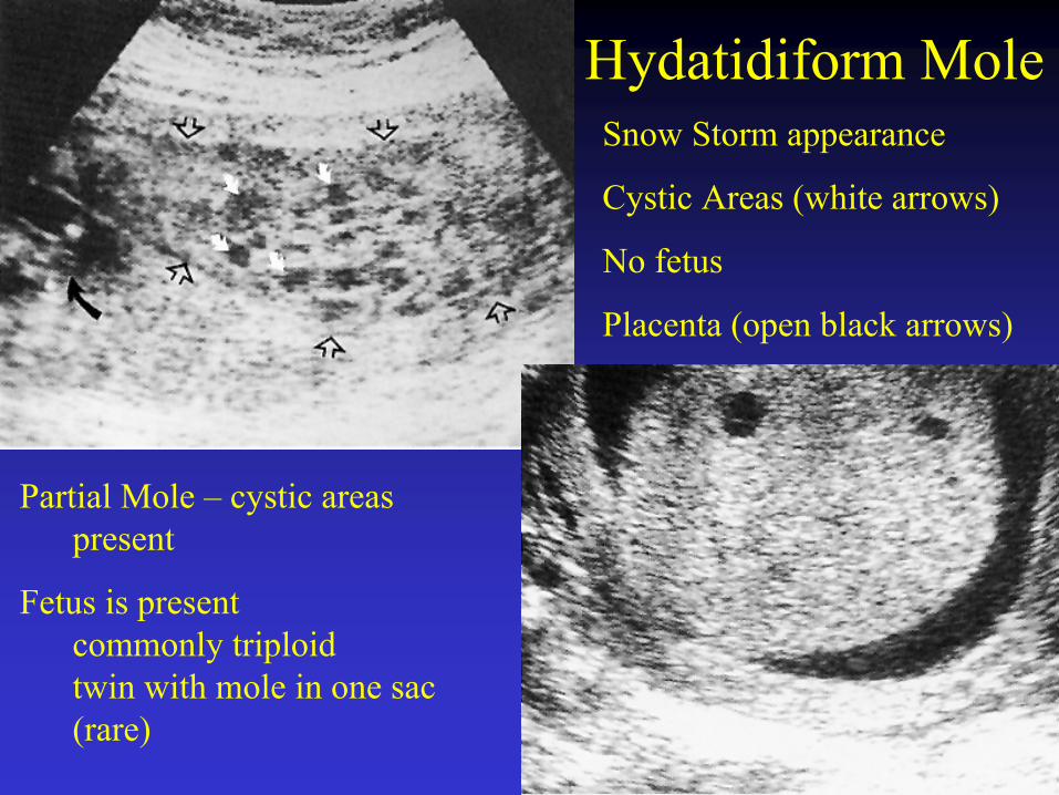
Hydatidiform MoleSnow Storm appearance
Cystic Areas (white arrows)
No fetus
Placenta (open black arrows)
Partial Mole – cystic areas present
Fetus is present commonly triploidtwin with mole in one sac (rare)
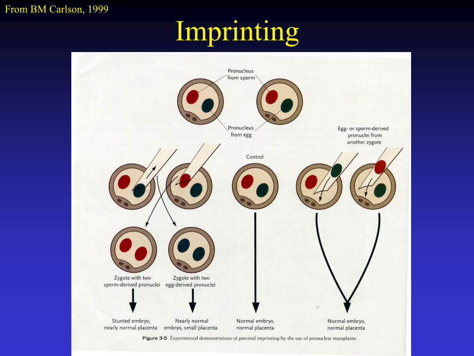
ImprintingFrom BM Carlson, 1999
Parental Imprinting
Identical genes derived from maternal and paternal DNA display differential expression
Selected genes are turned off during gametogenesis by methylation of certain bases
Imprinted patterns are not passed on to progeny, imprints erased during gametogenesis
Beckwith-Wiedemann syndrome - Igf2Long arm Chr 15 deletion
Angelman’s syndrome - Maternal deletionPrader-Willi syndrome - Paternal deletion
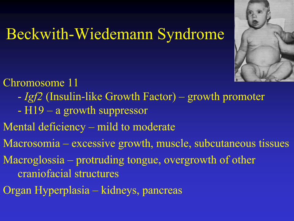
Beckwith-Wiedemann Syndrome
Chromosome 11 - Igf2 (Insulin-like Growth Factor) – growth promoter- H19 – a growth suppressor
Mental deficiency – mild to moderateMacrosomia – excessive growth, muscle, subcutaneous tissuesMacroglossia – protruding tongue, overgrowth of other
craniofacial structuresOrgan Hyperplasia – kidneys, pancreas

Angelman’s Syndrome
“Happy Puppet Syndrome”Maternal long arm of Chromosome 15 deletionSevere mental deficiency – marked delays in motor
milestones, absent speech, frequent laughter, frequent seizures
Puppet like gaitWidely spaced teethMacroglossiaDecreased occular pigment pale blue eyes
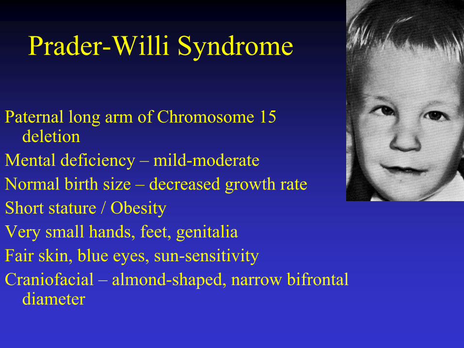
Prader-Willi Syndrome
Paternal long arm of Chromosome 15 deletion
Mental deficiency – mild-moderateNormal birth size – decreased growth rateShort stature / Obesity Very small hands, feet, genitaliaFair skin, blue eyes, sun-sensitivityCraniofacial – almond-shaped, narrow bifrontal
diameter
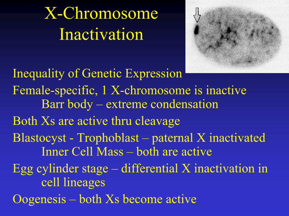
X-Chromosome Inactivation
Inequality of Genetic ExpressionFemale-specific, 1 X-chromosome is inactive
Barr body – extreme condensationBoth Xs are active thru cleavageBlastocyst - Trophoblast – paternal X inactivated
Inner Cell Mass – both are activeEgg cylinder stage – differential X inactivation in
cell lineagesOogenesis – both Xs become active
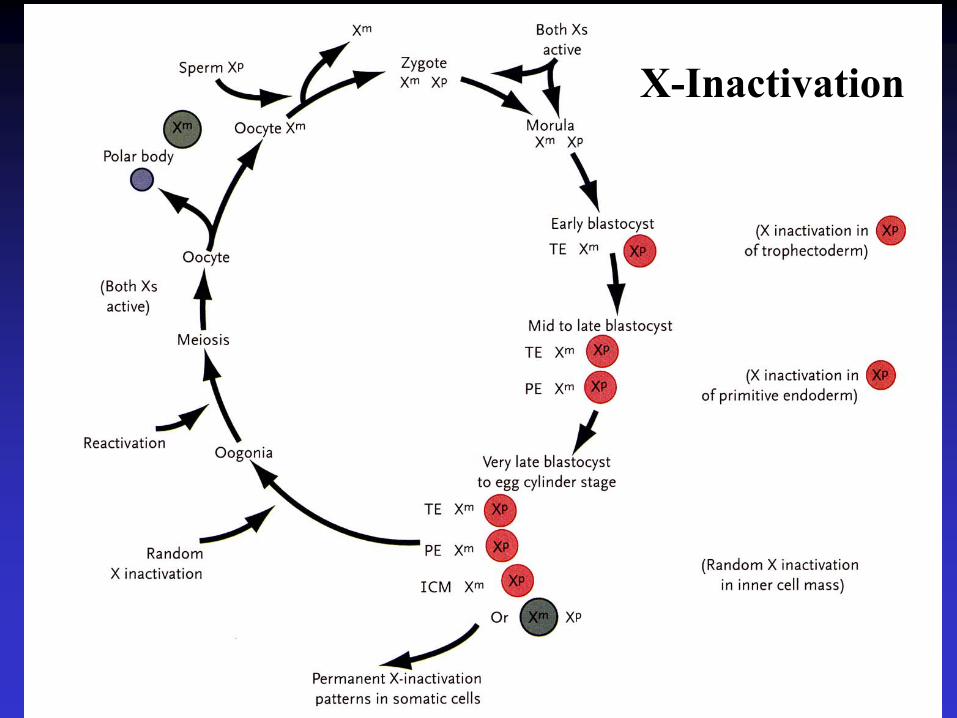
X-Inactivation

Regulative Development
Ability of an embryo or organ to develop normally after removal or addition of parts
Fate of cells is not irreversibly fixed – influenced by environment
Contrast Mosaic Development
Fate Mapping studies
Developmental Potency – Totipotency
Stem Cells

Tetra-Parental Mice
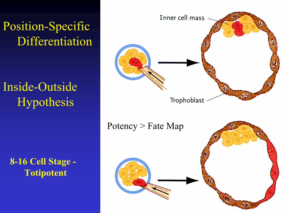
Position-Specific Differentiation
Inside-Outside Hypothesis
Potency > Fate Map
8-16 Cell Stage -Totipotent


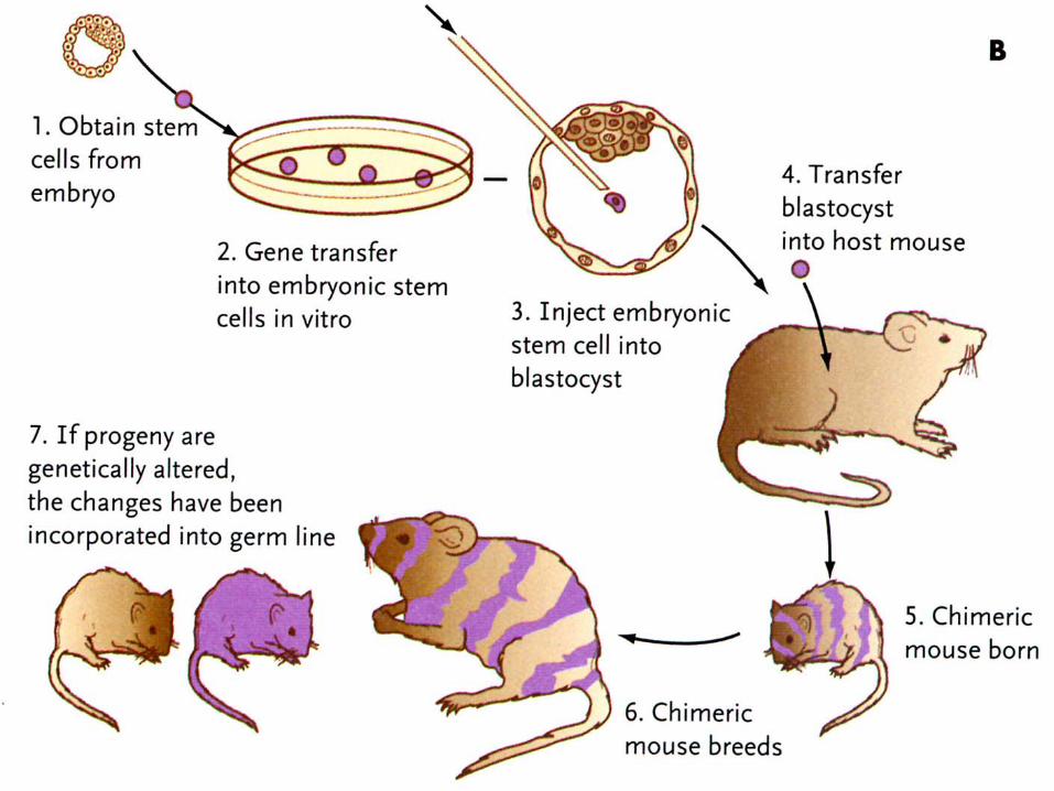
Transgenic Mice

CloningDolly