functions of the skeletal system __________________ (support for body, attachment for soft tissues)...
TRANSCRIPT
BUILDING MATERIAL

Functions of the skeletal system__________________ (support for body,
attachment for soft tissues)Storage of _______________ (calcium and
phosphate) Calcium most abundant mineral in body
(~2–4 lb)98% stored in bones
Blood ____________ production (all formed elements of blood)
Protection (delicate tissues and organs surrounded by bone)
____________________ (act as levers with skeletal muscles to move body)
QUICK REVIEW Six categories based on shape
1. ____________ bones Thin, roughly parallel surfaces Examples: cranial bones, sternum
2. Sutural bones (Wormian bones) Irregular bones formed between cranial
bones Number, size, and shape vary
3. ___________ bones Relatively long and slender Examples: various bones of the limbs
Six categories based on shape (continued)4. Irregular bones
Complex shapes Examples: vertebrae, bones of pelvis, facial
bones5. _______________ bones
Small, flat, and somewhat shaped like sesame seed
Develop in tendons of knee, hands, and feet Individual variation in location and
number6. Short bones
Small and boxy Examples: bones of the wrist (carpals) and
ankles (tarsals)
FUNCTIONS OF BONE Bones are important mineral reservoirs
Mostly _____________________________ but other ions as well
Calcium Most abundant mineral in body1–2 kg (2–4 lb)~__________% deposited in skeletonVariety of physiological functions
Concentration variation greater than 30–35% affects neuron and muscle function
Normal daily fluctuations are <10%
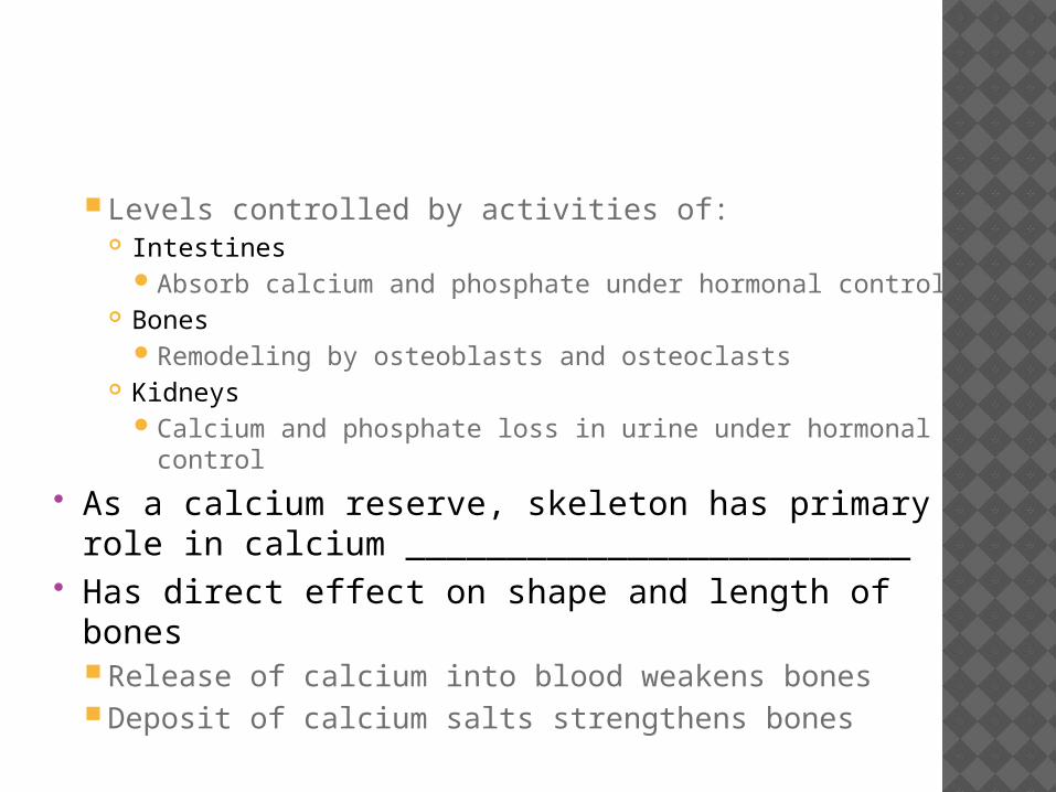
Levels controlled by activities of: Intestines
Absorb calcium and phosphate under hormonal control Bones
Remodeling by osteoblasts and osteoclasts Kidneys
Calcium and phosphate loss in urine under hormonal control
As a calcium reserve, skeleton has primary role in calcium _________________________
Has direct effect on shape and length of bonesRelease of calcium into blood weakens bonesDeposit of calcium salts strengthens bones
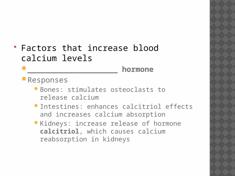
Factors that increase blood calcium levels____________________ hormone Responses
Bones: stimulates osteoclasts to release calcium
Intestines: enhances calcitriol effects and increases calcium absorption
Kidneys: increase release of hormone calcitriol, which causes calcium reabsorption in kidneys
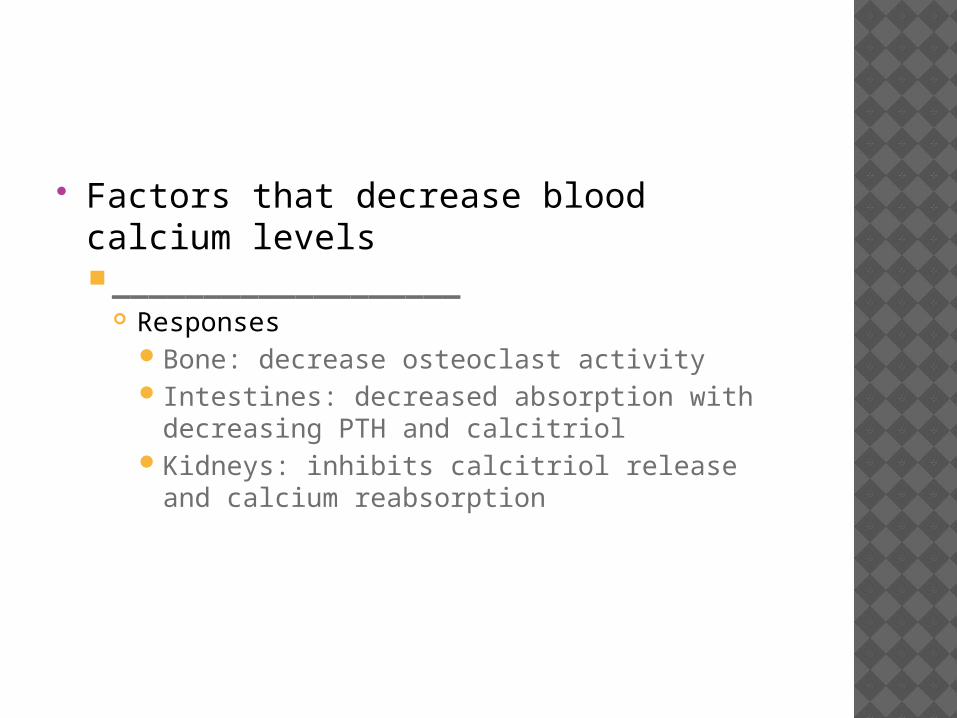
Factors that decrease blood calcium levels___________________
ResponsesBone: decrease osteoclast activity Intestines: decreased absorption with
decreasing PTH and calcitriolKidneys: inhibits calcitriol release and calcium
reabsorption

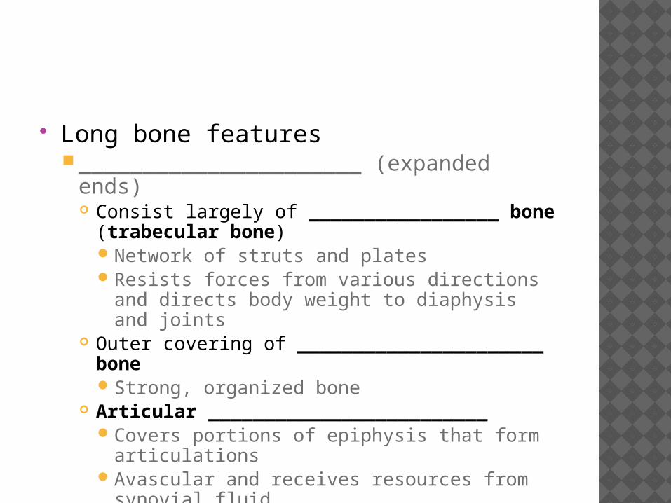
Long bone features______________________ (expanded ends)
Consist largely of _________________ bone (trabecular bone)Network of struts and platesResists forces from various directions and
directs body weight to diaphysis and joints Outer covering of ______________________ bone
Strong, organized bone Articular _________________________
Covers portions of epiphysis that form articulations
Avascular and receives resources from synovial fluid
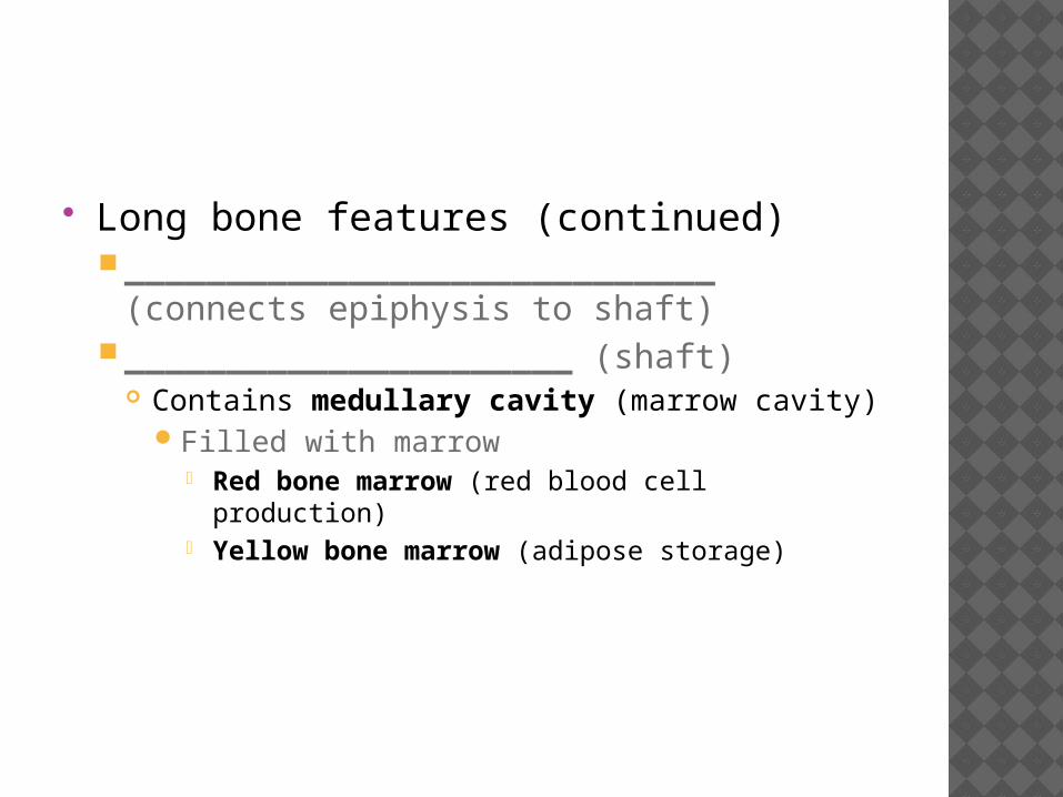
Long bone features (continued)_____________________________ (connects
epiphysis to shaft)______________________ (shaft)
Contains medullary cavity (marrow cavity)Filled with marrow
Red bone marrow (red blood cell production) Yellow bone marrow (adipose storage)
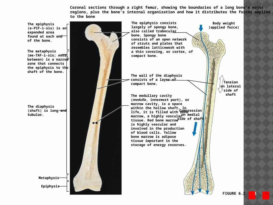
FIGURE 6.2 1 - 2
Coronal sections through a right femur, showing the boundaries of a long bone’s majorregions, plus the bone’s internal organization and how it distributes the forces appliedto the bone
The epiphysis(e-PIF-i-sis) is anexpanded areafound at each endof the bone.
The metaphysis(me-TAF-i-sis; meta,between) is a narrowzone that connectsthe epiphysis to theshaft of the bone.
The diaphysis(shaft) is long andtubular.
The epiphysis consistslargely of spongy bone,also called trabecularbone. Spongy boneconsists of an open networkof struts and plates thatresembles latticework witha thin covering, or cortex, ofcompact bone.
The medullary cavity(medulla, innermost part), ormarrow cavity, is a spacewithin the hollow shaft. Inlife, it is filled with bonemarrow, a highly vasculartissue. Red bone marrowis highly vascular andinvolved in the productionof blood cells. Yellowbone marrow is adiposetissue important in thestorage of energy reserves.
Compressionon medial
side of shaft
Tensionon lateral
side ofshaft
Body weight(applied force)
The wall of the diaphysisconsists of a layer ofcompact bone.
Metaphysis
Epiphysis

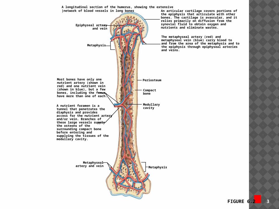
FIGURE 6.2 3
A longitudinal section of the humerus, showing the extensive|network of blood vessels in long bones
Epiphyseal arteryand vein
Metaphysis
An articular cartilage covers portions ofthe epiphysis that articulate with other bones. The cartilage is avascular, and itrelies primarily on diffusion from thesynovial fluid to obtain oxygen andnutrients and eliminate wastes.
The metaphyseal artery (red) andmetaphyseal vein (blue) carry blood toand from the area of the metaphysis and tothe epiphysis through epiphyseal arteriesand veins.
Periosteum
Compactbone
Medullarycavity
Metaphysis
Metaphysealartery and vein
Most bones have only onenutrient artery (shown inred) and one nutrient vein(shown in blue), but a fewbones, including the femur,have more than one of each.
A nutrient foramen is a tunnel that penetrates thediaphysis and providesaccess for the nutrient arteryand/or vein. Branches ofthese large vessels supplythe osteons of thesurrounding compact bonebefore entering andsupplying the tissues of themedullary cavity.
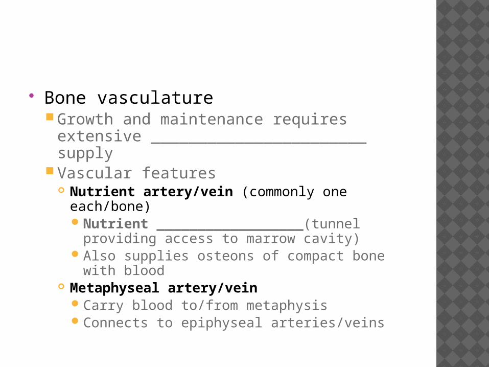
Bone vasculatureGrowth and maintenance requires extensive
_______________________ supplyVascular features
Nutrient artery/vein (commonly one each/bone)Nutrient __________________(tunnel providing
access to marrow cavity)Also supplies osteons of compact bone with
blood Metaphyseal artery/vein
Carry blood to/from metaphysisConnects to epiphyseal arteries/veins
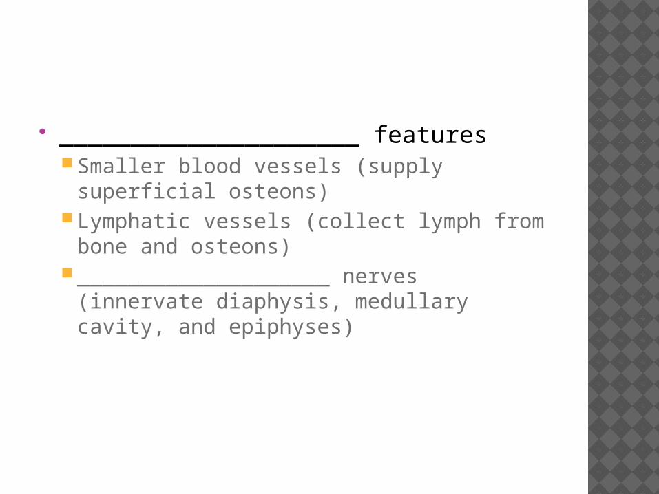
_____________________ featuresSmaller blood vessels (supply superficial
osteons)Lymphatic vessels (collect lymph from bone
and osteons)____________________ nerves (innervate
diaphysis, medullary cavity, and epiphyses)
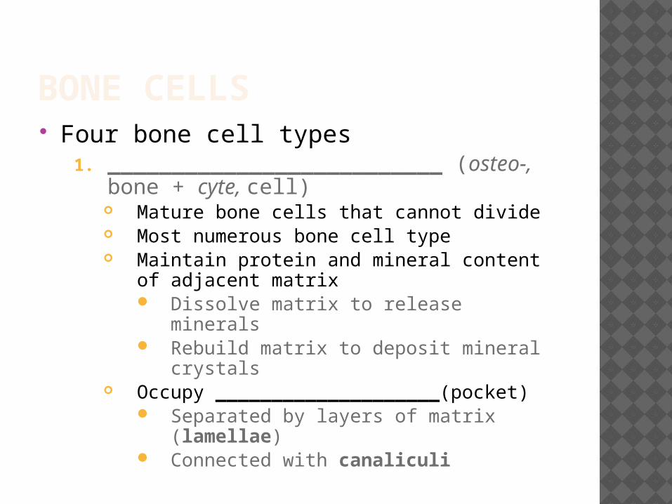
BONE CELLS Four bone cell types
1. __________________________ (osteo-, bone + cyte, cell)
Mature bone cells that cannot divide Most numerous bone cell type Maintain protein and mineral content of
adjacent matrix Dissolve matrix to release minerals Rebuild matrix to deposit mineral
crystals Occupy ____________________(pocket)
Separated by layers of matrix (lamellae)
Connected with canaliculi

2. ______________________ (blast, precursor) Produce new bony matrix (osteogenesis or
ossification) Begins with release of proteins and other
organic components to produce unmineralized matrix (= osteoid)
Then assists in depositing calcium salts to convert osteoid to bone
Become osteocytes once surrounded by bony matrix
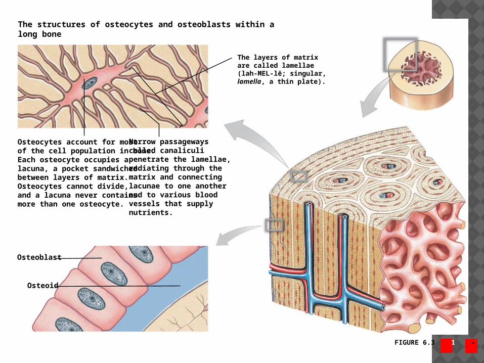
FIGURE 6.3 1 - 2
The layers of matrixare called lamellae(lah-MEL-lē; singular,lamella, a thin plate).
The structures of osteocytes and osteoblasts within along bone
Osteocytes account for mostof the cell population in bone.Each osteocyte occupies alacuna, a pocket sandwichedbetween layers of matrix.Osteocytes cannot divide,and a lacuna never containsmore than one osteocyte.
Narrow passagewayscalled canaliculipenetrate the lamellae,radiating through thematrix and connectinglacunae to one anotherand to various bloodvessels that supplynutrients.
Osteoblast
Osteoid
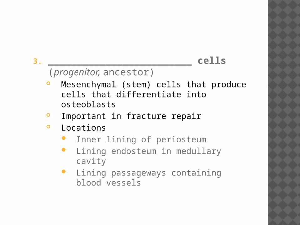
3. _________________________ cells (progenitor, ancestor)
Mesenchymal (stem) cells that produce cells that differentiate into osteoblasts
Important in fracture repair Locations
Inner lining of periosteum Lining endosteum in medullary cavity Lining passageways containing blood
vessels

4. ______________________ (clast, to break) Remove and remodel bone matrix Giant cells with ______________+ nuclei
Derived from same stem cells as macrophages
Release acids and proteolytic enzymes to dissolve matrix and release stored minerals = Osteolysis (lysis, loosening)
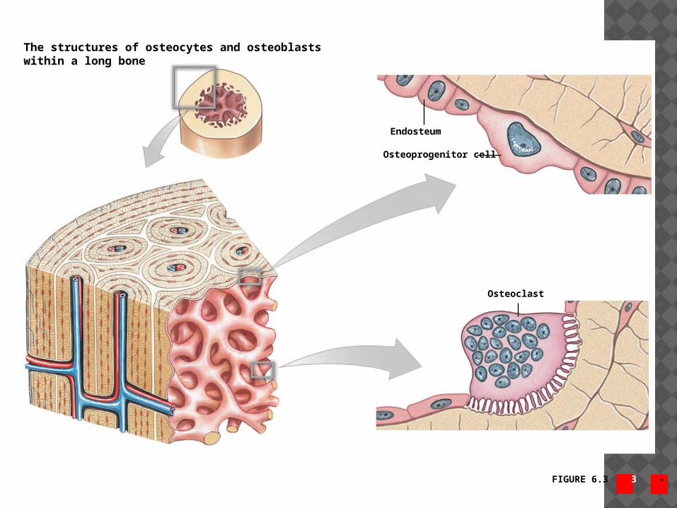
FIGURE 6.3 3 - 4
Endosteum
Osteoprogenitor cell
Osteoclast
The structures of osteocytes and osteoblastswithin a long bone
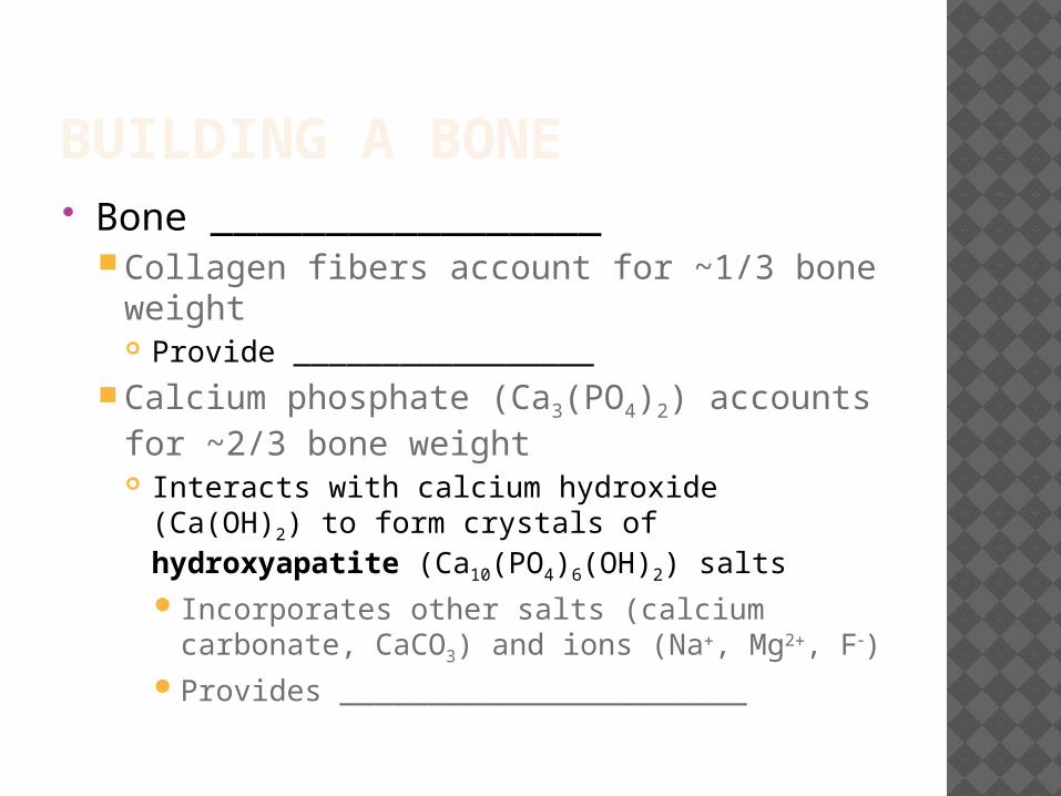
BUILDING A BONE Bone _________________
Collagen fibers account for ~1/3 bone weight Provide _________________
Calcium phosphate (Ca3(PO4)2) accounts for ~2/3 bone weight Interacts with calcium hydroxide (Ca(OH)2) to
form crystals of hydroxyapatite (Ca10(PO4)6(OH)2) salts Incorporates other salts (calcium carbonate,
CaCO3) and ions (Na, Mg2, F)Provides _______________________

_________________ boneFunctional unit is _________________
Organized concentric lamellae around a central canalOsteocytes (in lacunae) lie between lamellaeCentral canal contains small blood vessels
Canaliculi connect lacunae with each other and central canal
Strong along its length
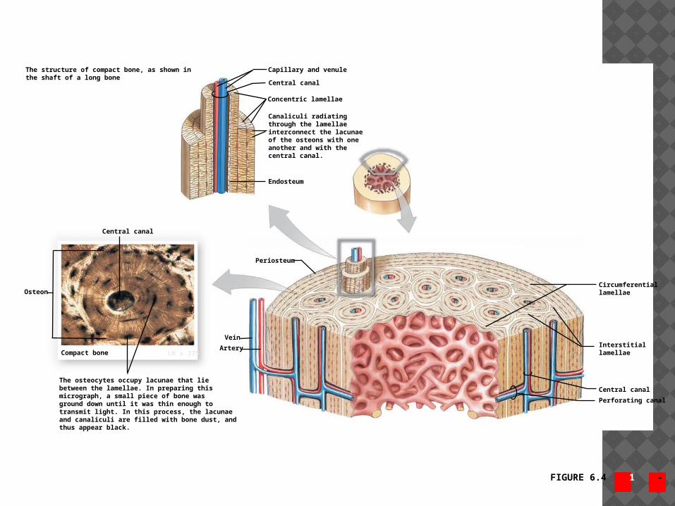
FIGURE 6.4 1 - 2
The structure of compact bone, as shown inthe shaft of a long bone
Capillary and venule
Central canal
Concentric lamellae
Canaliculi radiatingthrough the lamellaeinterconnect the lacunaeof the osteons with oneanother and with thecentral canal.
Endosteum
Periosteum
Central canal
Vein
Artery
Osteon
Compact bone LM x 375
The osteocytes occupy lacunae that liebetween the lamellae. In preparing thismicrograph, a small piece of bone wasground down until it was thin enough totransmit light. In this process, the lacunaeand canaliculi are filled with bone dust, andthus appear black.
Circumferentiallamellae
Interstitiallamellae
Central canal
Perforating canal
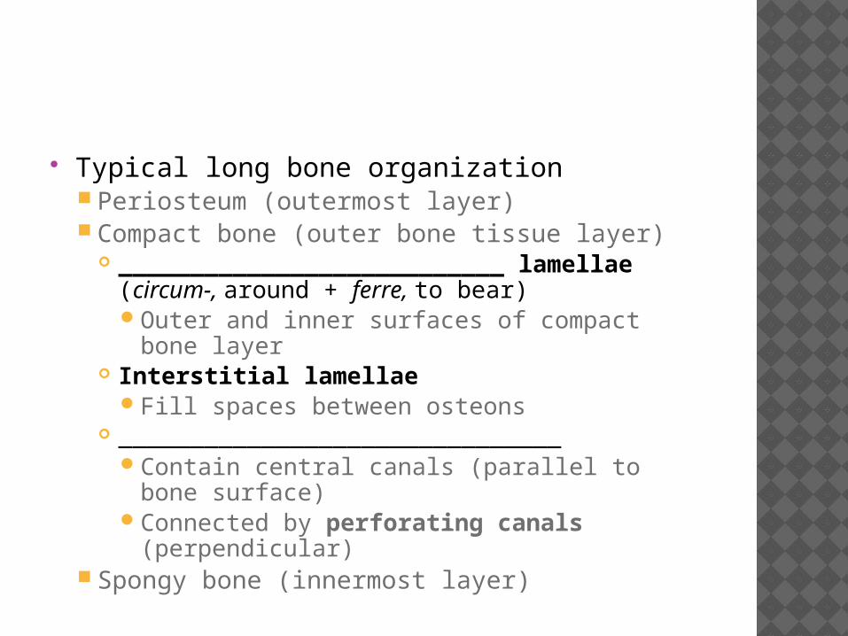
Typical long bone organization Periosteum (outermost layer) Compact bone (outer bone tissue layer)
___________________________ lamellae (circum-, around + ferre, to bear)Outer and inner surfaces of compact bone
layer Interstitial lamellae
Fill spaces between osteons _______________________________
Contain central canals (parallel to bone surface)
Connected by perforating canals (perpendicular)
Spongy bone (innermost layer)
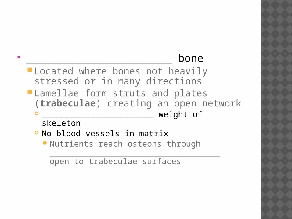
_______________________ boneLocated where bones not heavily stressed
or in many directionsLamellae form struts and plates
(trabeculae) creating an open network _______________________ weight of skeleton No blood vessels in matrix
Nutrients reach osteons through ___________________________________ open to trabeculae surfaces
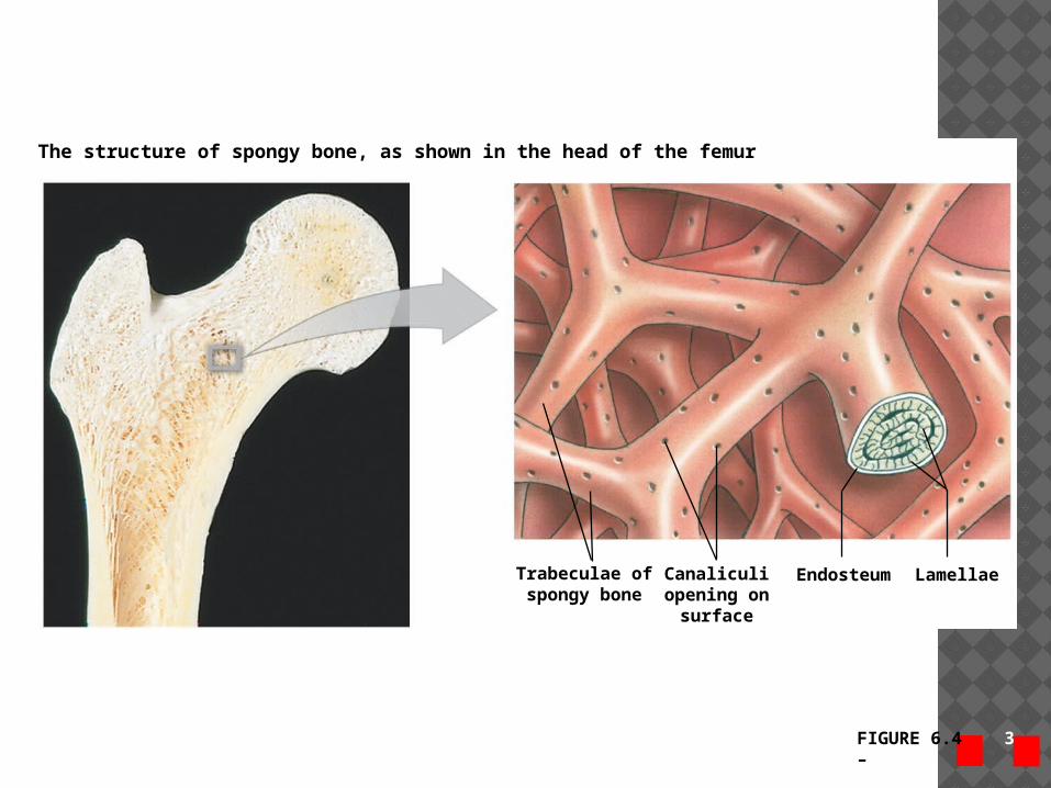
FIGURE 6.4 3 – 4
Trabeculae ofspongy bone
Canaliculiopening on
surface
Endosteum Lamellae
The structure of spongy bone, as shown in the head of the femur
_____________________ bone growth Increases bone diameter of existing bones
Does not form original bonesOsteoprogenitor cells differentiate into
osteoblasts that ____________ bone matrix under periosteum Adds successive _____________________ of
circumferential lamellae Trapped _____________________ become osteocytes
Deeper lamellae recycled and replaced by osteons
Osteoclasts remove matrix at inner surface to enlarge medullary cavity
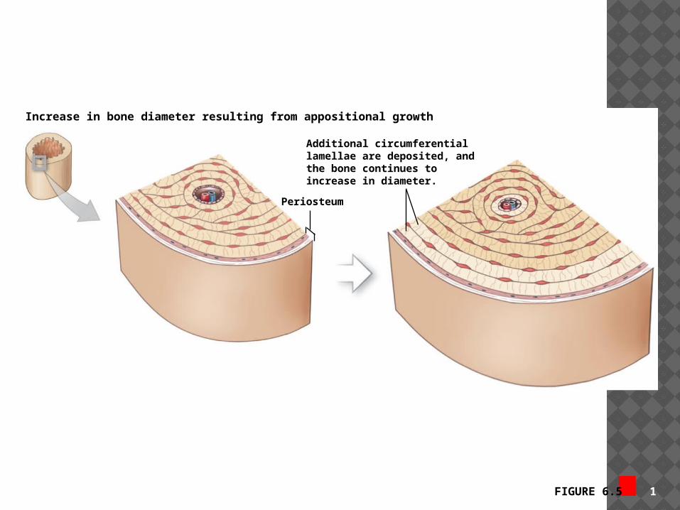
FIGURE 6.5 1
Increase in bone diameter resulting from appositional growth
Additional circumferentiallamellae are deposited, andthe bone continues toincrease in diameter.
Periosteum
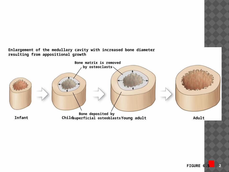
FIGURE 6.5 2
Enlargement of the medullary cavity with increased bone diameterresulting from appositional growth
Bone matrix is removedby osteoclasts
Bone deposited bysuperficial osteoblastsInfant Child Young adult Adult
_________________Two layers
1. _________________________ outer layer2. _____________________________ inner layer
Functions1. ________________ bone from surrounding
tissues2. Route for blood and nervous supply3. Actively participate in bone growth and
_____________________
____________________ fibers Created by osteoblasts in periosteum
cellular layer Strongly connect tendons, ligaments, and
joint capsules to bone through periosteum
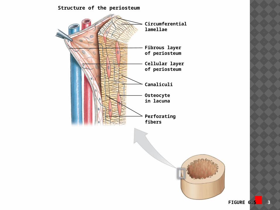
FIGURE 6.5 3
Structure of the periosteum
Circumferentiallamellae
Fibrous layerof periosteum
Cellular layerof periosteum
Canaliculi
Osteocytein lacuna
Perforatingfibers
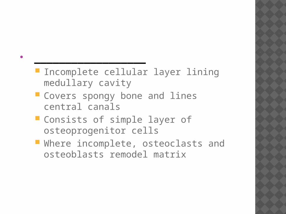
__________________ Incomplete cellular layer lining medullary
cavity Covers spongy bone and lines central
canals Consists of simple layer of
osteoprogenitor cells Where incomplete, osteoclasts and
osteoblasts remodel matrix
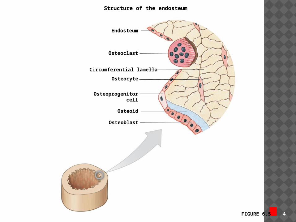
FIGURE 6.5 4
Structure of the endosteum
Endosteum
Osteoclast
Circumferential lamella
Osteocyte
Osteoprogenitorcell
Osteoid
Osteoblast
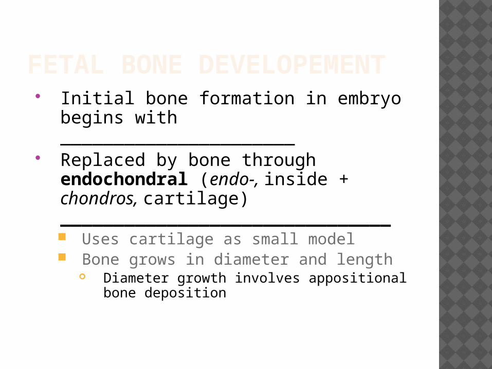
FETAL BONE DEVELOPEMENT Initial bone formation in embryo
begins with ______________________ Replaced by bone through
endochondral (endo-, inside + chondros, cartilage) _______________________________ Uses cartilage as small model Bone grows in diameter and length
Diameter growth involves appositional bone deposition
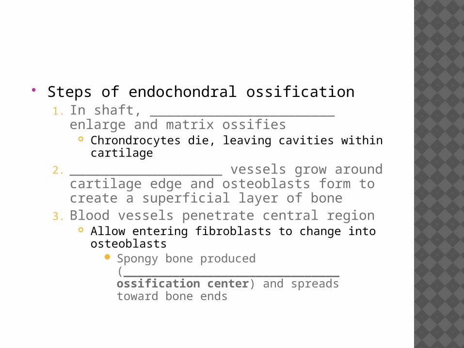
Steps of endochondral ossification1. In shaft, _______________________ enlarge
and matrix ossifies Chrondrocytes die, leaving cavities within
cartilage
2. ___________________ vessels grow around cartilage edge and osteoblasts form to create a superficial layer of bone
3. Blood vessels penetrate central region Allow entering fibroblasts to change into
osteoblasts Spongy bone produced
(_______________________________ ossification center) and spreads toward bone ends
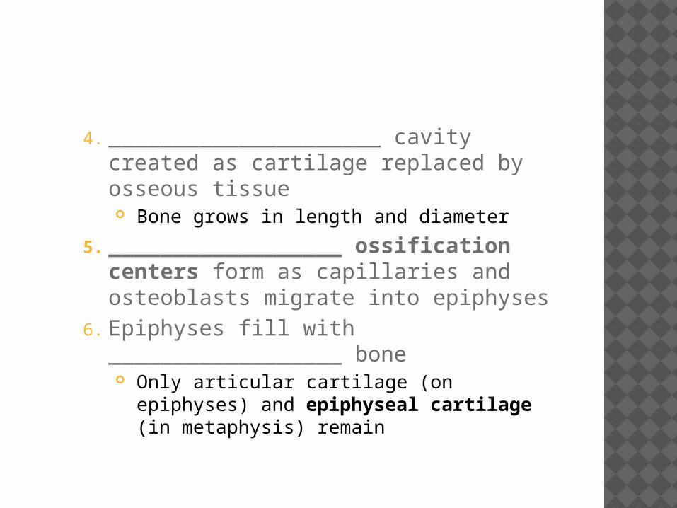
4. _____________________ cavity created as cartilage replaced by osseous tissue Bone grows in length and diameter
5. __________________ ossification centers form as capillaries and osteoblasts migrate into epiphyses
6. Epiphyses fill with __________________ bone Only articular cartilage (on epiphyses) and
epiphyseal cartilage (in metaphysis) remain
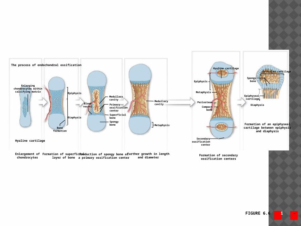
FIGURE 6.6 1 – 6
The process of endochondral ossification
Enlargingchondrocytes within
calcifying matrix
Hyaline cartilage
Epiphysis
Diaphysis
Boneformation
Bloodvessel
Medullarycavity
Primaryossificationcenter
Superficialbone
Spongybone
Medullarycavity
Metaphysis
Enlargement ofchondrocytes
Formation of superficiallayer of bone
Production of spongy bone ata primary ossification center
Further growth in lengthand diameter
Hyaline cartilage
Epiphysis
Metaphysis
Periosteum
Compactbone
Secondaryossification
center
Formation of secondaryossification centers
Formation of an epiphysealcartilage between epiphysis
and diaphysis
Articular cartilage
Spongybone
Epiphysealcartilage
Diaphysis
7. Bone grows in _________________ at epiphyseal cartilage Chondrocytes actively produce more
cartilage on epiphysis side Osteoblasts _____________________ replace
cartilage with bone on shaft side As long as both processes are equally active,
bone lengthening continues At puberty, hormones increase bone
growth and epiphyseal cartilage is replaced
Leaves _____________________________________________________in adults
INTRAMENBRANOUS OSSIFICATION Steps of intramembranous ossification
_____________________________ cells secrete osteoid matrix
Differentiate into osteoblasts Osteoid matrix becomes mineralized
Forms _________________________________ Bone grows out in small struts
(____________________________) Osteoblasts become trapped and
mature into osteocytes Mesenchymal cells produce more osteoblasts
Blood vessels enter and become trapped in developing bone
Further membranous bone development __________________ bone formed initially Remodeling around blood vessels forms
osteons of compact bone Periosteum forms, lined with osteoblasts Begins at approximately ____________ week
of embryonic development Examples:
Roofing bones of skull Lower jaw Collarbone Sesamoid bones such as patella
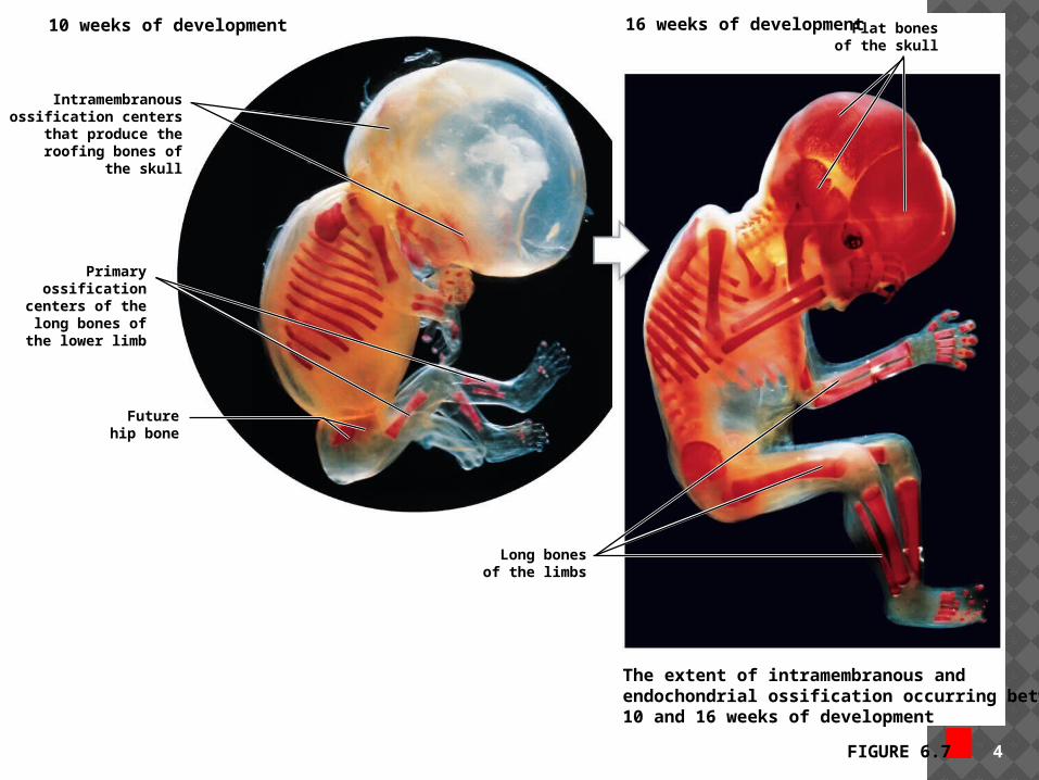
FIGURE 6.7 4
Futurehip bone
Primaryossification
centers of thelong bones of
the lower limb
Intramembranousossification centers
that produce theroofing bones of
the skull
Long bonesof the limbs
10 weeks of development Flat bonesof the skull
16 weeks of development
The extent of intramembranous andendochondrial ossification occurring between10 and 16 weeks of development
ABNORMAL BONE GROWTH Endocrine and metabolic problems can
affect the skeletal system
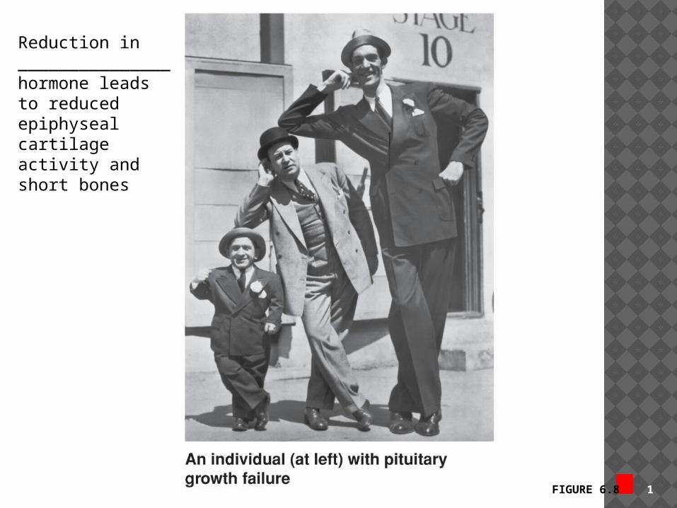
FIGURE 6.8 1
Reduction in _______________ hormone leads to reduced epiphyseal cartilage activity and short bones
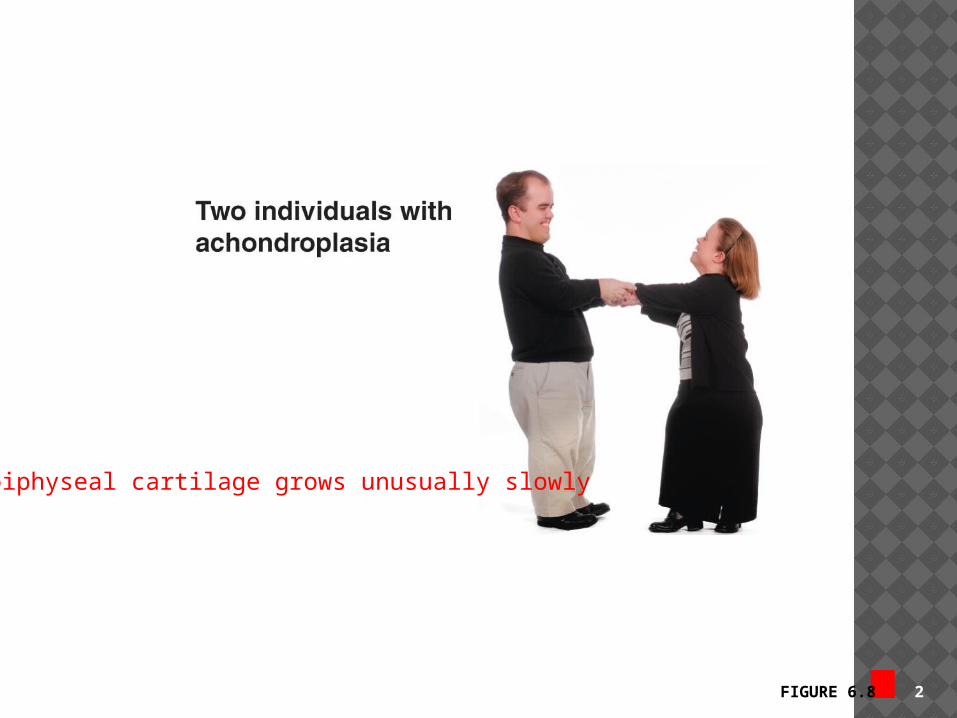
FIGURE 6.8 2
Epiphyseal cartilage grows unusually slowly
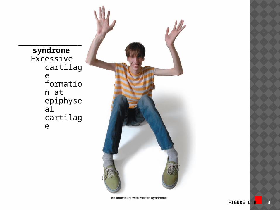
FIGURE 6.8 3
_______________ syndromeExcessive
cartilage formation at epiphyseal cartilage
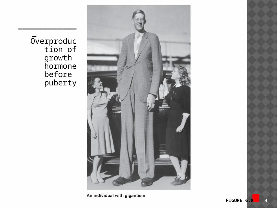
FIGURE 6.8 4
_______________Overproduct
ion of growth hormone before puberty
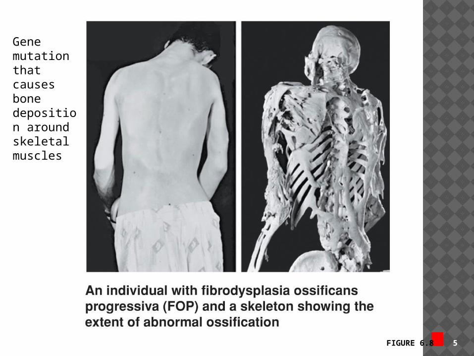
FIGURE 6.8 5
Gene mutation that causes bone deposition around skeletal muscles
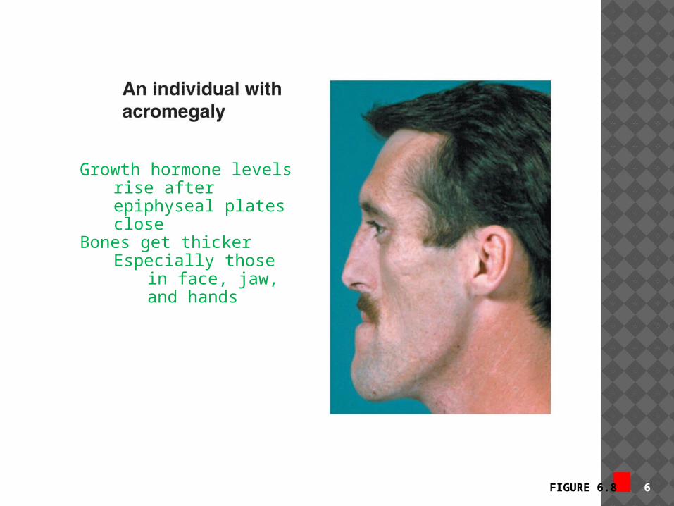
FIGURE 6.8 6
Growth hormone levels rise after epiphyseal plates close
Bones get thickerEspecially those in
face, jaw, and hands


FOR WEDNESDAY Joint
Cht 8 Movement
8.3-8.4 Labs
Pages 19-27